 | ||
| | | |
 |
 |
 |
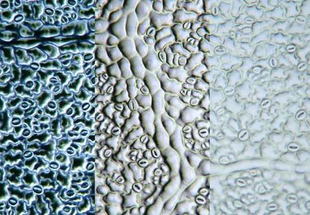
| This image of the surface of a leaf shows the differences in contrast between these types of illumination. Bright-field illumination has very limited contrast. This image clearly shows that it is very useful to experiment with contrast techniques. Oblique illumination gives a relief-like enhancement of contrast. In 19th century microscopes there was often an arrangement for oblique illumination. Early microscopists knew that this technique has great advantages! Dark-field illumination is also one of the most rewarding techniques. Objects smaller than the resolving power of the objective can also made visible simply because they light up! It is thought that Antony van Leeuwenhoek observed bacteria using a kind of dark-field illumination! The image on this page is of a cast of the surface structure of a leaf complete with nerves and stomata. With the aid of nail varnish it is easy to make perfect casts. Read more about this technique next month! 
Rheinberg illumination back to main article
| ||