The eyepiece reticle is widely used for measuring subjects on a microscope slide, (after first calibrating it with a stage micrometer). However, since the reticle is a finely divided scale or grid and often of a standard length (e.g. 1 cm) as well as flatfield, such a well defined subject can also be very useful as a subject on the stage.Eyepiece reticles aren't too expensive new and much cheaper than the stage micrometers, stage finders etc. However, if on a budget, a cheap source of eyepiece reticles can be the junk sales, or scientific surplus stores. Look out for the 'no name', damaged or other 'unwanted' eyepieces as these are often sold off for a few dollars. By holding them up to the light they can be checked to see if there's a useful reticle inadvertently being sold with it. I've obtained both my eyepiece reticles by this route. In my case I did want the eyepieces, so the reticles were a bonus!
(A note on reticle removal from the eyepiece. The reticle usually sits on the field stop within the eyepiece. The top lens mount of most eyepieces are designed to be removed to insert reticles, so should readily unscrew without releasing a pile of lenses! Gently tipping the opened eyepiece onto a lint free cloth will remove the reticle. But if in any doubt, it's best not to risk damaging a good eyepiece.
To use a reticle on the stage, place it carefully on a microscope slide. If used with a 40x objective, the scale engraving will probably need to face upwards as the objective may not be able to focus through the glass thickness. The reticle will need careful cleaning if returning it to the eyepiece as the scale is focused in the image field).
Having found some unwanted reticles, here's a few uses they can be put to in case of interest.
1) On-stage 'ruler'
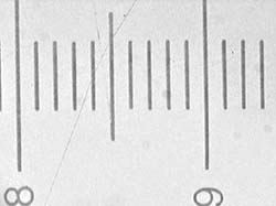
As remarked above, proper stage micrometers can be expensive. So if you have an eyepiece reticle, it's worth checking to see if has been engraved to a whole unit of length, they often are. Parts of the two reticles the author possesses are shown below. The linear scale is exactly 1 cm long (check with a good rule or with the vernier scale of the stage), with 100 sub-divisions i.e. of 0.1 mm. The square grid is exactly 1 cm square with 0.2 mm smaller squares.The eyepiece reticle won't match a proper stage micrometer for accuracy but can be fine for less rigorous measurements at low to medium powers.
The methods for using an accurate stage rule for subject measurement and field of view calculations (both visual and projected images) are described in Walter Dioni's excellent overview of size measurement in the April Micscape issue. The author also links to other contributor's Micscape articles which discuss measurement methods where a reticle could be used.
Field of view ca. 1.5 mm for a 3.5x objective when the image is projected onto a security camera CCD sensor.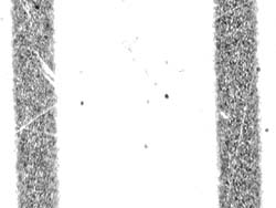
Even with a 40x objective, 0.1 mm rulings are just sufficient for estimating the field of view i.e. ca. 0.13 mm.A standard ruled eyepiece reticle if of known unit length gives a fine enough ruling for field of view measurements from low to medium power objectives. 2) Checking projected image quality
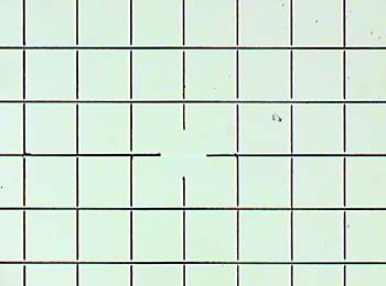
When setting up a microscope for image projection e.g. into a camera (either film or digital), for video or onto paper for drawing by camera lucida, it's very important that the field is flat, evenly lit and free from aberrations; in particular those that relay lenses or eyepieces may introduce if not well matched. (See Ted Clarke's recent article which illustrates the importance of matching optics for image projection).The grid eyepiece reticle is a particularly useful slide subject for checking for major faults in the projected image as the eye is good at spotting even slight geometrical distortions when compared against the rectangular border of the image frame.
The contrasty black lines against a bright background may also pickup any marked chromatic aberration. The essentially light background is also good for checking even illumination or hot spots in the optics train when a subject (the scale) is in focus.
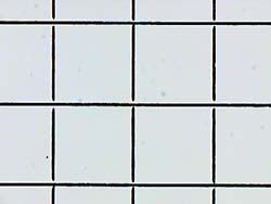
A 0.2 mm square grid reticle imaged using a Russian 3.5x planachromatic objective. Full area image capture (resized from 800x600 master).
A section from the master image capture. There's slight but acceptable aberration.Image quality check (for flatfield, distortion and even illumination) when image projected onto a security camera sensor with no microscope eyepiece (the author's current image capture route).
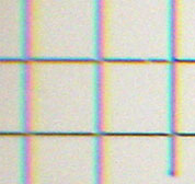
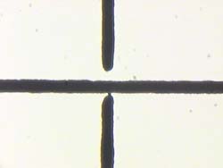
Very severe aberration is readily shown up in an image crop further out from the field centre captured left.Image quality check for the same 3.5x plan objective used above, but now used with a consumer fixed lens digital camera and relay lens which the author is currently assessing. Although the same central area crop, the poor matching of the optics is clearly shown by using a reticle as a subject. Using prepared slides of e.g. stained sections may not show such aberrations as clearly.
Russian 8x non-plan achromatic objective.
Russian 9x plan achromatic objective.Flatfield comparison between non-plan and plan objective for an image projected directly onto the CCD sensor without a microscope eyepiece. A CCD sensor often captures only the centre portion of a visual image so a non-plan objective as here can often be quite sufficient. The differences are more noticeable for the visual image.
4) Assessing parfocality and parcentrality of objectives
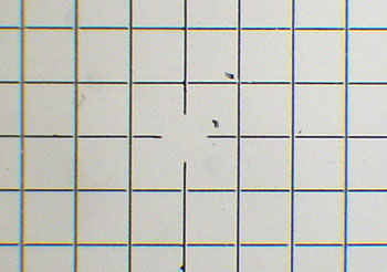
Depending on a microscope's quality and how well matched the optics are, the parfocality and parcentrality of objectives can be less than perfect, and it's unlikely to be adjustable on a budget microscope. A gridded reticle can be useful for noting if and to what extent the focus and centration changes across the mag. range. Any suitable prepared slide with well defined features can of course be used to assess this, but reticles are as practical as any.For example, I know that when changing up from the 20x to 40x objective on my Russian Biolam with pre-DIN objectives, the image moves to the left of field centre, (see images below). So I know which way to compensate with the mechanical stage if following a moving subject across the field of view when moving up and down the mags. I've also noted which way the focus changes by using the graduated fine focus knob, as tiny critters can swim up and down in the water film and be lost when changing mags even for perfect parfocal and parcentral optics.
Russian 40x NA 0.65 objective.
Full image capture of centred reticle.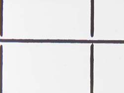
Russian 20x NA 0.40 objective.
Full image capture. The centred reticle at 40x has moved to right of centre at 20x.When checking for parcentrality, the author prefers to centre on the highest objective mag and work down, to minimise errors centring by eye at lower mags. With a reticle with repeating features, pick a feature that can be uniquely defined (e.g. in this case the small join at cross section), otherwise confusion can occur when identifying the centred feature at a lower mag. Contribution by Dave Walker, comments welcomed.