These are a further selection of old slides that I've been enjoying recently and which I hope may be of interest.
I chose two of the four slides because they have quite a considerable 'depth' and have proved difficult to photograph. So I have used these for my first try with the popular CombineZ3 freeware program written by Alan Hadley to create photomicrographs with improved depth of field. Other microscopists have found the program useful e.g. see Walter Dioni's current series of articles on mountants.
All microscope images were taken
with a 'C' mount security camera body using a 'Snappy' box to capture stills
with my Russian Biolam microscope/objectives without eyepiece.
|
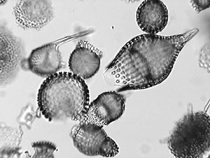
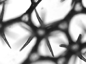
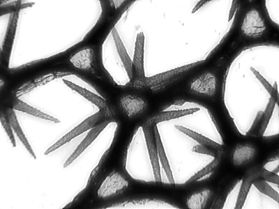
| This is one of three slides by this
mounter which I possess (a recent eBay acquisition). The other two in the
same slide style are of radiolaria from Haiti and the Bahamas. I haven't
found any references to this mounter (French?) and would be interested
to hear from readers who have any information on him.
The cleaned strew of radiolaria are beautifully presented; not too dense and very little debris apart from other interesting silica items such as sponge spicules. I'm not sure what the mountant was, but its uniformly yellow on all three slides rather than oxidising from the edges so it may be a high RI mountant of this colour. I enjoy viewing these by focussing through with a 10x objective but the depth of field in one plane is very small for photography. The left image above shows a single focus image capture (Russian 10x/NA0.3 objective). The image right was created in CombineZ3 from 12 frames taken by focussing through the slide at six division intervals (= 12 micron) on the graduated fine focus of my Russian Biolam. I was impressed with the resultant 'first pass, no tweaks' image. I used the 'Average' routine with default settings in preference to the standard routine with a little post image sharpening. The program offers plenty of parameter tweaks to improve images if desired. Incidentally the video still capture method is particularly suited to creating images for CombineZ as the video captures are saved by default in the required bmp format. The high quality video out allows accurate focus monitoring and control on a TV monitor (I use a PC monitor with a TV card). Focusing may be easier with this setup than with some digicams which can be difficult to focus accurately in required plane with a microscope. |

|
| This is an attractive papered slide
by an unnamed mounter. The T/S of lily has been mounted with a black fill-in
of some sort. It shows the interesting stellate clusters of needle-shaped
calcium oxalate crystals (raphides) that grow within the lily. A Google
search brought up a wide variety of interesting links to e.g. research
projects studying the various roles of calcium oxalate in plants; a deterrent
for herbivores is one role of raphides discussed. Yellow water lily grows
in my garden pond so it will be interesting to section some fresh material
when it's in full growth in summer to see if they contain raphides.
With a 10x objective this requires focussing through 300 microns to view the clusters, (1½ full turns of the fine focus knob on my Biolam), so I was interested to see how CombineZ would cope. The limited areas in focus in one plane is shown in the image left above. The image right was created from 15 frames taken at 20 micron intervals (10x/NA0.3 objective), again created with the default 'Average' routine in CombineZ3. This is an early imaging attempt but was again impressed at how it creates a good 'first pass' image without undue artefacts. Tweaking the parameters may improve the detail in the raphides and reduce the slight artefacts. |
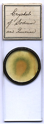 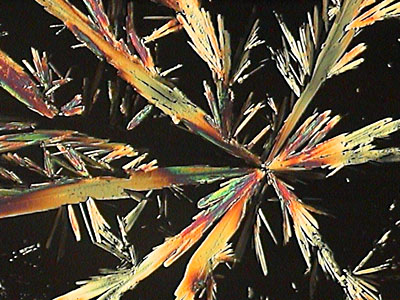 |
| This is by an unnamed mounter but
is a generous mount of these crystals to enjoy under crossed polars. I'm
not entirely sure what was meant by 'sodium and quinine', as I couldn't
find any references to sodium derivatives; quinine sulphate is one of its
commoner salts.
The right-hand image is part of the mount taken with the Russian 3.5x planachromatic objective with crossed polars. An interesting summary of quinine (structure, history, natural occurrence and uses) can be found on the University of Oxford's (Dept. of Chemistry) web site in their 'Molecule of the month series'. |
|
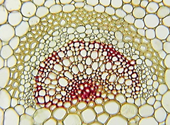
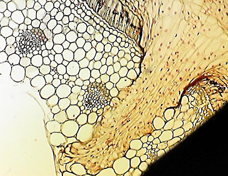
| This is a slide by the well known
mounter W H Darlaston. The section is unfortunately at the top right edge
of the mount and partly obscured by the ringing, I'm not sure if this slipped
through 'quality control' or can sections move over the years?
The stained section neatly shows the ingress of the parasitic plant (dodder) into the host bean plant. (Image top right above, 3.5x objective). The dodder is the yellow denser tissue growing into the bean from the top right of image. The lower image shows part of the conducting tissues of the host bean in another part of the section, (taken with the Russian 10x/NA0.3 objective). Dodder (Cuscuta epithymum) occurs wild in the UK but is not that common where I live. I've only seen it in Devon growing on gorse. It's easy to overlook when not in flower as it has thin pink stems with tiny scale like leaves and climbs over the larger host. It has attractive clusters of pale pink flowers. |