EXTREME ANNULAR ILLUMINATION
Frithjof A.S. Sterrenburg, Netherlands
Email: fass AT wxs DOT nl
Browsing through some issues of Micscape, I found the article by Walter Dioni (Nov. 2003) on the revered onion pellicle with several fine images including one with what he calls critical circular oblique illumination after Sterrenburg. I also found a note by one of the Walker brothers asking for references to the oblique illumination affair, plus a description of Osamu Okus experiments with oblique illumination and polarized light for visualizing fine diatom structure, about which I had contacted that author some time ago. Here are some notes on these subjects.
Polarized light
As for polarized light and diatoms, there is a clear reference to it on page 143 of Beck (1938). I began using this technique in the Sixties and can well remember a rather heated discussion with a contemporary authority who rashly stated it would not work because the diatom frustule is not made of a birefringent material I referred to it in Sterrenburg 1975 and 1978 and youll also find it mentioned in Chapter 8 of the Microscopy Primer on this website.
Note that even a single polar, inserted into the filter ring of the condenser, will also markedly enhance the visibility of fine diatom structure. There is an azimuth effect, slowly rotate the polar or the stage while examining the image for the best result.
Pseudo-phase contrast
Of course, oblique illumination has been used for a very long time; from the letters of Anthony van Leeuwenhoek its clear that he must have observed the phenomenon. After all, with his technique of observation, it was almost unavoidable that he sometimes aimed his contraption slightly off-centre to the light source and as he was a keen observer, he will have noticed the effect immediately. Annular (a better name than circular) oblique illumination was used by the Victorians, who even appear to have had a sort of inside-out iris diaphragm although Im not sure that was indeed on sale then.
Regarding the extreme form of annular oblique illumination, where only the marginal zone of the objective is filled with light, Victorian microscopists already accidentally stumbled upon it when they forgot to stop down an oil-immersion objective while using a wet darkfield condenser. However, I do not think that an explanation of the effect (a form of zero-absorption phase-contrast caused by phase shift in the wave-front at the periphery of the objective) was presented prior to Sterrenburg 1975, where I showed a photomicrograph illustrating the effect, also shown in Fig. 51 of the Microscopy Primer on this website) and Sterrenburg 1978. Actually, my choice of the term pseudo-phase contrast at that time was not a happy one; true PC is obtained, although the phase shift is not precisely defined and there is no absorption. Certain diatomists then were wary of my optical trickery, claiming that artefacts were lurking around the corner, not realizing that this would equally apply to phase-contrast. The only artefact, as with PC, is a slight halo-effect.
Hungarian Rhapsody
A 3D-condenser was described by Zselionka et al. (1958). In exciting English, this paper discusses a plastic supercondenser (sic), which is claimed to raise the performance of achromatic objectives to that of apochromats, to permit 2 or 3 times higher magnification (?) than ordinary condensers, to render cardioid darkfield condensers superfluous for dry and even for immersion objectives and in general, to cast doubt on the reliability of all phase-contrast studies carried out at that time The description of its principle is cryptic rather than informative, but it was actually produced by a Hungarian manufacturer (whose name I cannot supply), as I examined such a condenser in the Eighties. Annular illumination was obtained by grinding down the internal (convex) surface of the top lens and painting the flat central portion black. There were 3 interchangeable top lenses. Unfortunately, there was no Hungarian manual so I may have missed something. In particular, it sported a lightly silvered glass disc whose effect I was then unable to observe. Of course it yielded annular illumination or darkfield with low power objectives, but from my notebook I conclude that I found it neither plastic, nor a supercondenser.
An example
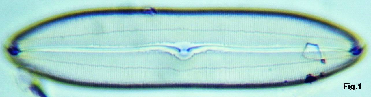
To illustrate the full pseudo-PC effect, here is an example taken with the old LOMO 40x water- immersion, optically identical to the pre-war Zeiss D*. Ive seen a remark somewhere on Micscape that this objective is not much better than a dry 40/0.65, because its NA is only slightly higher (0.75). If you examine this objective, youll see that this is not true, the image in normal brightfield and darkfield is far superior to that of a 40/0.65, tack-sharp and with much higher resolution. The reason: amazingly, LOMO understated the NA, which I once measured as near 0.85! In addition, water immersions are much less sensitive to coverglass thickness, of course.
The images are of a delicate and finely structured Caloneis species and were not digitally processed except for cropping. In brightfield (Fig. 1), the striae are resolved, contrast is rather low at fully illuminated aperture; with peripheral annular illumination (Fig. 2) contrast is greatly enhanced and some striae are partially resolved into dots; with a true phase-contrast objective of high quality (Leitz 40/0.75 apo PV, Fig. 3) contrast is very high but resolution is inferior as its NA is lower.
LOMO 40x NA0.85 water immersion objective with strictly central brightfield.
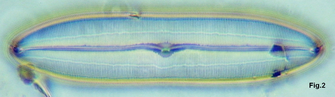
LOMO 40x NA0.85 water immersion objective with peripheral annular illumination.
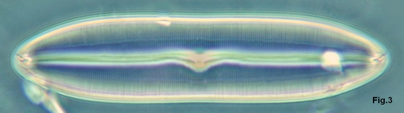
Leitz 40x NA0.75 apo ob jective with phase contrast.
Comments to the author, Frithjof Sterrenburg, are welcomed.
References
Beck, Conrad (1938), 'The microscope, theory and practice.' R. & J. Beck Ltd., London.
Sterrenburg, F.A.S. (1975). 'Leidraad bij de microscopie.' Kluwer, Deventer.
Sterrenburg, F.A.S. (1978). 'Enhancing the visibility of diatoms.' Microscopy 33, July-December 1978.
L. Zselionka, F. Kiss and I. Barabas. 'A plastic supercondenser.' Bulletin of Experimental Biology and Medicine, 46/3, Sept. 1958.
Microscopy UK FrontPage
Micscape
Magazine
Article
Library
Published in the April 2010 edition of Micscape Magazine.
Please report any Web problems or offer general comments to the Micscape Editor .
Micscape is the on-line monthly magazine of the Microscopy UK website at Microscopy-UK .
© Onview.net Ltd, Microscopy-UK, and all contributors 1995 onwards. All rights reserved. Main site is at www.microscopy-uk.org.uk .Published in the March 2010 edition of Micscape Magazine.