|
|
(An
article in next month's Micscape will discuss the use of the filters,
stops used etc. described below.)
|
|
|
(An
article in next month's Micscape will discuss the use of the filters,
stops used etc. described below.)
It was a time when all year round was spring, because spring
was not a state of the weather, but of the heart. It was the time when
hours of wonder were trapped in the yellowish sheets of the biology books at the town hall
library. | |
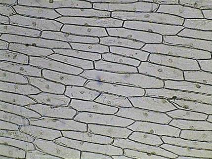
| In these books resided the onion cells, with their hexagonal architecture and round dotted nuclei, and those of Elodea canadensis, with the small circles which imitated the chloroplasts, and even more: there it was the drawing of the rectangular cell of the staminal hair of Tradescantia (Which were the staminal hairs? What was Tradescantia?) with the broad shaded outline of the cytoplasm bridges and the small arrows showing the direction of the current of the cyclosis. | |
| To see the cyclosis moving slowly and constantly in the fine transparent lines of the cytoplasm through the cell of Tradescantia it required almost 20 more years in the life of the young person that I was. | |
| Tradescantia was an exotic plant which I only knew much later. | |
| The drawings had the crudeness of a broad outline in black ink, and the simplicity of the diagrammatic image... but it also had the fairy-like suggestion of the nature they imitated. Moreover when I skim over some ten pages more, a plate equally rough and flat summarized the artists concept of the life in a water drop. And these images caused my heart to accelerate its emotive beats. | |
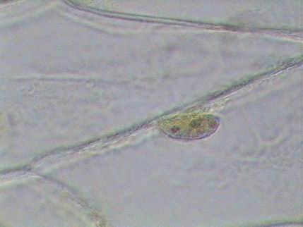
| There it was the chalice of Vorticella, with its rolled up stalk and well adhered to its base on Spirogyra. Paramecium, Colpoda cucullus, and the dazzling Actinophrys sol, all of them locked up by the circular black border imitating the microscopes field of view. | |
| All the year was spring, and the days were of dreams which always centered around the toy microscope in the nearby store. | |
| Meanwhile the hand magnifying glass allowed me to approach my dream; spiders were preserved in small vials with alcohol, butterflies wings were spread on the mounting board, and the flowers of the 'blue bells' (Ipomea cairica) which covered the fence in the front of my house displayed their sexual secrecies under the relentless Gillette blade and the sewing needles of my grandmother, fixed in improvised wooden handles. | |
| It was therefore unforgettable that day of July when my mother put in my hands the small case of dark cardboard. Out of it my hands supported the 20 cm high wonder. The arm and the stage articulate with the foot where the small mirror sparkled. Black enamel contrasted with the brilliant and suggestive chromium of the tube to which the two small and mysterious lenses were attached. | |
| That was the day of the onion skin. | |
| I discovered the slide and the coverslip, the wet mount and the iodine tincture staining, which I hastily stole from the family medicine-chest. | |
| Finally my left eye discovered, through the 120 powers, the true aspect of the vegetable cell, its tender color, and its depth, the aspect of the cytoplasm, the central vacuole, and the shapes and multiple positions of the nuclei. Oh, wonder! The iodine also showed that the nucleoli really existed. | |
| I never forgot nor forsook the humble onion cell. It always was my rule to measure many of my techniques of working. | |
| Today, sixty years on, I want to offer to the
onion cell the homage which I believe
that it deserved, by showing "Cinderella" in its new evening dresses. | |
(COL - circular oblique illumination) | |
| Today, so much like yesterday, I use an amateur's microscope, although it is no longer 20 cm high. | |
| What has changed is my capacity for seeing many small details and to record what I see. Not my feeling of the miracle when I see these invisible things. | |
| And what has also changed through time is the manner in which the men of science investigate the cell, including the new microscopes, which only reach some fortunate amateur's hands. | |
| Here is a selection of images taken with an amateur's microscope using some of the various methods developed over the years by other amateurs. | |
| And as a splendid corollary, we can show the results produced by three professional methods, two of which are brought to us by the kindness of two very advanced amateur friends. | |
| It seems very important to me to make clear (as many of you have surely discovered) that the visual images observed with the techniques I use here and discuss in a companion article, are much more intense, and give a feeling of depth and color which are not faithfully registered by the recorded images. | |
| Rheinberg filters for example give in my microscope many nuances and contrasts which my camera is unable to record. | |
| Except for the field diaphragms, used in bright field over the front lens of the lamp to obtain the critical illumination with each objective, all the other filters, diaphragms, and stops (obturators) are placed in the condensers filter tray. | |
| Two of my examples were captured in bright field and electronically processed and after-stained with PhotoPaint. |
|
|
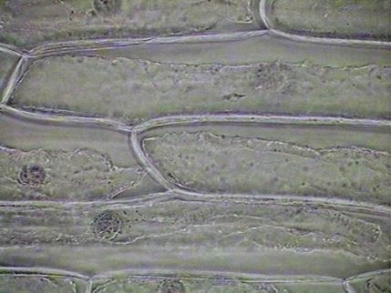
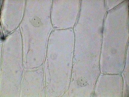
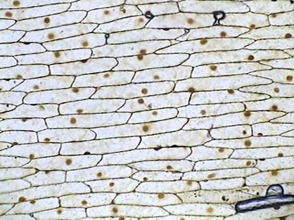
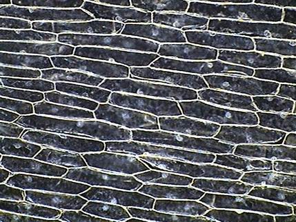
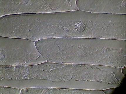
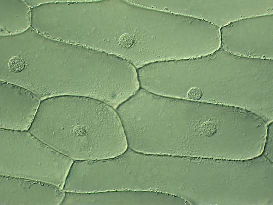
| x10 objective - critical illumination, live, mounted in water, without filters or stains |
As left, x40 objective. |
|
|
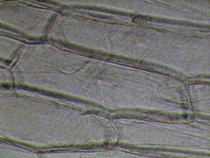
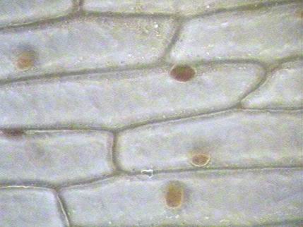
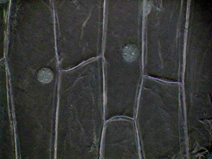
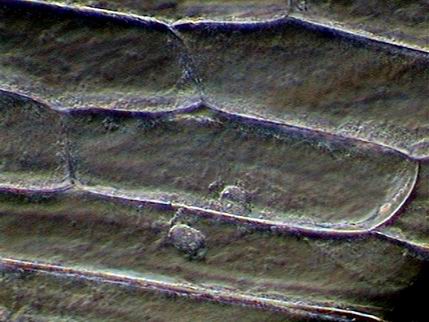
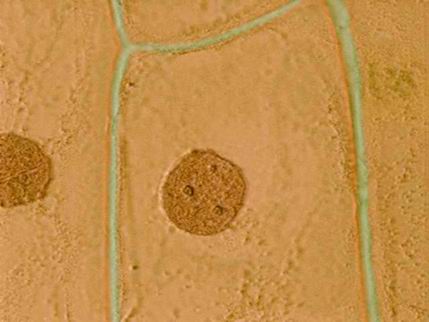
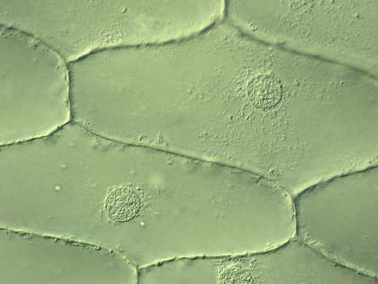
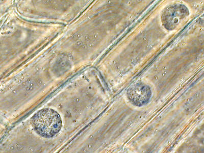
| x40
- live, the surface of the living cell, critical illumination |
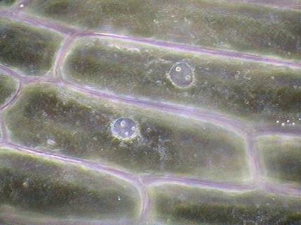
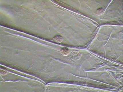
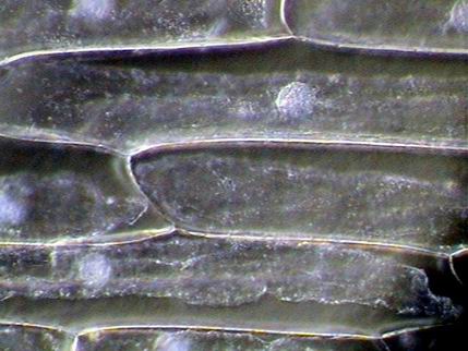
x 40
- live, the nuclei seem to float in the cytoplasm. COL-D2 diaphragm |
|
|
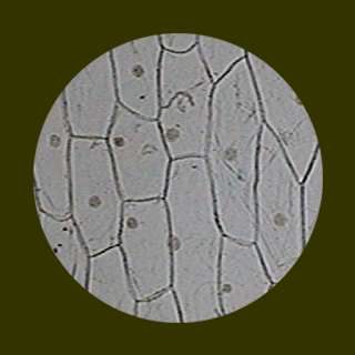
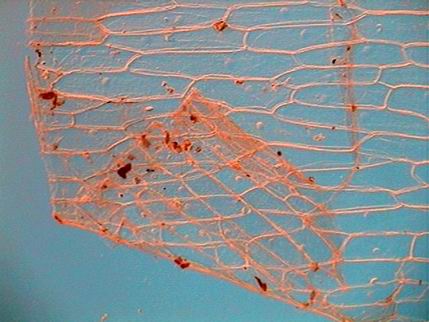
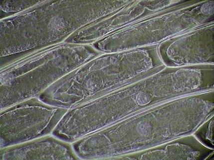
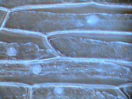
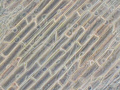
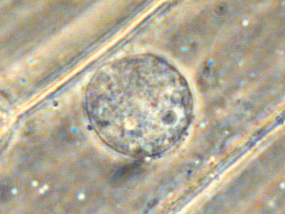
| x10 -
onion skin thin layer - Rheinberg: blue 12.5 mm center, red exterior ring |
10x objective
- stained with Iodine, critical illumination
|
|
|
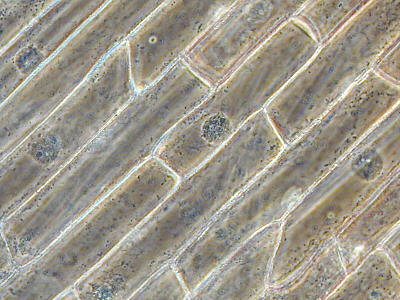
| x40 - Iodine tincture- COL-J diaphragm |
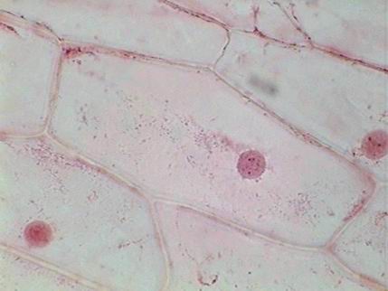
40x
- Fixed with AFA. Mounted in glycerin. Stained with Allura Red |
|
|
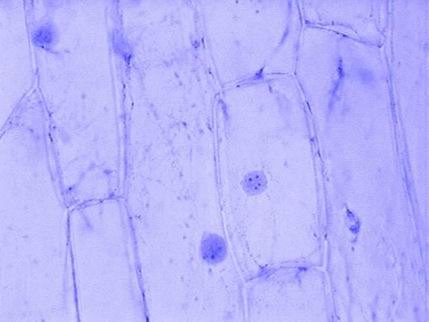
| 40x AFA Mounted in glycerin, Methylene blue - in excess - but I find this image beautiful |
x10
- live, dark field, opaque disc of 12.5 mm |
|
|
| x40 - dark field. Central stop 20 mm in diameter. Mounted in glycerin. |
x40
-
DIC of the poor man. Central stop of 15 mm, and oblique marginal illumination. Disc somewhat moved to accentuate the oblique illumination. Mounted in
glycerin. |
|

|
| x40
-
Critical marginal circular illumination, after Sterrenburg, 15.5 mm dark
stop adjusted to cover 95% of the objective's aperture. |
x40
- Another similar image of oblique ilumination |
|
|
| x40
- live, Mathias arrow. Another false DIC. |
x40 live, with the squared end of the Mathias arrow !! |
|
|
| x40
- Tortured in a concentrated saline solution. Wet mount - COL-C |
x40- idem. Crystal transparencies. COL-C, backed by a cyan filter. |
|
|
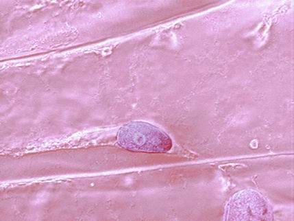
| COL-J, side view of a nucleus and its 4 nucleoli, mounted in glycerin. |
100x Emboss effect
and electronic staining in PhotoPaint, mounted in glycerin. |
|
|
| x100
- Oblique illumination. No other added effects. Electronically stained in Photo Paint. Mounted in glycerin. |
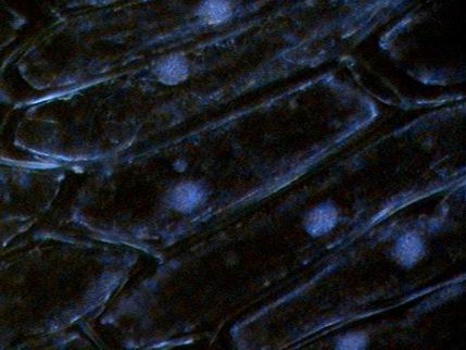
x40
- Wet mount, live. 15 mm opaque stop backed by a dark blue disc. The picture imitates a DAPI fluorescent image. |
|
APPENDIX
There is a photomicrograph of onion skin, which I do not like too much, examined with the Hoffmann Modulation
Contrast technique at www.microscopyu.com/smallworld/gallery/contests/1980/12thlarge1980.html Here I present to you the kind contribution of Steve Durr, an exceptional photomicrographer (very well equipped, one cannot doubt it; visit his website to see incredible examples of creatures of the micro world) which also illustrates the effect that Differential Interference Contrast (DIC) as applied to the humble onion skin. Apart from these there is only one other very small image on the Web of onion skin taken with DIC. "Walter . You were correct when you said this is one of the first objects that most microscopists view, I remember very well many years ago looking at onion skins trying to find a real live nucleus. The photographs were taken with a Nikon Coolpix 4500 digital and the lower mag is x312 while the higher mag is x500. I do not possess any higher objectives in DIC. Best wishes Steve." |
|
|
| Original at 312x, but here reduced |
Original at 500x, reduced here. |
| Paul James,
an artist and mastercraftsman of microscopy, innovator, and enthusiastic advocate
of the COL
diaphragm contributes these four very beautiful images captured with
his Zernike Phase Contrast
outfit. I can say that these pictures are exceptional. There is not on the Web another Zernike
phase example of onion skin. According to his notes the pictures were taken with a Wild microscope with phase fluorite objectives. They have been captured at 100x, 200x, 400x and 1000x, with a digital Nikon Coolpix 800 of 2.1 megapixels and reduced to 400 X 300. |
|
|
| 100x |
200x |
|
|
| 400x |
1000x |
The onion skin is beautiful, isnt?
Please report any Web problems or offer general comments to the Micscape Editor.
Micscape is the on-line monthly magazine of the Microscopy
UK web
site at Microscopy-UK