|
Galleries of Old Image Reprocessed. Part 2
Richard L. Howey, Wyoming, USA |
Once again, I have been idly browsing through some old file of old images and I would like to remind you that the selection is indeed random. Do try to think of this as simply a gallery and not an exposition with a theme. I will not include images which I have recently taken nor will I include any crystal images, at least not in the early galleries, since I have recently written a good deal about crystals. (Although, as it turns out, I may make one or two exceptions, as usual.) Also, I will keep my comments at a minimum, just enough to help orient you regarding the subject matter of the image. So, this is just a kind of ramble from one thing to another with no particular order.
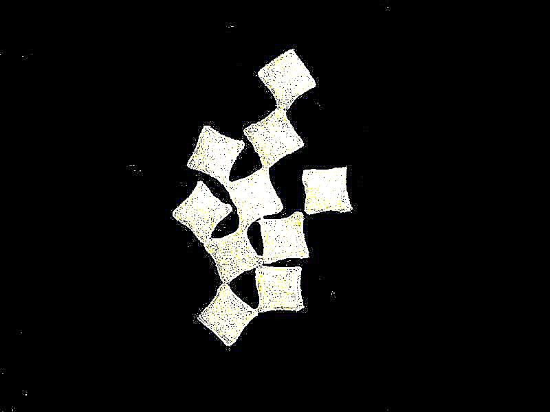
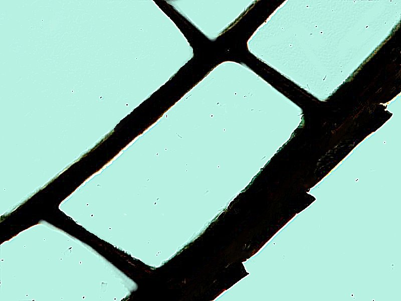
In the previous gallery, we looked at some diatoms, those wonderful marvels constructed of silica; that is, they’re mostly glass. Sometimes they aggregate in unpredictable ways as is the case with these very small “square” diatoms.
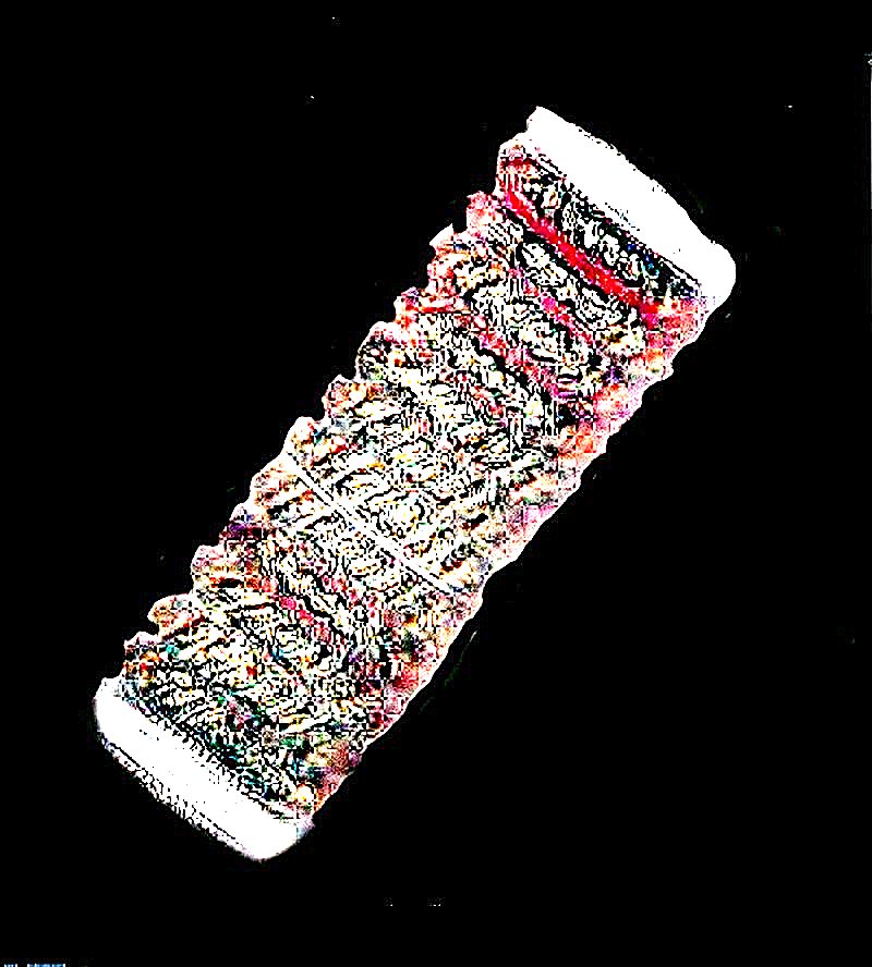
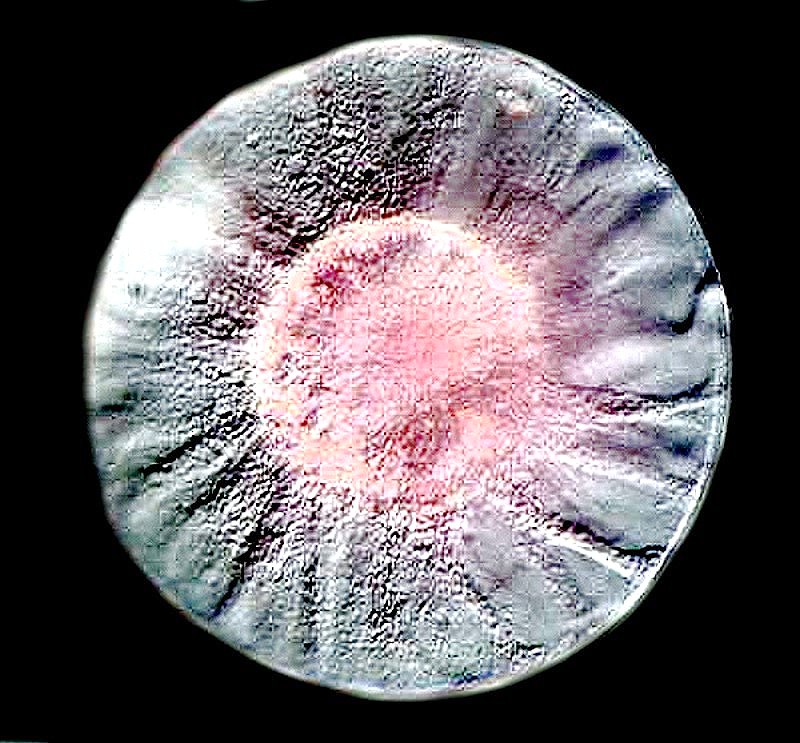
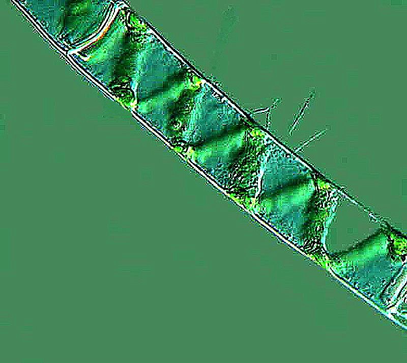
In other instances, they take on surprising configurations as is the case with this cylindrical form. I’ll show you a side view and then a top view.
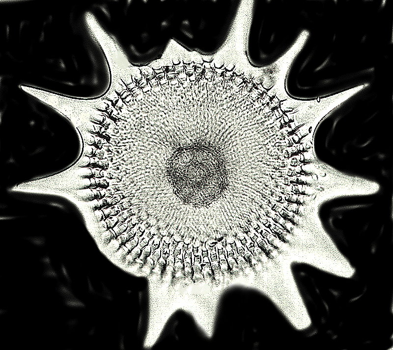
While we’re looking at things predominately silica, let’s take a glance at an unusual radiolarian (although, in truth, they’re all unusual).
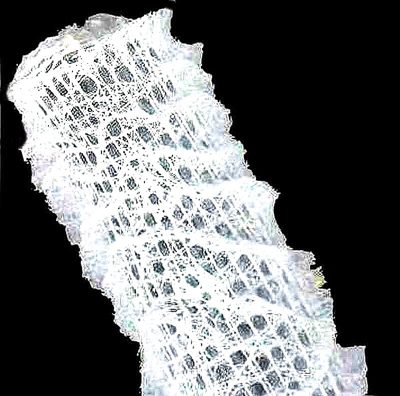
Oh, why not splurge and go on a silica binge. Next up, a closeup of a section of a glass sponge called Euplectella aspergillum. The intricacy of the lattice work is amazing and it becomes even more amazing when one realizes that it is composed of thousand of tiny spicules “glued” together.
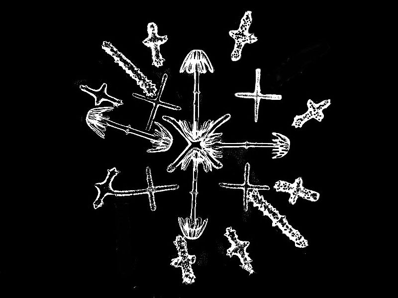
The image below is a cluster of spicules from a different species of glass sponge, Hyalonema.
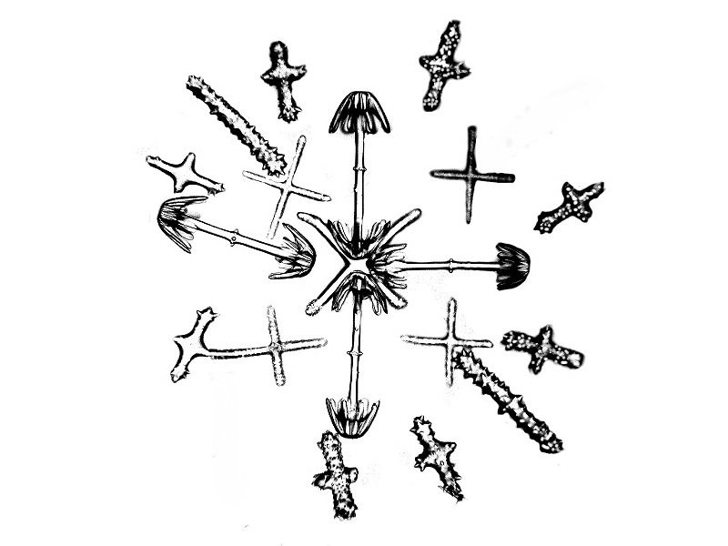
This is the same image, but this time using darkfield.
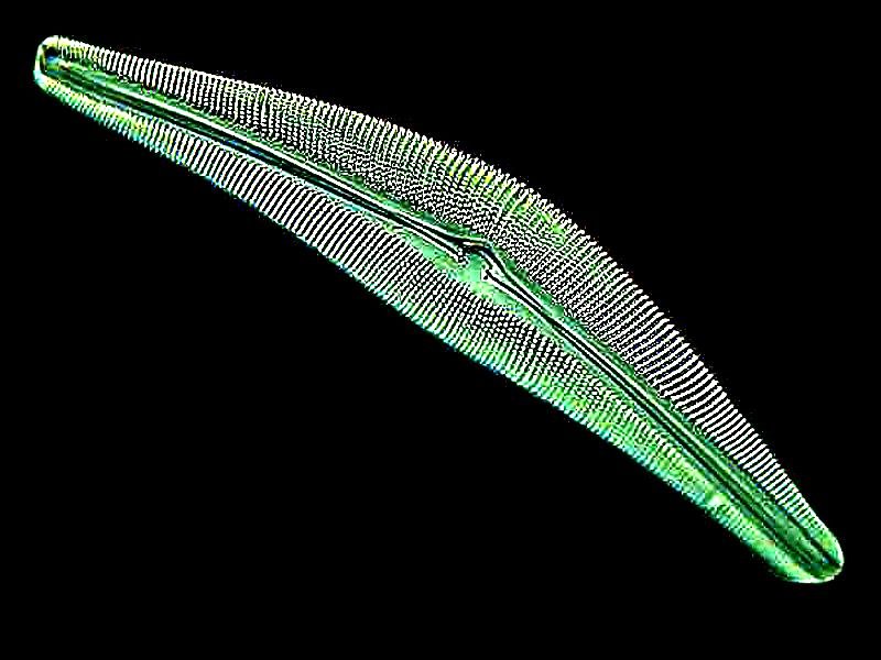
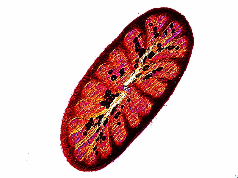
Let’s leapfrog back to take a look at a really splendid diatom called Cymbella. Each of the little “dots” is a pore through which protoplasm streams, thus providing a slow, gliding motion to the organism.
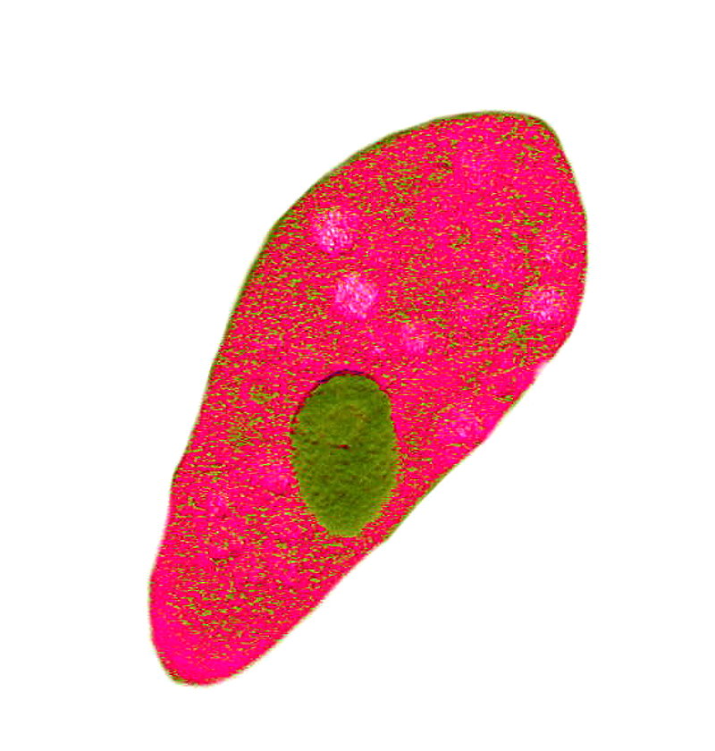
Now, a selection of protozoa, beginning with one of the most familiar, Paramecium. The first image is one stained with a Trichrome stain which clearly demonstrates the macronucleus.
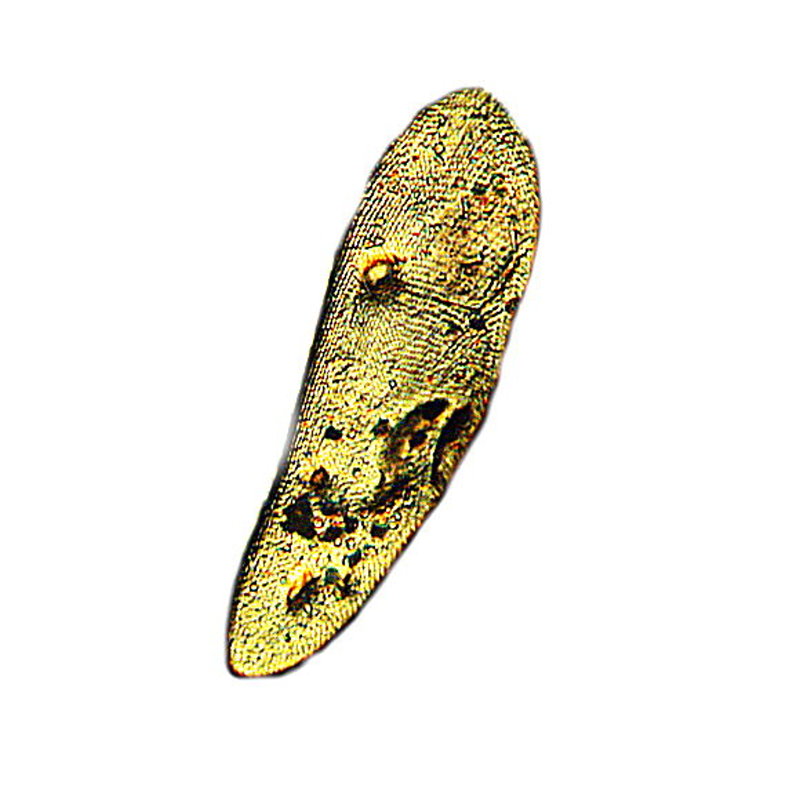
This next image is a Paramecium which has been dried and treated with a very dilute solution of the biological stain Nigrosin to show the surface structure of the pellicle.
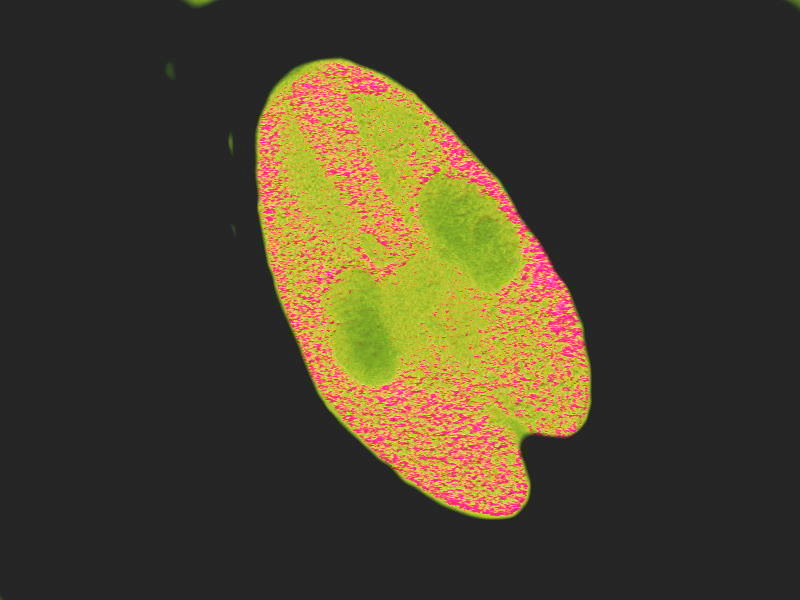
The third image is one in which again the organisms have been treated with the Trichrome stain and here we see 2 Paramecia conjugating which is a reproductive process which as it progresses, involves the exchanging of micro-nuclei and then separation and genetic repair and “revitalization”.
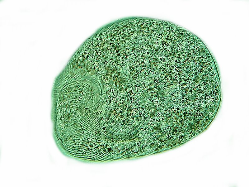
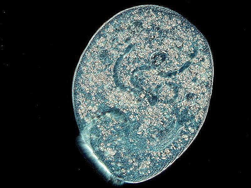
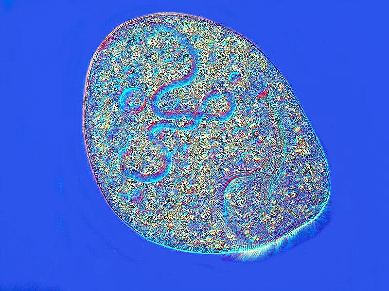
Reproduction in protozoa can be extraordinarily complex. Let’s take a look at one process in a large ciliate named Climacostomum. When a suitable partner is not available, some ciliates go through a process called autogamy in which they internally reorganize and repair their genetic material. First, I’ll show you an image of the organism without any special illumination so that you have an idea of what it looks like ordinarily.
Then, 2 images using “optical staining” achieved through Nomarski Differential Interference Contrast. These clearly show the macronucleus curling around inside the organism as the autogamy takes place.
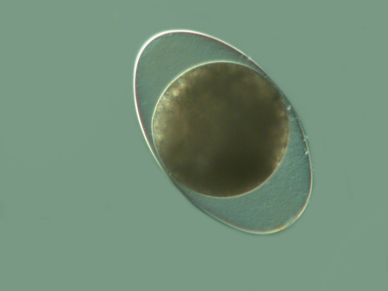
When conditions become undesirable, Climacostomum forms cysts which are resistant to adverse conditions and can help the organisms survive until more favorable conditions return. Below is an image of such a cyst.
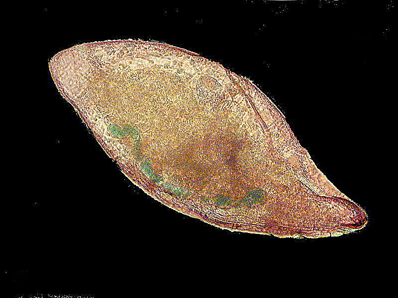
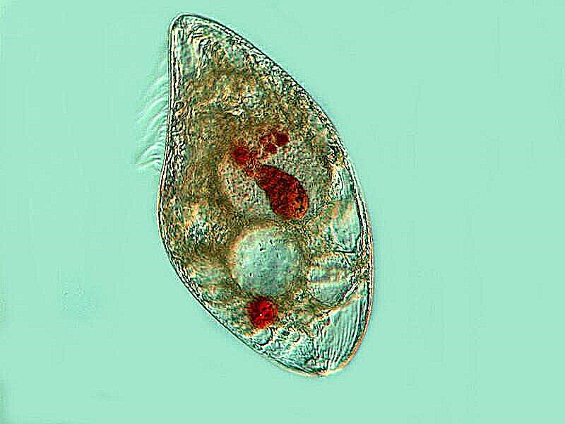
Protozoan behavior can also be amazingly complex. Let’s consider the remarkable Blepharisma which the distinguished marine biologist and protozoologist, Arthur Giese, devoted an entire book to.
First a view of an “ordinary” Blepharisma.
Blepharisma is notorious for its unseemly behavior toward members of its own species; when things get sparse food-wise, they can turn cannibalistic and engulf others thus turning them into cannibal giants. Here’s an image of one and the red lumps are Blepharisma that have been devoured and are slowly being digested in vacuoles.
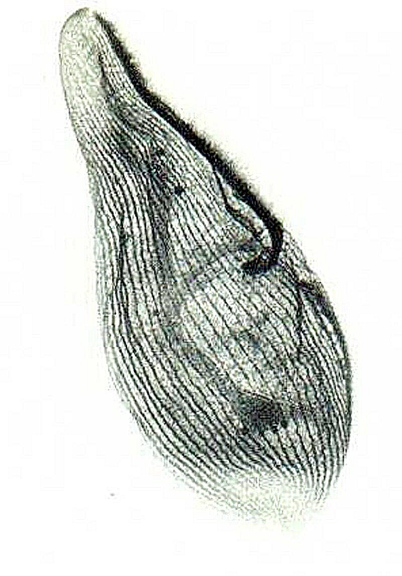
These ciliates and a number of others have a complex network of fibrils which compose what is called the “silver-line system”. Using a complex and demanding technique using organic silver, the “Protargol technique”, shows this system beautifully.
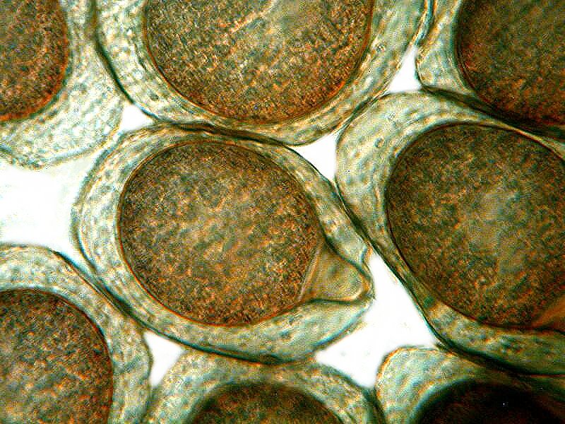
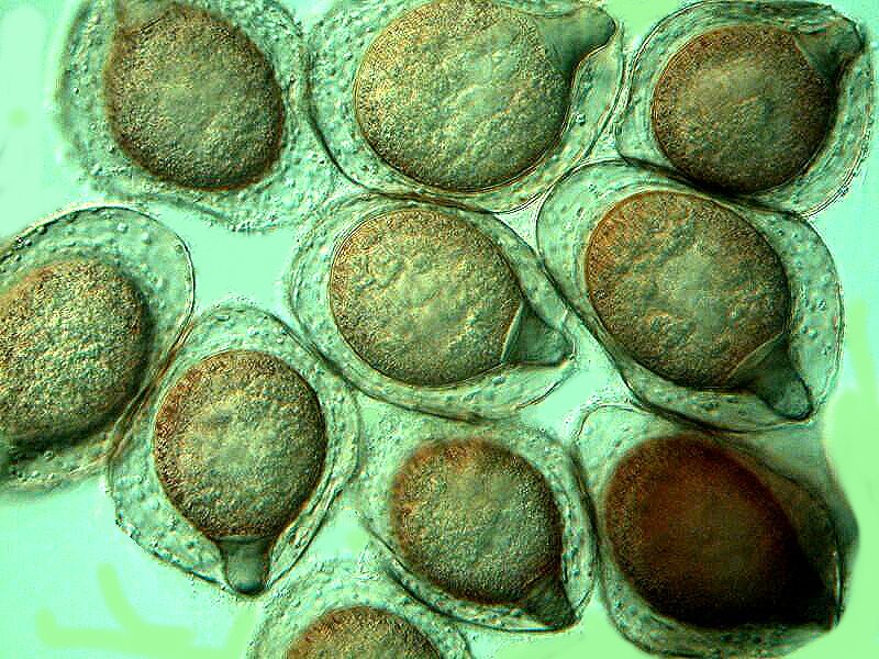
As we saw with Climacostomum, some ciliates can go into survival mode by forming cysts. This is certainly true for Blepharisma and the cysts are very distinctive. I’ll show you 2 images of their cysts.
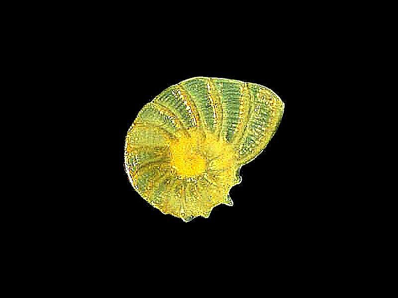
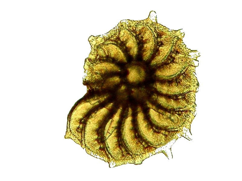
While we’re looking at protozoa, let’s examine a few foram (foraminifera) “shells”. These are primarily composed of Calcium carbonate and are often opaque, thus hiding the internal complex chambers. I devised a technique using a drop of oil (usually immersion or cedar oil), to enhance internal visibility. The 2 examples below use that technique.
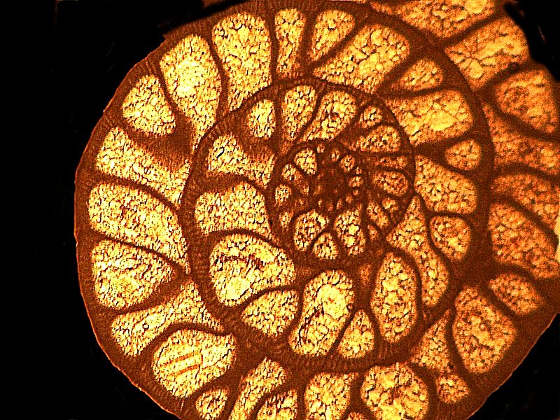
Some of the fossil forams that are imbedded in a matrix are suitable for sectioning and can reveal some of the inner beauty of these shells. Below is such an image of a foram called Pseudoschwagerina.
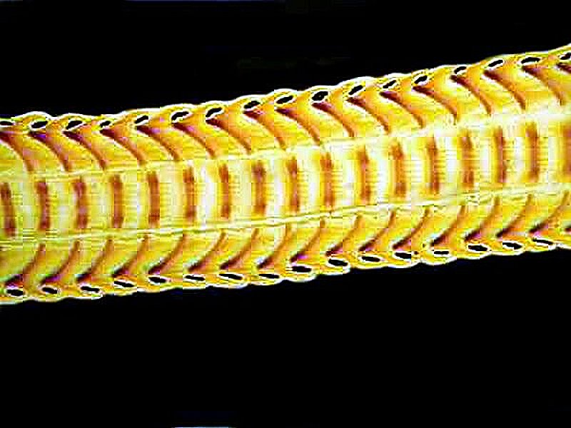
While we’re looking at extraordinary calcareous structures, how about a quick glimpse of a snail tongue, known as a radula. There are rows and rows of teeth here and the band of tissue containing them is moved back and forth by muscles to scrape food (algae) off of surrounding strata.
While mineral materials compose a significant number of biological structures, nonetheless, as we might expect, organic materials far exceed them. So, let’s look at a few examples. To begin with, consider hair; it’s primarily keratin which is a fibrous protein.
I’ll show you 3 examples; the first is a cross section of a hair from a peccary (you can Google it); the second is a strew of hairs from a prairie dog, and the third I collected, at great risk I might say, from a tiger when I was hunting one day at a zoo and asked the keeper for a sample. I won’t repeat what he said here so, I had to resort to buying a small packet of tiger hair online. Well, there was a risk that it wasn’t real; give me a break.
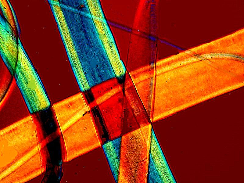
Another example of dense protein composition can be found in dragonfly wings. They are structured like a series of little windows. I’ll show you a closeup of a tiny section.
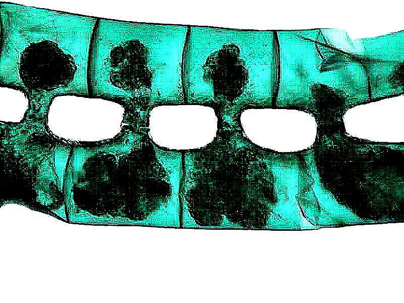
Finally, a brief excursion into the world of plants. First 2 images of a splendid, common, and easily collected algal form called Spirogyra. As you can see, there is a spiral arrangement of the chloroplasts.
And this second image shows the filaments conjugating–remarkable!
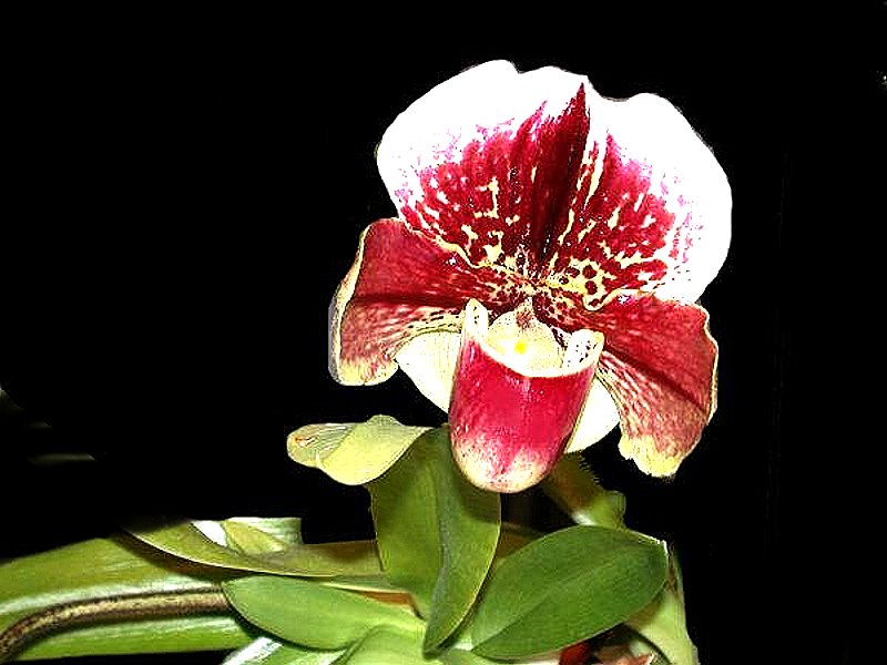
Here’s a photo which I took of a Lady Slipper orchid which I managed to maintain for several years. The first year, it only had one flower, the next there were 3 and then thereafter 5. It was a wondrous plant. This is a closeup of a single bloom.
In conclusion, 3 images. This first one is of a miniature shrub.
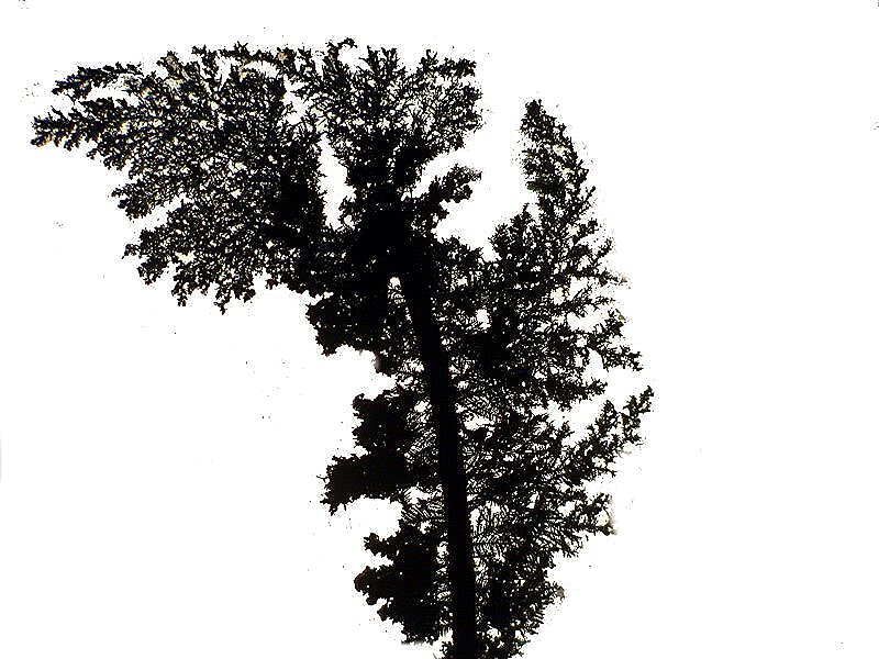
The second is another shrub of the same species which is windswept and therefore leaning to the left.
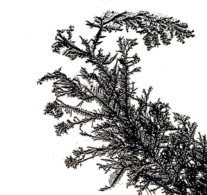
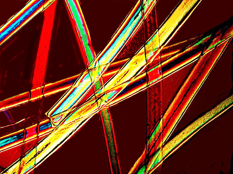
Oops! I guess I made a mistake. These are arborescent silver crystals which form when I place a tiny bit of copper wire in a solution of Silver nitrate, as you can see in this last image.

I hope you had some fun looking at this eccentric collection of images and I’ll keep browsing in my files to come up with yet another gallery. In the meantime, write something, take some pictures, and send it or them off to Micscape.
All comments to the author Richard Howey
are
welcomed.
If email software is not linked to a browser, right click above link and use the copy email address feature to manually transfer.
Editor's note: Visit Richard Howey's new website at http://rhowey.googlepages.com/home where he plans to share aspects of his wide interests.
Microscopy UK Front
Page
Micscape
Magazine
Article
Library
Published in the April 2022 edition of Micscape Magazine.
Please report any Web problems or offer general comments to the Micscape Editor .
Micscape is the on-line monthly magazine of the Microscopy UK website at Microscopy-UK .
© Onview.net Ltd, Microscopy-UK, and all contributors 1995
onwards. All rights reserved.
Main site is at
www.microscopy-uk.org.uk .