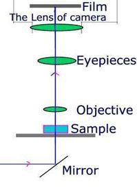
Method 1: SLR lens in place.
This
is the most difficult and worst method of the three. Please look at the
optical pathway and the photo created is shown in the other image below:
 |
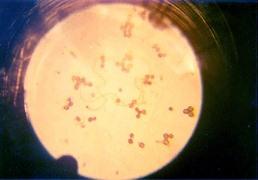
(150x) |
You
can see that the field of the photo is small and in the photo there is
an unsightly black ring. This method is very difficult to do. You must
use a single lens reflex camera and focus your subjects under examination
on the frosted glass screen of the camera very carefully. It is not a good
way to take micrographs.
Method 2: SLR lens and microscope eyepiece removed.
This
is a better way to take micrographs. You can see both the optical pathway
and the effect created below:
 |
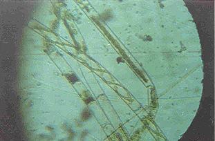
(150x) |
This method can give the images a higher definition. But there are some disadvantages: The magnification is low and the field is still small all the same. It is not a good way to take micrographs either, but the method can enable you take photos of small plants or flowers with low magnification.
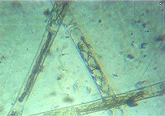
Method 3: SLR lens only removed.
This
is the best way to take micrographs with all kinds of optical microscopes.
It can give photos of high magnification (up to the highest magnification
of your microscope), the images are very clear and fills the frame of the
film. It is widely used by most microscopists all over the world. You can
see both the optical pathway and the effect created below:
 |
(300x) |
About the camera, you had better use a single lens reflex or a digital camera because they are easy to focus your subjects. Remove the lens from the camera and fit in an extension tube. You can buy the extension tube from most camera shops or make one with cardboard.
The third method is very nice to use for all microscopists, whether a professional microscopist or an amateur.
I hope this message will be helpful for you.
Many thanks to all my friends for their help.
All
comments to the author
Wan
Yu are welcomed.
Editor's notes: Many thanks to Wan Yu for sharing this article. Wan has started his own web site to promote microscopy as a hobby in China.(The connectivity to the Chinese host of this site is rather variable).
One
way of minimising the vibrations Wan remarks on, is to use an old enlarger
stand as a firm independent support for the camera. See Ted
Clarke's article in the August 2001 Micscape.
Microscopy
UK Front Page
Micscape
Magazine
Article
Library
© Microscopy UK or their contributors.
Please report any Web problems or
offer general comments to the
Micscape
Editor,
via the contact on current Micscape
Index.
Micscape is the on-line monthly magazine
of the Microscopy UK web
site at Microscopy-UK