|
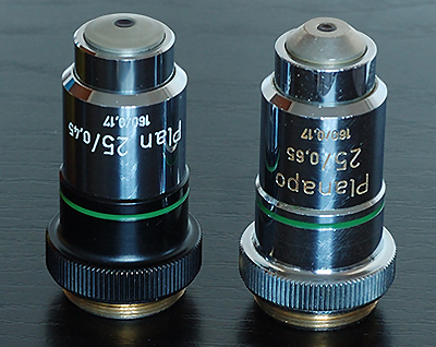
Comparing two
pairs of Zeiss objectives - notes on the effect of delamination
by David Walker, UK
|
1)
Comparing a badly delaminated 40x NA0.6-1.0 planapo oil with a
pristine near equivalent.
2)
Comparing
a delaminated and damaged 25x NA0.65 planapo with a 25x
NA0.45 planachro.
Two
examples of essentially the same Zeiss objective, the 40x NA0.6-1.0
planapo oil immersion, passed through my brother Ian and my hands over
the past year. But they had one major difference, one was extensively delaminated
and the other was in excellent condition.
The
tendency of some Zeiss microscope optics of a certain vintage to delaminate
will be very familiar to users and purchasers of Zeiss kit.
I'm not a Zeiss aficionado, just a user of the optics, so can't offer any insight
myself into the causes of this very well discussed phenomenom. See links below
for discussions of delamination.
My
own interest in delamination was very much a practical one and curiosity; here was an opportunity
to see how the two objectives compared in performance with typical brightfield
subjects
both visually and for image capture with my Nikon D50 digital SLR. Given the often large price
difference between a pristine objective and one with declared extensive delamination
it was also interesting to see if the quality loss could be tolerated if I was
purchasing the cheaper objective.
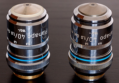
The
two objectives being compared, (serial nos. 5195400 and 5022152) the righthand one has the delamination.
They are not totally identical in external design; the lefthand example has a white
ring on front, the 'lock up' feature and design differences at objective rear so possible
there may be internal design differences (comments from Zeiss users welcome);
but same spec in mag and NA so as close as the author is likely to have as
an opportunity to assess the effect of delamination.
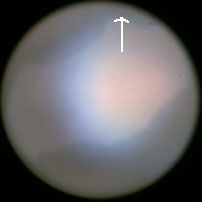
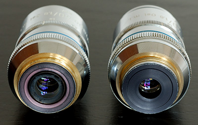
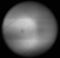
Views
of the back of the objective sequentially focussing down. Delamination was occurring
on three different optical planes, the first two were major and across
central field, the third marked was slight edge delamination not clearly shown.
Note
on testing methods for delamination:
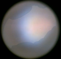
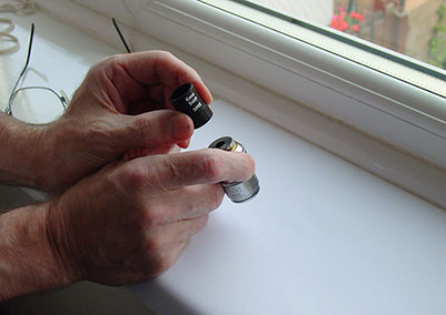
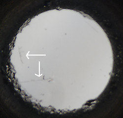
1)
A quick and easy method for checking an objective that I've found useful is
shown in the image right below. The objective is held close to and pointed at a well illuminated
white paper sheet and the back of the objective inspected with a good quality
5x-10x hand lens by focussing carefully from front element through to back element—complex objectives could have a number of potentially delaminating surfaces. Apart from very minor edge defects this method
should pick
up any significant delamination. An unstructured illuminated surface is
required as easy to overlook faults if the objective pointed at e.g. a cloudy
sky. This method could be used if in a dealer's
showroom or at a club sale if more extensive means of checking aren't available.
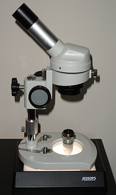
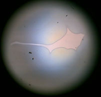
2)
A low power stereo microscope with good focus range is a more rigorous way of
inspecting objectives. A simple low power (ca. 20x) dissecting microscope as shown above
left with bottom illumination, in this case by a light box, will also suffice. If
the objective has a flush or protruding front element, great care needs to be
taken if placing the objective on a surface. To be safe, it's quite easy to
support the objective by hand just above the surface while inspecting it or annular disc support
could be devised.
3)
A phase telescope or microscope with focussing Bertrand lens can be used with
objective focussed on a blank area of slide with condenser iris fully open and
correctly focussed to ensure whole aperture of objective is illuminated. Ensure the inspection
method can focus
throughout the 'optical depth' of the objective as this isn't always the case. My own preference to check for delamination is to always to use method 1 first, then inspect
the damage more carefully by 2) or 3).
Notes
on digital imaging:
Microscope - Zeiss Photomicroscope III, achromatic-aplanatic
condenser. Camera - Nikon D50 DSLR using monocular tube. Parallax focusing by
focussing an eyepiece in bino' head to match camera focus. 'Projection' eyepiece
- Zeiss Kpl 10x W on 5mm collar. Flash used exclusively as described in this
article. RAW images. Resized using Fred
Miranda 'WP Pro' v1.1' plug-in for Photoshop (better resizing algorithm
than Photoshop's), 'low sharpening' setting used in plug-in to compensate for
softening of an extensive resize. Saved as low compression jpegs. Further
work-up as described below for each image sequence.
Comparing the badly delaminated 40x NA 0.6-1.0 planapo oil with a
pristine near equivalent.
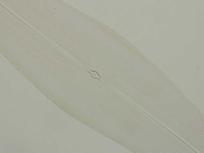
Diatom
- Pleurosigma angulatum: (Klaus
Kemp 'Test plate 8 forms'. Hyrax mountant.)
This diatom species is
a classic test subject and particularly useful for 40x objectives. A good achromatic 40x
NA0.65 can resolve the frustule detail in brightfield but benefits from contrast
enhancement like oblique. A high NA 40x planapo should readily resolve detail
in brightfield but the image quality of this low contrast subject can suffer
if there's any problems.
The
'good' 40x NA1.0 planapo objective in brightfield. Resized crop of 'out of camera' image.
The
'delaminated' 40x NA1.0 planapo objective in brightfield. Resized crop of 'out of camera'
image with slight tonal balance change to match exposure of image above.
This image reflects the visual image, i.e. noticeably lower contrast and a less
neutral 'muddy' hue but the resolution is essentially still there.
The
'delaminated' 40x NA1.0 planapo objective in brightfield. As above except the condenser iris
was stopped down more (to ca 60% of field) than for 'good' objective. This brings
back the contrast to some extent, as the diatom was readily resolved the
resolution didn't suffer unduly.
Histology
- rat ileum, iron haematoxylin and eosin, Numount, NBS course slide, prepared
by author.
I've
never found imaging of histology subjects very easy or satisfying, there's often no sharply
delineated features to catch the eye to give an impression of sharpness, but
had a go anyway. The damaged planapo again gave lower contrast images but quite
acceptable when stopped down a bit. Colour accuracy seemed affected as seen
in the images below.
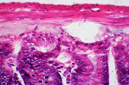
The
'good' 40x NA 1.0 planapo objective in brightfield. Resized crop of 'out of camera' image.
Depth
of field is small at this mag.
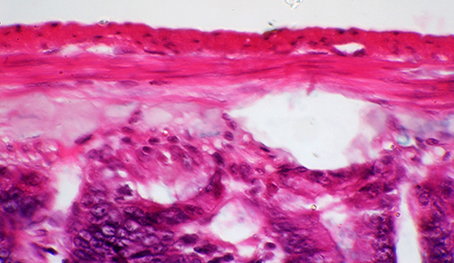
The
'delaminated' 40x NA1.0 planapo objective in brightfield. Resized crop of 'out of camera'
image with
tonal balance change to match exposure of image above.
Zeiss
40x NA0.65 planachro, good condition.
Comparing
a delaminated and damaged Zeiss 25x NA 0.65 planapo with a Zeiss 25x
NA 0.45 planachro
Out
of interest two Zeiss 25x objectives were also compared. The Zeiss 25X NA 0.65
planapo I possess has extensive delamination in two optical planes and also
the front element is badly cracked. Knowing the optical performance would be
impaired, I was interested to see if it still had merits in use cf a Zeiss 25x
planachromatic in good condition.
Above:
Left - Zeiss 25x NA 0.45 planachro, in good condition; right - Zeiss 25x NA
0.65 planapo, damaged as shown below.
Above -
Zeiss 25x NA 0.65 planapo: Left front element, two bad cracks arrowed and edge
chipping, the lower crack is worse and deeper than shown in the image above
left. Middle and right images of elements from rear; delamination across the
field was present in two different optical planes.
Brake
fern, T/S rhizome, stained. Biosil slide.
This was a colourful, contrasty
slide to assess a typical prepared stained subject. In the images below I have
compared the optimised images attained from each objective, using normal image
work-up procedures i.e tonal balance adjustment, colour cast removal of white
background (if any) and resize. This was a fairer comparison than 'out of camera'
images, where exposure differences can overlap objective differences. (My
flash gun changes power in factors of two so too big a jump to exactly match
exposures. I need some finely graded neutral density filters.)
Visually,
the damaged planapo gave noticeably lower contrast and duller images than
the planachro and planapo image was not as visually pleasing but still competent. With this sort
of subject the higher resolution isn't readily noticed. But as the images below
show, the worked up digital images aren't dissimilar. The slight differences
are to some extent due to a less than perfect match of tonal balance adjustments.
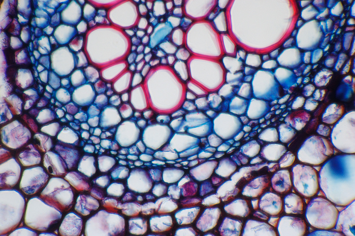
Zeiss
25x NA0.65 damaged planapo.
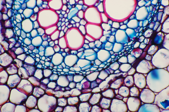
Zeiss
25x NA0.45 planachro in good condition
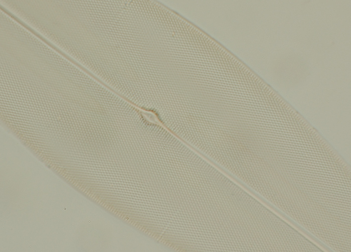
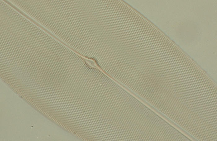
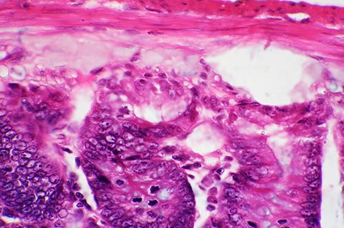
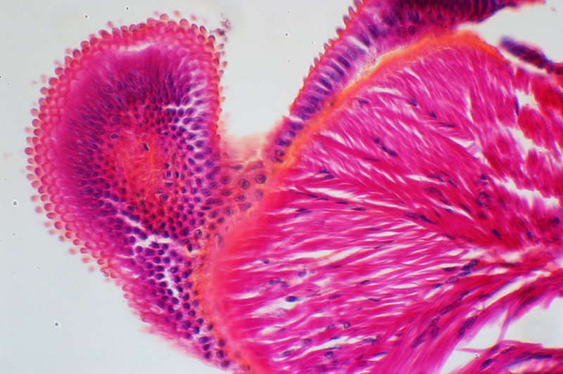
Octopus
tentacle suckers, V/S, stained, Biosil slide
As
for the stained botany section above, visually the damaged planapo gave a duller less
contrasty image but still fine for showing detail and benefited from slightly
more stopping down than usual to ca. 60%. After image work up the finer subject differences
are less marked although the planachro image looks more 'punchy'. Note that exactly the same tonal balance and focus is tricky
so image comparisons can't be too rigorous.
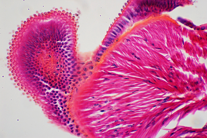
Zeiss
25x NA0.45 planachro in good condition
Zeiss
25x NA0.65 damaged planapo.
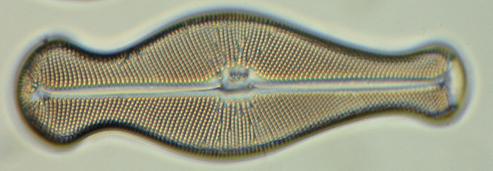
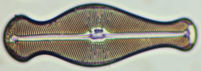
Diatom
- Didymosphenia geminata (Klaus Kemp '100 forms' type slide, mountant
Zrax)
This subject was chosen to see if the better resolution of the
planapo was still a benefit albeit lower contrast. The planachro does resolve
this diatom but the image differences cf the planapo are more noted; the planachro
gives the thickening typically seen of lower NA objectives.
Zeiss
25x NA0.65 damaged planapo.
Zeiss
25x NA0.45 planachro in good condition.
Overall
comments:* As expected and as noted by other microscopists (see
link 1), the contrast suffers most noticeably in visible brightfield work with
the delaminated objectives assessed above. If a planapo is being bought to enjoy the finest
visible brightfield image I think a badly damaged one could disappoint especially
for subjects with colour, but the above were quite badly damaged and still gave
competent images with some stopping down. The two damaged objectives tested though could
well have sold for a few tens of pounds cf the Ł100-200 for a good condition
planapo, so if digital imaging was of more interest, the normal image work up
with corrections for tonal balance, colour cast etc may give
images not far off those for a pristine objective except for the more critical
work. A planachro of the same mag could be considerably more expensive than
a damaged planapo and the resolution could still exceed the planachro, so worth
keeping an eye out for what comes up for sale if the price is right. (*For
brightfield work, not for techniques like DIC.)
Comments to the
author
David
Walker
are welcomed.
Links:
1)
The UltraPhot
Shop FAQ page - Spike Walker, the noted photomicrographer and Zeiss microscope
expert has a valuable summary of potential causes of the delamination of
Zeiss microscope optics and its consequences. (Towards bottom of web page link
given.)
2)
Yahoo
'Real Microscopy' forum - discussions from 'delamination' keyword search,
where a number of microscopists shared their views and experiences of objective
delamination.
3)
So
a scratch ruins an objective?? A surprising revelation gleaned from a chance meeting
with an abused optic - Paul James shows how an objective
can have a surprising amount of visible cracks and scratches on front element
and still give reasonable images.
Acknowledgements:
With
thanks to Klaus Kemp
of www.diatoms.co.uk
for making and supplying such excellent diatom test and type slides and to John
Wells, Biosil for other prepared slides.
© Microscopy UK or their contributors.
Published in the July 2006 edition of
Micscape.
Please report any Web problems or offer general comments to
the
Micscape
Editor
.
Micscape is the on-line monthly magazine of the Microscopy
UK web site at
Microscopy-UK
© Onview.net Ltd, Microscopy-UK, and all contributors 1995
onwards. All rights reserved.
Main site is
at www.microscopy-uk.org.uk
with full mirror
at www.microscopy-uk.net
.