|
How To Milk A Weed: Part II by Richard L. Howey, Wyoming, USA |
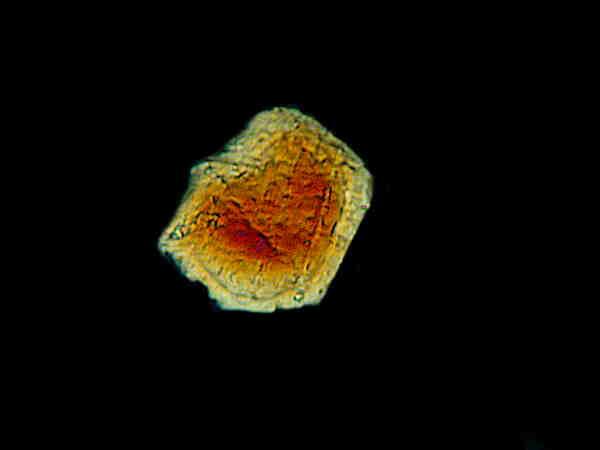
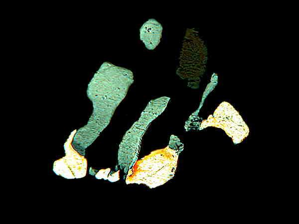
Last time we examined some weeds and yard plants and, in this second part, we’ll be taking a look at some houseplants. We have a rather unusual Ficus (or, at least that’s what we were told. It was a gift from my wife’s brother.) We have had it for over a decade now and there is an ongoing battle with mealy bugs, which my wife heartily abhors. I think they must like its milky sap. I found a leaf that had yellowed and plucked it off. Immediately, at the end of the stem, white fluid began to appear. I took it upstairs and made three smears. For those of you who haven’t done this before, the procedure is very simple and is identical to that used to make blood smears. Place a small drop on a clean slide and then using another slide, angled at about 45 degrees, pull it across the first slide thus forming a thin film of the liquid across the central half of the first slide. If, however, you have quite fibrous leaves, stems or berries that you are working with, they may not so readily give up their “precious bodily fluids”. (See the film “Dr. Strangelove or How I Learned to Stop Worrying and Love the Bomb”) In these cases when you have to slice and dice the plant material, you will likely also end up with bits of plant tissue on the slide as well. If you wish to use a cover glass for medium to high magnification, then using a pair of fine forceps, remove the visible pieces and then use the smear technique.
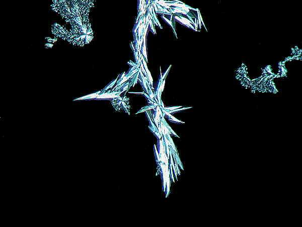
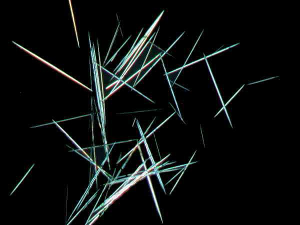
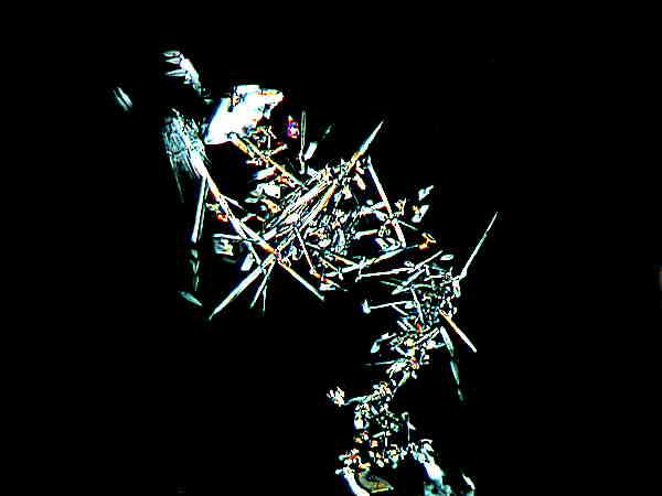
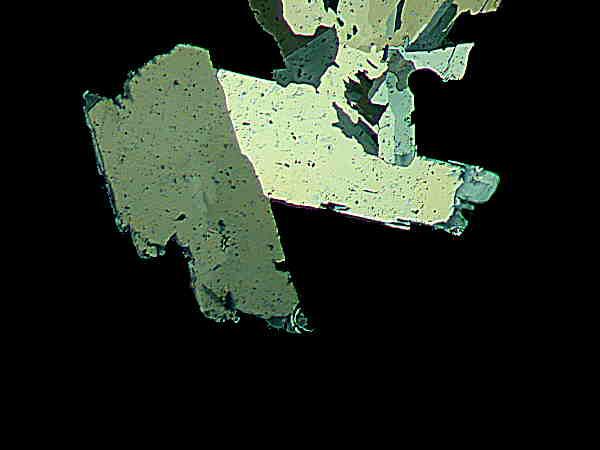
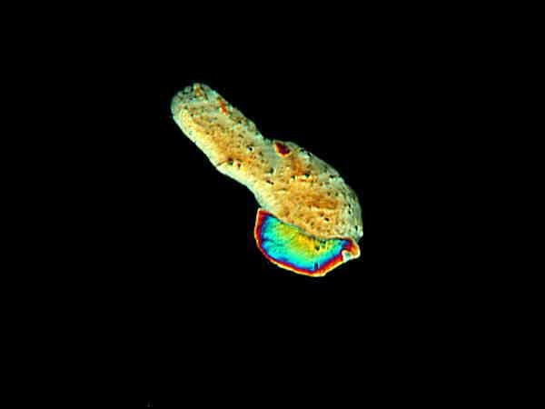
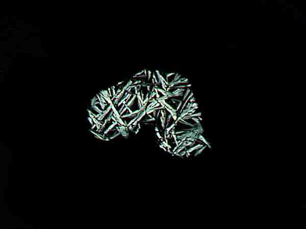
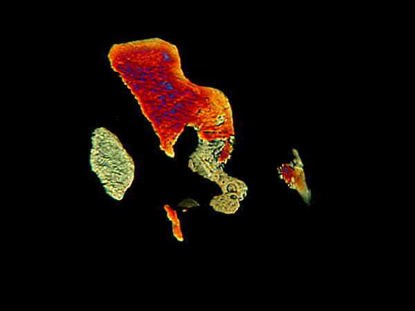
I found this plant-flea-infected Ficus fascinating (I love alliteration). As it turns out, the crystals are quite striking. I’ll show you 3 images.
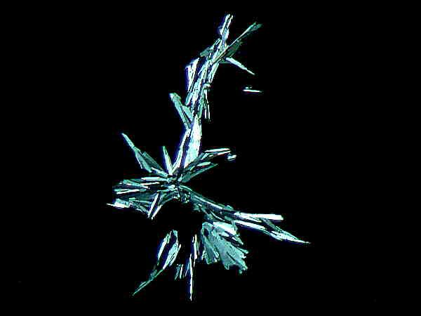
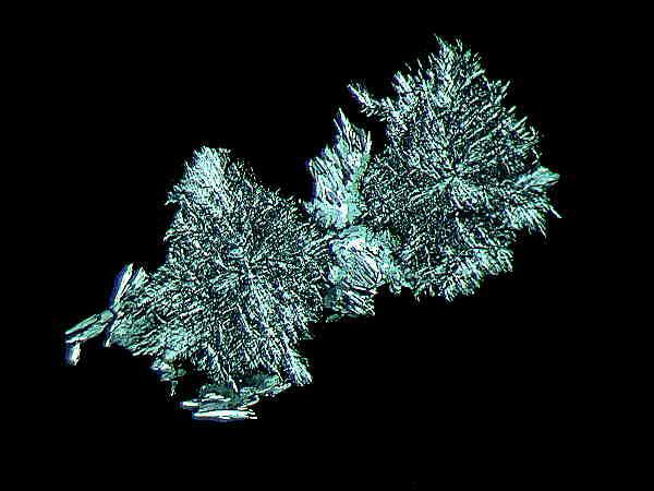
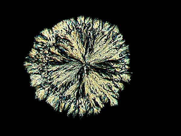
In the first two images, the aggregation of acicular (needle-like) crystals is evident. In the third image, however, there is a clumping and the crystals are much smaller.
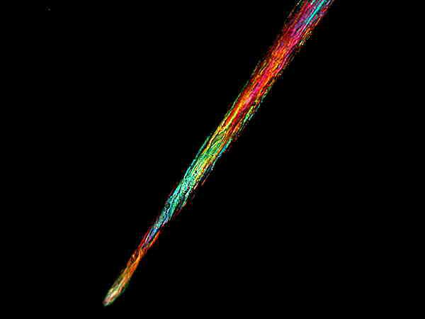
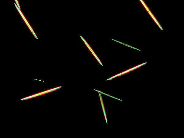
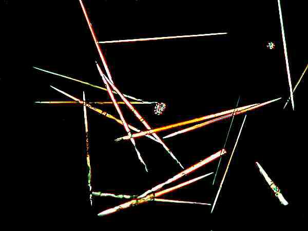
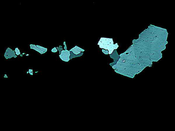
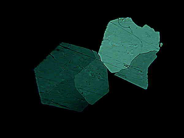
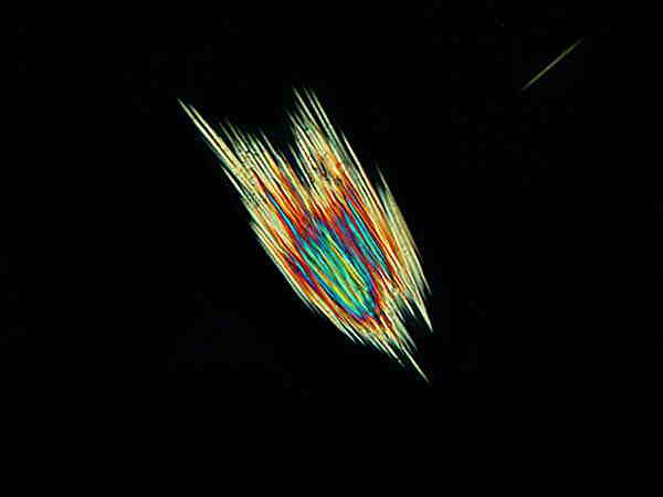
I mentioned in Part I of this essay that acicular crystals are quite common. In a windowsill, I have a small specimen of the succulent Haworthia. Here are 2 images of its crystals.
The acicular character of the crystals is quite clear and notice that, under polarization, some have that bluish-white luminescence that is typical of the Ficus crystals, whereas others have pastel red, blue, or purple coloration.
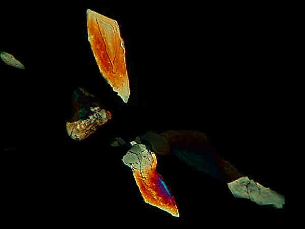
Finding a variety of crystal forms in a single plant is not unusual and sometimes the differences are quite dramatic. I purloined a piece of a “leaf” from one of my wife’s 837 Zygocactus plants hoping that she would not notice that it was missing. I exaggerated slightly; actually she only has about 20, but they, like the Hoyas, can soon begin to take over rooms so that it seems like there are more than there really are. In truth, my wife is very generous about making sacrifices for my eccentric microscopical pursuits. The Zygo smear turned out to be especially interesting.
This image makes me think of a crystalline, hominid skeletal simulacrum trying to capture a butterfly and, no, I haven’t been taking any exotic drugs.

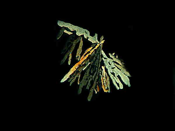
This one makes me think of a alien, crystalline space station rotating out in the vast, black cosmic emptiness. The next two images–still from the same slide–are radically different.
Obviously this last image is of an alien spacecraft flying off to dock at the crystalline space station. (I’ve got to quit watching reruns of Star Trek!) The intriguing issue is that on the space of a single slide, with sap from the same leaf of one plant, we find these extraordinarily different crystal formations. This is also true for some other plants as we’ll see in a bit.
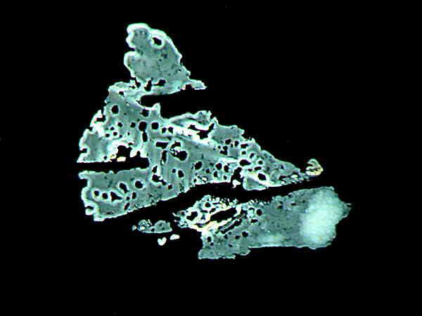
I took a sample from a small house cactus (not a zygocactus) which has no genus or species information on the label. The smear revealed no crystals of interest, but the spines are spectacular.
When I looked at this under higher magnification, I found hints of intriguing internal structure which I hope to investigate later.
The smear I made from a Geranium leaf produced only extremely minute, very boring crystals except for these 2 “moon rocks”.
As I looked at these, I had a sense of deja vu and then it struck me–I had gotten a similar result with my dandelion smear–small, boring crystals and 1 “moon rock”.
As I thought about it, I realized what these “moon rocks” were–tiny sand grains from the soil surrounding the plants! One has to be constantly on the alert not to deceive oneself by misinterpreting artifacts, random inclusions, or airborne invaders. In our household, the intruding items I most often find on slides are mold spores, butterfly scales, cotton fibers, dust mites, and cat hair. Nature is always playing tricks on us and is quite happy to help us play tricks on ourselves.
One of the tricks that nature may play on us in our quest for crystals in plant juices is to form crystals only out at the very edge of the slide where we might not look. Another possibility is that no crystals will form on one slide and then on the next, from the very same bit of plant, give us a lovely batch.
I encountered just this sort of abuse when I was looking at 2 smears from the charming houseplant popularly know as “string of hearts” (Ceropegia woodii). The first slide–nada, zilch, nothing and I nearly tossed the second slide without even looking at it. I’m very glad I didn’t, because look at what I found on the second slide.
I know that I have been personifying nature here, but that is something which humans often do, men in particular I think, and especially with automobiles and boats. However, paranoid creatures that we are, it does sometimes seem that there are perverse forces at work to keep us from unraveling nature’s secrets or, as Heraclitus put it much more succinctly: “Nature loves to hide.”
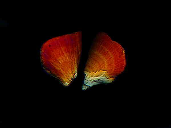
I’ll give you another example. I have a nice little Leopard plant (also known as Violet Squill–Scilla violacea). Again, I made up 2 slides and as I scanned the first, I found lots of acicular crystals.
I had rather expected that and so was not disappointed at all. However, as I scanned the second slide, I came across what I have dubbed the “clam shell” crystal.
I was quite taken with the vibrant red and then got curious regarding this crystal’s appearance in only partially polarized light.
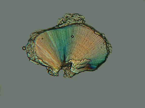
Here you get a clearer idea of why I call it the “clam shell”. Around the edges, bits of plant tissue are still visible, so this is clearly not a sand grain or some other artifact, but rather a crystal which occurs within the plant itself.
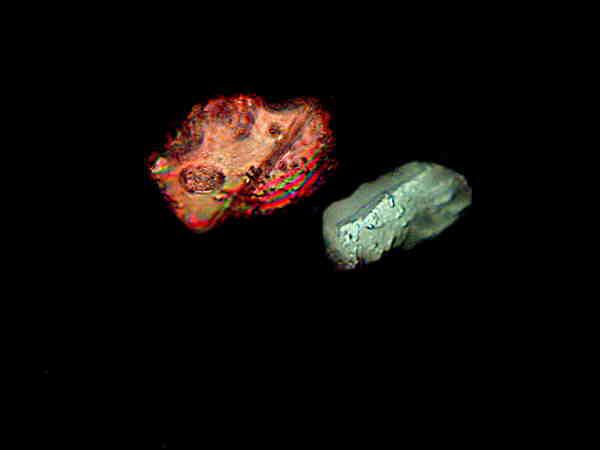
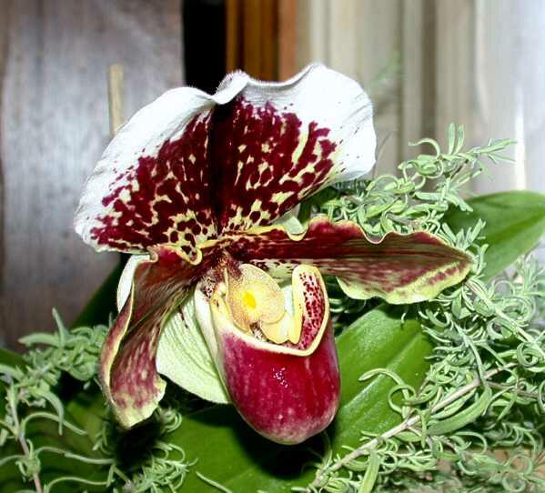
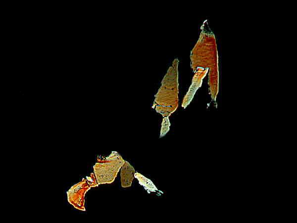
Some plants simply have no discipline and will produce a variety of crystals that seem to have no relationship to each other. I have a large Lady Slipper orchid which produces 1 to 3 blooms a year. The wonderful part is that the blooms will last over 2 months.
The leaves, like that of the iris, are tough and fibrous, but I managed to scrape enough juice for a couple of smears. On the first slide I found some typical acicular crystals.
Notice that there are also 2 very small, roughly disk-shaped crystals.
Then I came across an elongate crystal, but with blunt ends.
This one might be described as a little minuet of pastels.
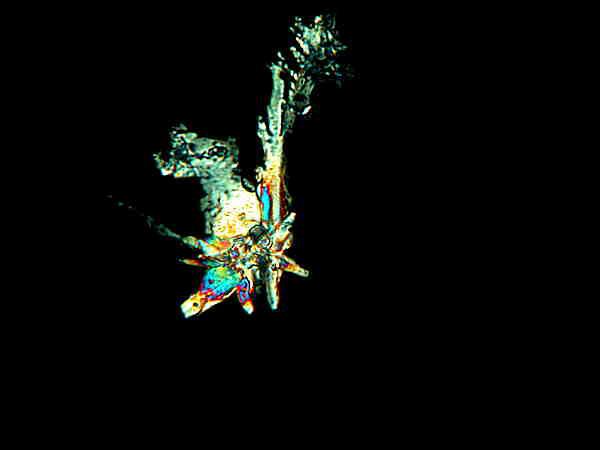
The second slide was such a surprise that I went back and double-checked to make sure that I hadn’t misread the slide label. Here are 3 crystals from the second slide.

The ancients knew about the deviousness of nature, for in virtually every mythological pantheon there is a trickster figure. I guess we shouldn’t be surprised about orchids being a bit quirky and whimsical, since there is such an enormous variety of them and they can be found everywhere from tropical rainforests to alpine meadows.
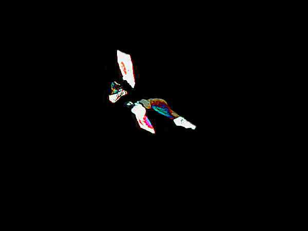
However, there are other plants that are even more playful when it comes to crystal displays. I suspected from the outset that insectivorous plants would be unorthodox simply because they are such bizarre organisms in so many other respects. I have a small cluster of cobra lilies with slender “pitchers” about a foot high.

They did not disappoint. They provided these odd blue and gray abstract paintings. I’m going to copy these and paint them on 12 foot by 18 foot canvases and sell them to the Museum of Modern Art for a fortune.
However, for sheer inconsistency, irrationality, and feminine madness (Mother Nature’s, of course–please no feminist e-mail diatribes), the sundew, Drosera, takes the prize. I expected something unusual, because of the oddity of the plant and the fact that it has one of the strongest glues in all of nature, but I didn’t expect everything from a 2 lobed acicular bundle to Miro paintings. In order to show you graphically what I mean, I’m going to use 6 images.
Who would have thought? A miniature botanical art gallery hidden in the juices of a plant that eats gnats and flies!
Finally (I know you’ve been waiting for that word), the crystals of a plant which Freud and I have diagnosed as being profoundly schizophrenic–the Daylily. I shall show you just 2 images (which is appropriate as an illustration of its bipolar personality).
A lovely disk comprised of countless numbers of extremely small acicular crystals.
However, on the second slide, these acicular crystals “decided’ to do something quite different.
Not only is the shape radically different, but the range of color as well. Nonetheless, one can clearly see the “needles” composing the aggregate and one antisocial crystal in the upper right-hand corner refusing to join the group.
As I sit here at the kitchen table scribbling away with my fountain pen, I am looking at a vase of rich, pink roses on the table and wondering whether or not their juices contain crystals. I suspect it will be several months before I forget to remember to wonder about this with every plant I encounter. Next summer my curiosity may forcefully reassert itself and I might have to write yet another sappy article on crystals.
All comments to the author Richard Howey are welcomed.
Microscopy
UK Front Page
Micscape
Magazine
Article
Library
Please report any Web problems or offer general comments to the Micscape Editor.
Micscape is the on-line monthly magazine of the Microscopy UK website at Microscopy-UK