The continuing developments in TFT, LCD and LED technology has prompted a variety of papers by workers assessing their use in optical microscopy for multi-mode lighting techniques. In some cases the traditional substage condenser is supplemented e.g. by creating programmable 'filters' (1,1a), in others the condenser is replaced. In 2015, Dr. Kevin F Webb (2) published a fascinating paper describing the use of LED rings to replace the condenser to offer a variety of contrast enhancement techniques including phase, darkfield, annular oblique, Rheinberg and mixed. His work used an inverted microscope where a particular cited benefit was the increased working distance between subject and illumination to allow better access for e.g. electrophysiological probes. In the Conclusion he noted their likely wider application including for upright microscopes.
Many hobbyists like
myself
who have built up an upright microscope system over the years as budget permits are fortunate to have condensers which offer a range of techniques (in my case for a used Zeiss Photomicroscope III). But this may not be the case and many of the potential benefits which Webb proposes may also be applicable to the hobbyist's use of upright microscopes. This includes trying to source increasingly hard to find condensers for older microscopes or if one or more phase rings in a condenser don't match
phase objectives owned. Webb's approach* seems to offer a cost-effective route for the hobbyist seeking one or more techniques for their upright microscopes—a ready supply of cheap rings are available, the so-called 'halo rings' or 'angel eyes' being used in many modern car lamps.
* Dr. Webb used both an off-the shelf chip on board (COB) LED ring and an Aura Phase Contrast Illuminator (Cairn Research)—the latter uses concentric LED rings and a design based on Webb's work.
I was keen to explore their use on my Zeiss Photomicroscope and how they performed compared with Zeiss condensers. In particular their use for Rheinberg and coloured phase as coupled with the microscope's base lamp, they potentially offer independent control of the light intensity for both colours, which is trickier with conventional condensers. The Photomicroscope system shares many modular parts with other Zeiss models such as the Standards so trials may be more widely applicable.
Car lamp LED 'halo rings' - sizing, power requirements and sourcing
Webb's work carefully reports all the sizes and working distances tried which was valuable in selecting two sizes likely suitable for the smaller working distances on an upright cf an inverted model.
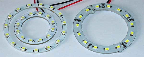
Sizing (refers to OD of the board). Left - 40 mm (12 x SMD) and 60 mm (15 x SMD) in one style and the two used predominantly. Right - a 60 mm in a more robust format. The latter was found to give more stray light in darkfield mode so the beam characteristics may vary between designs. The SMDs are flat, vertically facing and appear to be lensless—a typical spec sheet describes
them as having a 'yellow diffuse flat mould'. 40 mm is the smallest have seen but other sizes up to 120 mm in 10 mm incremental sizes are available. The number of SMDs used increase with diameter.
Rating - Full board 12V. The boards use the so-called '1210' also known as the '3258' SMD LED (see www.lighthouseleds.com for a typical data sheet). Each typically rated at 3 - 3.2V so they are likely wired in series of three with dropping resistors and each set in parallel across the supply (4 SMD resistors are on each board).
Current demands are modest at ca. 75 mA for the 40 mm and ca. 90 mA for the 60 mm i.e. battery use is possible. I measured 3.1V across each at 12V so may be near their max rating, in which case overdriving could be inadvisable.
Sourcing - Widely available on eBay, Amazon Marketplace and from car part suppliers. Typically cheaper direct from Asia but check feedback of seller. Those above were £1-89 a pair for the 40 mm and £2-19 a pair for the 60 mm both including free shipping from China.
Intensity control - All the rings tried lit up at ca. 7V across the ring with good even control of intensity as the voltage / current was increased to the max rating of 12V.
Various approaches to powering LEDs are recommended and have been widely discussed. I prefer the simple variable voltage / current lab power supply with a dropping resistor. The accurate V/I readouts are useful when understanding their mode of action and repeating settings. If not owned, the PSU is likely the most expensive part of the project but could prove valuable in other roles.
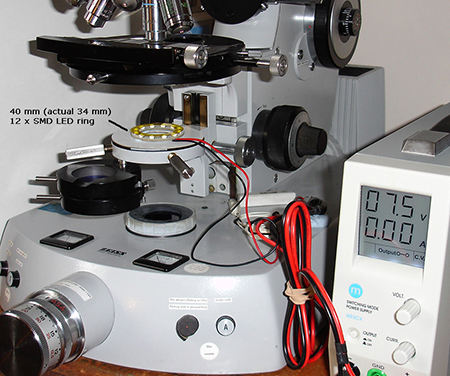 In use on a Zeiss Photomicroscope III
The suitability and ease of use of LED rings on an upright microscope may vary widely between models. Those with built-in lamps would also offer the full control of lighting / colours for Rheinberg and colour phase as described later. The large Photomicroscope was ideal with its easy access to the substage and long travel focussing condenser mount.
Right. The 40 mm ring (actual 34 mm) 12 x SMD ring in use at the focal plane to match the Zeiss phase ring II. Annular coloured acetate filters are easy to make with compass cutters and sit on top. The PSU is a Maplin N93CX 20V/5A switched mode supply that I bought secondhand a few years ago for ca. £45 and is also used for both smaller tungsten lamps and high power LEDs for fluorescence. It can be set at either
a constant current or voltage limit to avoid overdriving LEDs.
A suitable resistor in series is usually important for this mode of use and the one shown (the white block) is a 10W I use for power LEDs in fluorescence work but the wattage required is very modest for the rings. The rings have dropping resistors on the boards so could probably be run across a supply without an external resistor.
Phase
By changing its focal plane below the substage, the nominal 40 mm diameter ring matched all the phase rings in the Zeiss 160 mm tube length range. For phase I, the ring sat on the field iris housing, phase II and III (immersed objectives) were 70 mm and 32 mm from the stage respectively. The back focal planes of typical objectives are shown below.
Of the subjects studied using both the Zeiss phase condenser and LED ring I could see little difference between the phase imagery using dry objectives. Webb showed this to be the case in his paper up to the very highest NA objectives (i.e. an Olympus 100/1.65). The classic fresh buccal epithelial cell smear is compared below, as an example. The limited studies to date with immersed objectives suggest a lower contrast result with LED rings that was hard to recover in post-capture editing, see the diatom example below. This may be an imaging related problem using my Canon 600D DSLR body. Flipping between the two setups quickly to establish if they differed visually was not easy.
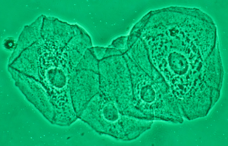
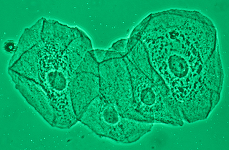
LED ring Condenser
Fresh buccal epithelial smear in phase (same acetate filter used for each). Zeiss 40/0.75 Neofluar (for use with a II phase ring). Left - using an LED ring as 'condenser', right using a Zeiss phase condenser. At ISO100, exposures were 13 sec for the LED at full rating and 0.5 sec for the condenser at reduced 100W lamp power. (Canon 600D body, raised Zeiss 10x Kpl W eyepiece as relay lens.)
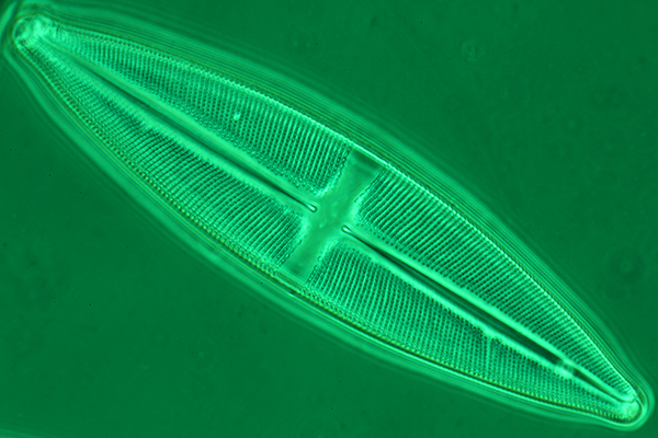
Above. Zeiss phase III ring, achromatic-aplanatic condenser.
Zeiss 63/1.25 Neofluar phase III oil, water immersed under slide.
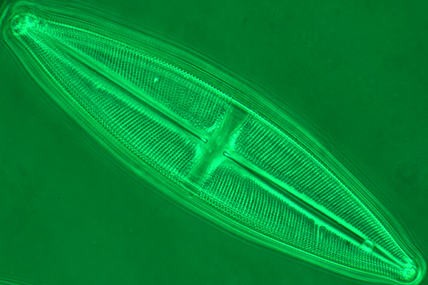
Above. 40 mm LED ring alone.
Zeiss 63/1.25 Neofluar phase III oil.
Above. Diatom Stauroneis phoenocenteron (Klaus Kemp 8 form test plate.). The same green Lee Filter for both. Condenser image out of camera, lower adjusted for similar tonal balance but still differed in micro-contrast.
As Webb notes in his paper, the LED rings do not face inwards (as they do in stereo ringlights). The wide beam angle of the LED is thus being exploited at different focal planes to match a required NA. This is evident by the more oval appearance of each LED in the back focal plane as focus the ring more closely (see images below). A disadvantage is that it is not a very efficient use of the LEDs as light intensity off-axis will fall off. The LED rings were bright enough for visual work but there was little in reserve. This would make them less suited for freezing action in live subjects as long camera exposures are required.
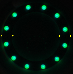
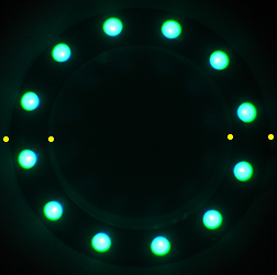
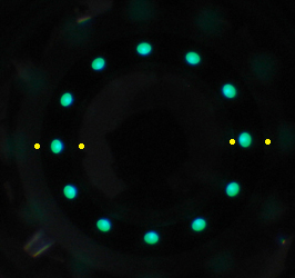
Back focal planes of Zeiss phase objectives using the 40 mm LED ring at different focal planes to match the phase plate.
Left to right - 10/0.22 phase I, 16/0.40 Neofluar phase II, 63/1.25 Neofluar phase III oil. The yellow dots show the boundaries of the phase plate.
Although the LED ring is closest to the stage for the phase III ring, the LED profile is smallest as exploiting off-axis lighting. This has implications for the efficiency of LED rings used in this way.
Circular oblique lighting (COL) and darkfield illumination
By focussing the 40 or 60 mm ring progressively closer to the stage, first COL (annular illumination) and then darkfield could be achieved with appropriate objectives. Darkfield is available on some of the Zeiss phase condensers which offer the 'D' position or by using their dedicated darkfield condensers. COL can often be achieved by matching a phase ring or 'D' setting with the appropriate NA objective. But the LED rings offers a ready way of accomplishing this for certain objectives if a suitable
condenser isn't owned.
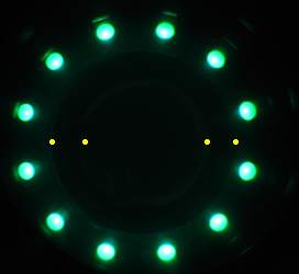
Back focal plane of Zeiss phase 16/0.40 Neofluar phase II objective with LED ring focussed near outer edge of full aperture for COL lighting. The yellow dots show the boundaries of the phase plate.
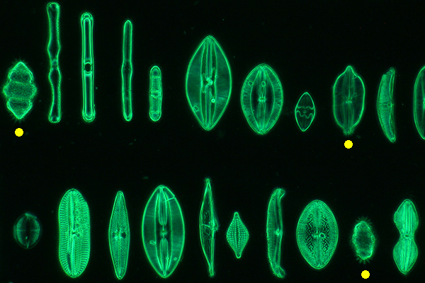
Above. Darkfield image of part of an arranged diatom slide (by Klaus Kemp, Maria Madre fossil marine forms) using the 40 mm LED ring and the Zeiss Neofluar 16/0.40 objective. An artefact was noted for parts of the subject as they became increasingly out of focus, shown with the yellow dots, i.e. stellated light tracks. With conventional darkfield methods an even out of focus image is more typically seen. If this effect is not specific to my microscope / camera, this may limit this approach for stacking of images, in my 'stacking' trials the software identified the light tracks as part of the image in focus.
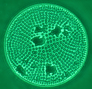
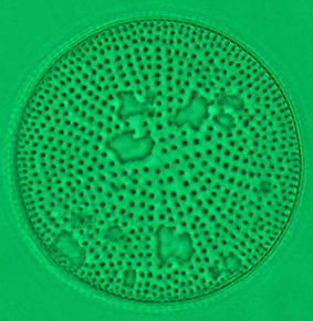
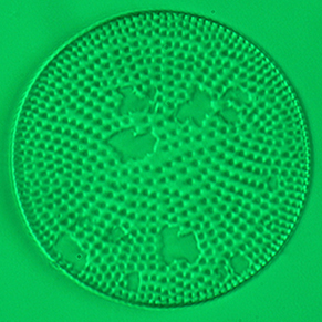
Above. Selected diatom from the above Maria Madre arranged slide by Klaus Kemp. Zeiss 40/0.75 Neofluar phase.
Left and middle. 40 mm LED ring alone at different focus planes create from left - phase, middle - circular oblique (COL).
Right. Brightfield using slight off-axis oblique using a Zeiss achromatic-aplanatic condenser.
Rheinberg
Traditional Rheinberg filters inserted within or just below the condenser in a filter tray, usually require high precision matching of the shaped, often small colour pieces to ensure no light gaps, as well as ideally leaving no traces of their immobilisation e.g. glue. One of the benefits of LED rings for Rheinberg is that because the emitters are discrete, high precision filters are not required without compromising the effect as shown below. A filter
on the lamp base provides the background
colour, in this case green.
Unlike normal Rheinberg filters which use a single light source*, the intensity of the background (with lamp PSU) and outer colour effects (with LED PSU) can be separately controlled for obtaining the often critical correct balance between the two. (*With conventional methods using a single light source, selective use of cross polarising filters are typically used for independently controlling intensity but adds complexity to the filter manufacture.)
LED ring only: For Rheinberg, I initially used the LED ring on the condenser mount, but the background light from the internal lamp only covered a small area of the back focal plane (BFP) of the objective for higher NA objectives, giving a 'stopped down' look for the part image contributed by the background. This is not ideal as a cited feature of Rheinberg is to fill the full aperture of the objective with the background colour.
LED ring plus LWD condenser for background: The 40 mm ring sat neatly on the condenser top of the LWD condenser and this provided better background coverage for an uncorrupted image. The use of an LED light ring with a LWD condenser as shown, essentially adopts for modest NA objectives the same approach as the very neat custom made condenser for Rheinberg described by Paul Martin in his Modern Microscopy article published in 2014 (4) for use with high NA objectives. He used a light ring lit by fibre optic cable with outbound filters in the cable line but retained a high NA condenser and immersed where needed.
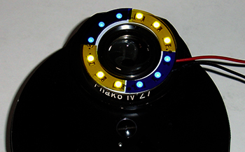
Above. Rheinberg setup. The 40 mm LED ring sits neatly around the LWD Zeiss condenser and with top lens removed provides the required NA for lower power objectives at the plane of focus required for a Rheinberg effect with the filters. The green background is provided by a filter sitting on the lamp base of the microscope.
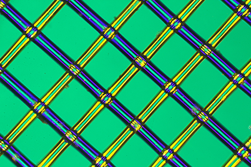
Above. A teabag (used by Dammann Frères for their 'Cristal' range of teas) made from a fine clear plastic weave 0.25 mm mesh rather than the more typical random weave of a paper based teabag. Leitz NPL Fluotar 6.3/0.20 objective using the same colour sectors as shown above.
Colour phase
Colour phase is a well established technique, see for example René van Wezel's May 2004 article 'Colour phase contrast'. The conventional approach requires carefully made phase plates and as for Rheinberg, selective use of part polar filters if also require independent colour intensity control. The same approach used above for Rheinberg can be adopted for colour phase with the same simple filters, except the LED ring is focussed
to match the annular plate of a phase objective and the microscope lamp with filter of choice providing the colour background.
Trials to date have been more successful visually than photographically and limited to lower NA objectives. The Canon 600D DSLR body used did not always faithfully record the subtle colour shading differences of some subjects seen visually. (This potential limitation of consumer digital cameras has also been noted by René van Wezel in his colour phase work and by Paul Martin in his Rheinberg studies.)
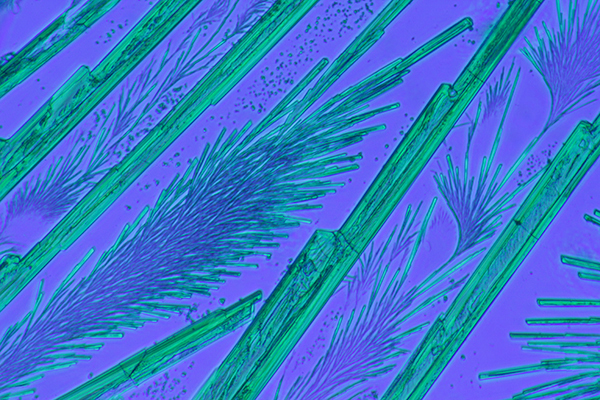
Above. Colour phase Zeiss 10/0.22 objective. Barium platinocyanide antique slide by unnamed mounter. Two dark blue filters on the microscope lamp gave the background and two dark green on the LED ring for phase.
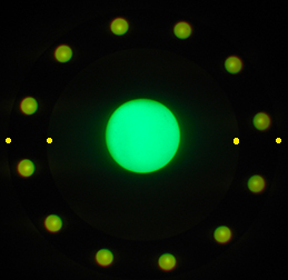
Above. The back focal plane of the Zeiss Neofluar 16/0.40 phase objective set for colour phase. The yellow dots show the boundary of the objective phase plate. Yellow filter on LED ring for phase, green filter on microscope lamp for the background. Without a condenser the microscope lamp is not occupying a good proportion of the back focal plane of the objective. This becomes more marked for higher NA objectives giving a 'stopped down' look to the image contributed by the background. A similar problem occurs for Rheinberg if a LWD condenser isn't used in combination. Lenses tried to date below the substage to try and correct this have been unsuccessful to date. (Webb in his work with inverted microscopes used a central LED with diffuser for the background but I would prefer to make use of the microscope lamp to keep the setup simpler.)
Observations to date
I have only tried more traditional methods to date but a variety of both mixed and unconventional approaches could be explored. For example, the easy of access to the LED rings allows the emitters to be selectively shaded off or filters added to modify a conventional technique (as K F Webb suggested in his paper using inverted microscopes).
Some thoughts on their potential for the hobbyist on an upright microscope from my trials with the Zeiss Photomicroscope are offered below.
Potential value if phase / darkfield condenser is owned.
- creating an illuminated annular ring for a phase objective if correct ring not available. (My three Zeiss phase condensers don't offer the number I ring and have had to make one for use to date).
- ready creation of Rheinberg and colour phase for modest NA dry objectives without the need to make precision filter stops or phase plates.
- independent control of the light intensity of both the central stop and outer ring allowing the desired balance to be found for different subjects that is particularly important in techniques such as Rheinberg and colour phase. (With conventional methods using a singe light source, selective use of cross polarising filters are typically used for controlling intensity.)
Potential value if phase / darkfield condensers not owned.
- cheap, readily available route to create the correct phase ring for one or more phase objectives.
- easy to create other techniques which a condenser would usually offer such as darkfield, Rheinberg and mixed techniques e.g. colour phase.
- may suit older microscope models where the correct legacy phase condensers are hard to source and/or expensive.
Potential disadvantages of car lamp LED rings cf condenser use from trials to date.
- intrinsically inefficient cf conventional microscope light source as using off-axis illumination from the flat SMD LED emitters.
- the rings tried at their full rated voltage of 12V were bright enough for visual phase although less so at higher powers and require long camera exposures which would not suit studies of live subjects. (Unless an LED flash mode could be developed where the emitters were used briefly much more intensely as found in smartphone 'flash'.) Conventional phase condensers can use an electronic flash source.
- on my microscope, in darkfield mode the rings gave stellated light artefacts from out of focus areas that may make stacking of images more difficult.
- if using the microscope lamp alone for the background colour for either Rheinberg or colour phase, it may only partially fill the back focal plane of the objective, giving a 'stopped down' look for that part of the image. Sitting the LED ring on a low powered condenser can negate this for more modest NA objectives.
An additional role of LED rings could be in education. It is easy to appreciate how the LED rings are acting as a light source (with or without filters) and how at different focal planes their position in the back focal plane of the objective changes to create the various techniques. With conventional condensers with their hidden optics and often filters, how they are acting may not be as obvious.
In my own hobby level studies, ready creation of Rheinberg and colour phase are the most appealing use of the rings and an option to explore other techniques which Webb notes such as 'relief contrast' and who cites the work of Piper (2,1a). As I own conventional Zeiss condensers, they will of course perform optimally in true Köhler mode and efficient use of the light source for conventional phase, darkfield and off-axis or annular oblique. But for microscope owners without suitable condensers because of e.g. budget constraints, difficulty in sourcing the correct legacy condenser or owning phase objectives without a matched condenser ring, the LED rings offer a wide variety of options at very little cost and they are great fun to use.
Comments to the author David Walker are welcomed.
Note added near completion of article. Facebook discussions in the Amateur Microscopy group of Webb's paper in July 2016 (3) show that hobbyists have been actively exploring the potential of LED rings for some time, in particular on inverted models and stereos, prompted by Kevin Webb's presentation to the Quekett Microscopical Club in 2014. It would be interesting to hear from fellow enthusiasts their experiences on using the rings on other makes / models of upright microscopes to build up a database for readers of what rings are best suited for different systems.
Acknowledgements
Thank you to Dr. Kevin Webb for sharing his work on LED rings with inverted microscopes which inspired the above trials on an upright microscope.
Thank you to Dr. Webb and Paul Martin for email communications. Any errors are solely mine.
Thank you to Ricardo Y Tsukamoto for highlighting the paper by K F Webb in the Yahoo Microscope Forum, May 22nd 2016.
Thank you to Klaus Kemp for his amazing skills in preparing arranged slides of diatoms.
References
1) e.g. C Zuo et al, Programmable colored illumination microscopy (PCIM): A practical and flexible optical staining approach for microscopic contrast enhancement, Optics and Lasers in Engineering, 2016, vol. 78, 35-47. Uploaded by the authors to ResearchGate.
1a) Jörg Piper and co-workers have also been very active in this area.
2) K F Webb, Condenser-free contrast methods for transmitted-light microscopy. Journal of Microscopy, 2015, 257: 8–22. doi:10.1111/jmi.12181. Open access.
3) Carel Sartory comment dated July 27th 2016 on the Facebook 'Amateur Microscopy' page. Thank you to Carel for sharing this.
4) Paul Martin, Rheinberg Illumination: A Fresh Approach to High Magnification Color Contrast, Modern Microscopy, dated August 26th, 2014.
URL: www.microscopy-uk.org.uk/mag/artaug16/dw-LED-rings.html
Uploaded August 14th 2016.