|
An Assortment of Marine Invertebrates From the Philippines Part 2: Other Echinoderms by Richard L. Howey, Wyoming, USA |
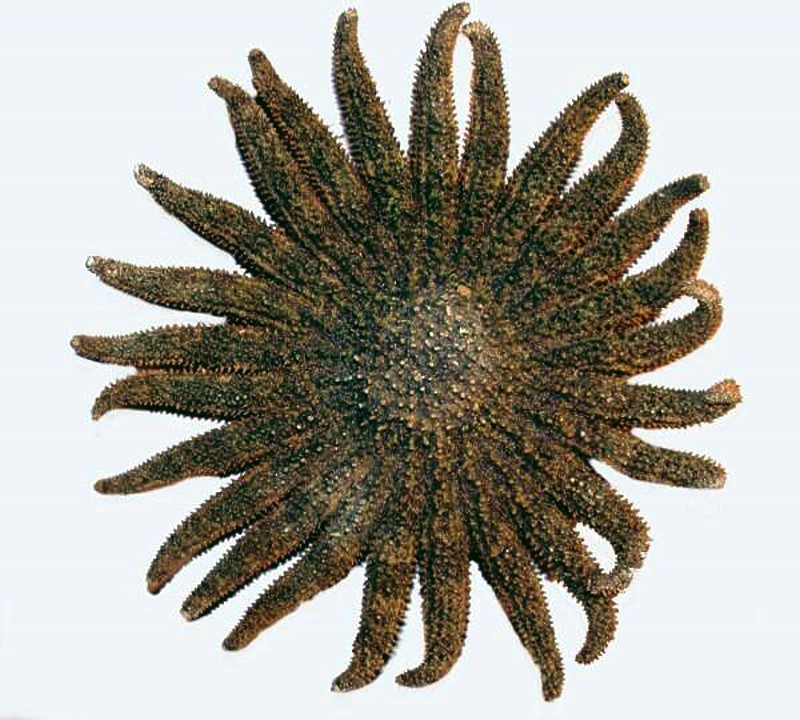
In Part 1, we looked at a variety of echinoids; in this part, I want to look at a variety of other organisms which I have gotten over the years from the Philippines beginning with a few starfish, which are, of course, not fish and not all of them are star-shaped. They are, however, relatives of the echinoids in that both groups are echinoderms. Let’s start with a real anomalous villain which for starters has, as you will see, 14 arms, but specimens can have up to 23 and can measure up to 15 inches in diameter. They show an astonishing variety of colors and are strikingly beautiful for such destructive organisms.
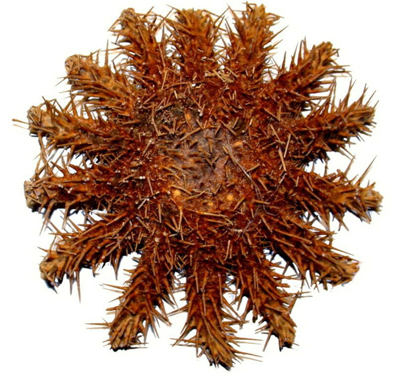
Here is a dried specimen which I have had for some time; it is brown and not at all impressive, except in terms of its size and masses of venomous spines, thus its popular name, the Crown-of-Thorns.
And now, let’s look at some of the really impressive specimens photographed in life - see this link.
These bizarre creatures can, under certain conditions, appear in enormous numbers and since they feed on coral polyps, they produce enormous damage to the coral reefs. Extensive research programs have been underway for some time trying to find means of controlling these pests and limit the massive damage they are doing. This is no easy task for as we know, you can’t just chop them up–many echinoderms are very good at regenerating and where you start with one and cut it up into several pieces you may end up with 2 or 3 or more new ones. Poisoning them is counterproductive since that also affects the coral reefs and the other wildlife around them. In some areas, divers have tried to clear and preserve small areas, but this has to be done with caution since the spines contain a toxin which in contact with the skin produces significant pain, blistering, and even ulceration.
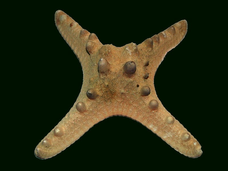
Here’s another fluke; Protoreaster nodosus, also known as the knobby starfish, is very common in certain parts of the world and typically has 5 arms, but here’s a specimen with only 4 and it has also lost most of its color.
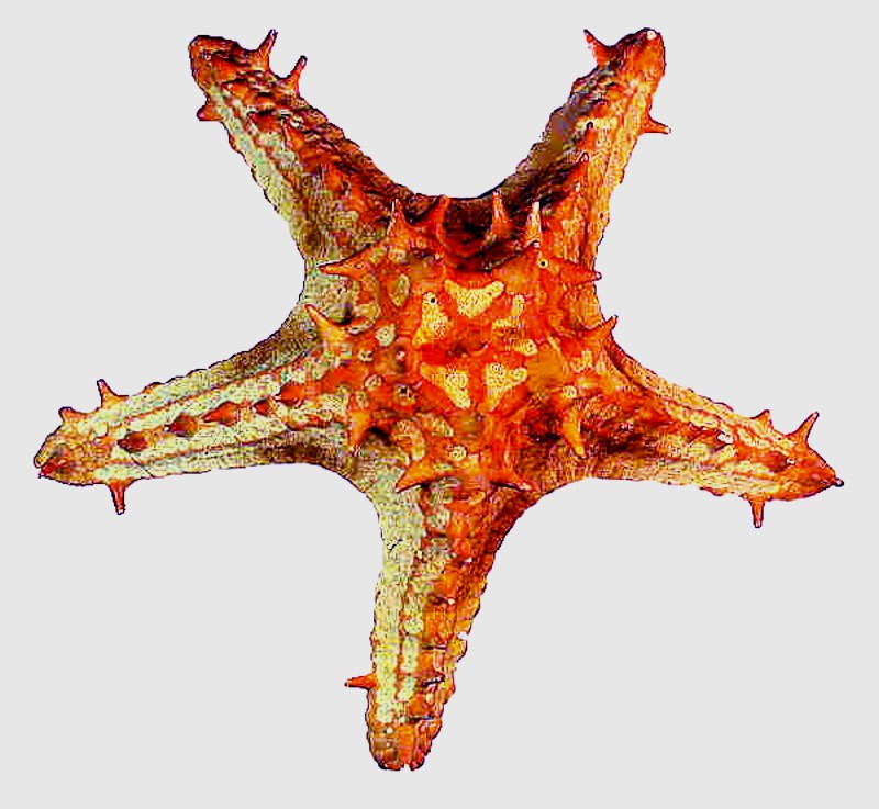
And here is a different species of the same genus and this one has the typical pentamerous symmetry and has retained some of its color.
A striking starfish is Linckia laevigata with its deep blue pigmentation and, yes, it’s natural, not dyed. However, I discovered accidentally that if left in sunlight, it will bleach out.
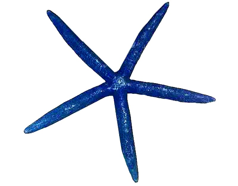
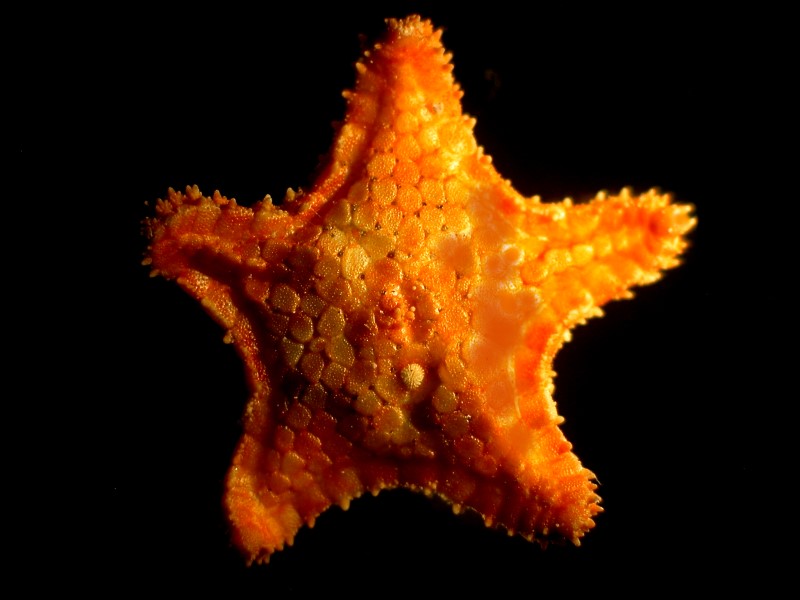
As you can see, there is a great deal of morphological variation just in these 4 examples. These are all of substantial size, the smallest being the one with 4 arms which is 3 inches across, Linckia 6 inches, the large Protoreaster 8 inches, and the Crown-of-Thorns 13 inches and here’s another sizeable one, “sun star” (Heliaster) with 25 arms.
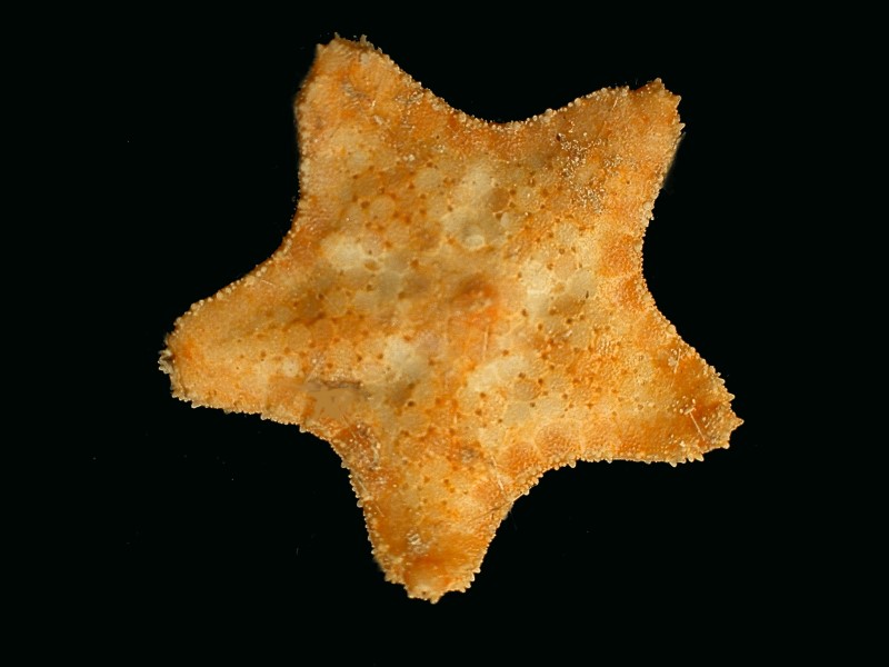
However, in one shipment, were a number of really tiny specimens and by that I mean only about 1 inch across. This little orange and yellow sea star reveals that the aboral surface consists of a series of small plates and just below the center is a white, round structure. This is the madreporite, that is, a filter into the water vascular system which controls the tube feet.
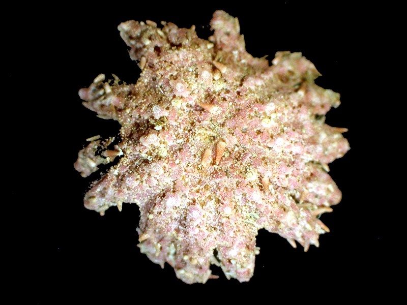
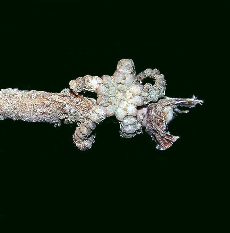
Next is what could be taken for a blob of debris, but turns out to be a multi-armed starfish with small spines scattered around the surface.
At first glance, this next example looks rather like the earlier orange star, but it quickly becomes evident that it is quite different; there are no plates and the surface looks to be much more granular in character.
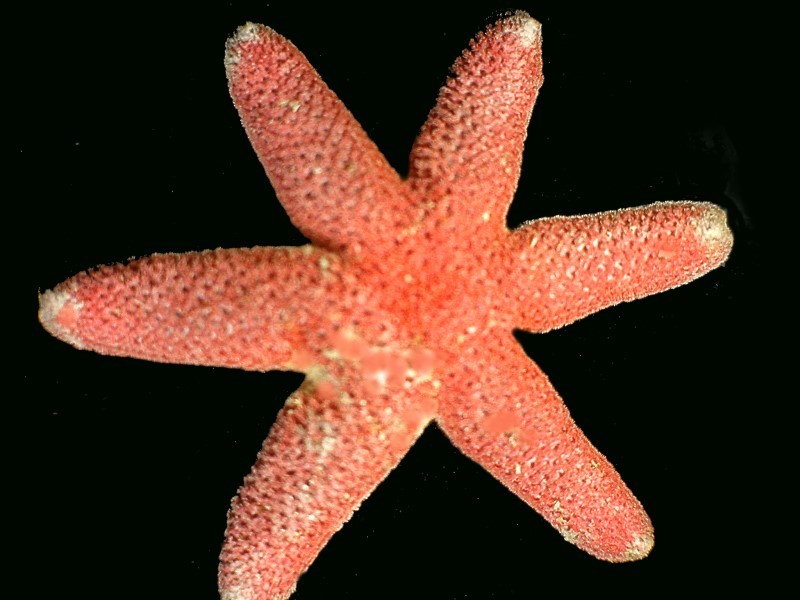
Then we come across a cheerfully red starfish that looks like what a starfish should be–except it has 6 arms–Oh, Mother Nature, will you never stop toying with our patience! What next, NO arms??
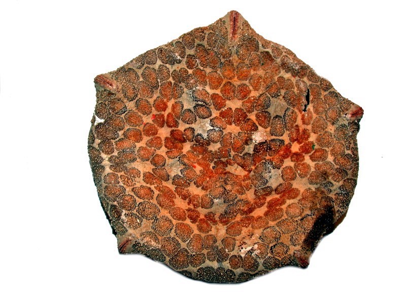
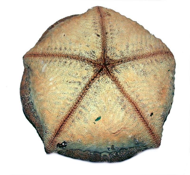
Well, O.K., sort of. I’ll show you 2 images of Culcita, the “cushion” starfish. The first is an aboral view and the second of a different specimen is the oral view. Each is about 4 ½ inches across.
Quite unusual looking creatures. Next thing you know, we’ll be finding specimens with tiled surfaces, tessellation. And, indeed we shall; first, I’ll show you the oral side of a small starfish and here we see the intricate patterns that are repeated over and over.
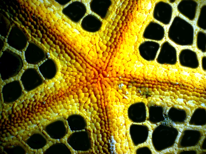
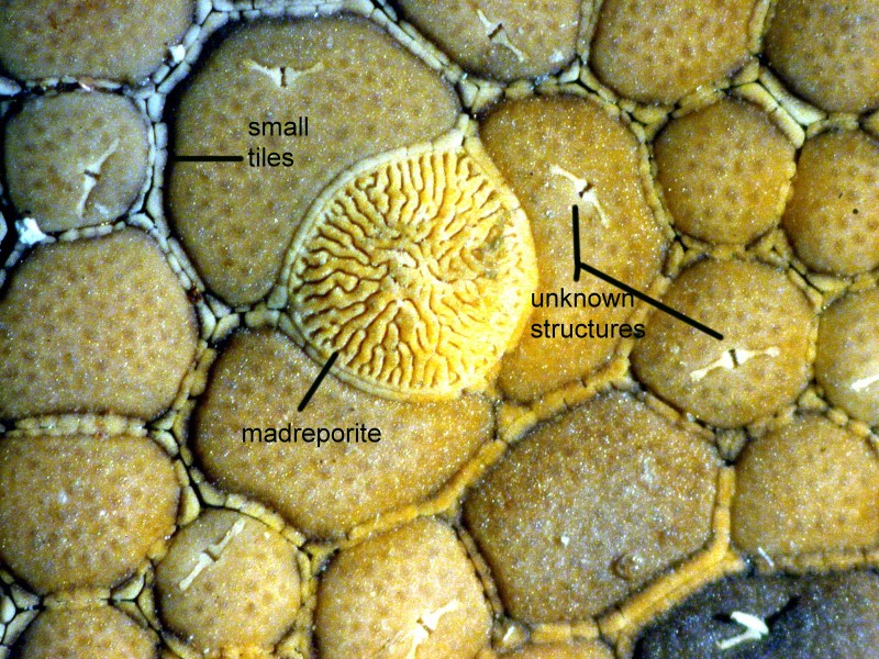
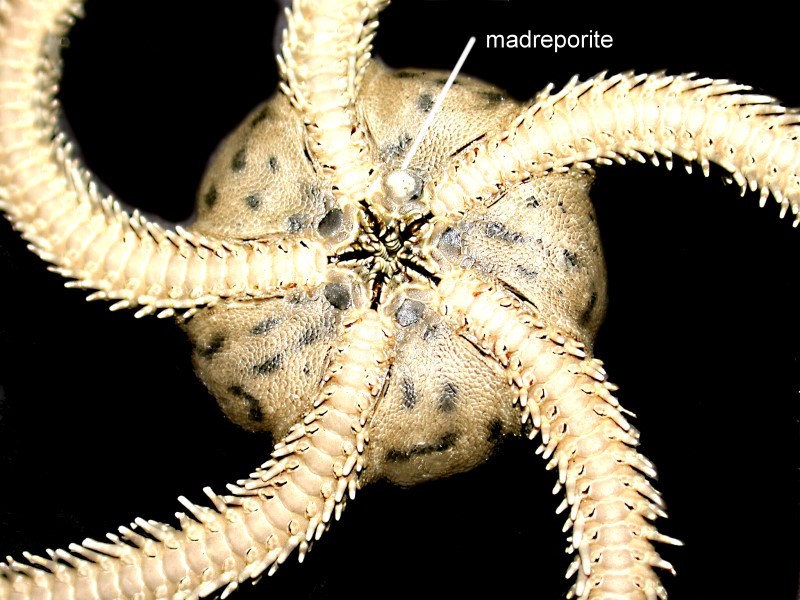
Now, if we flip over to the other side (aboral), we find in the central disk, the madreporite surrounded by tiles (plates) of relatively substantial size. The madreporite is, as mentioned earlier, that filter which looks like a bit of coral and keeps debris from entering and creating blockages in the water vascular system which controls the extension and withdrawal of the tube feet.
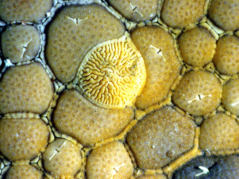
Note that around each of these larger plates, there are large numbers of small plates outlining them. There is also a tiny odd structure on some of the large plates which looks a bit like a bird with wings outstretched. I have been unable to find any references to such a structure in my examination of the literature but, surely, it must have been described before. I will show you the same image again with labels to the small tiles, the madreporite, and this odd structure.
Now, here’s a bit broader view showing a portion of the central disk and also the tiling along the edge of the disk leading down into the arm.
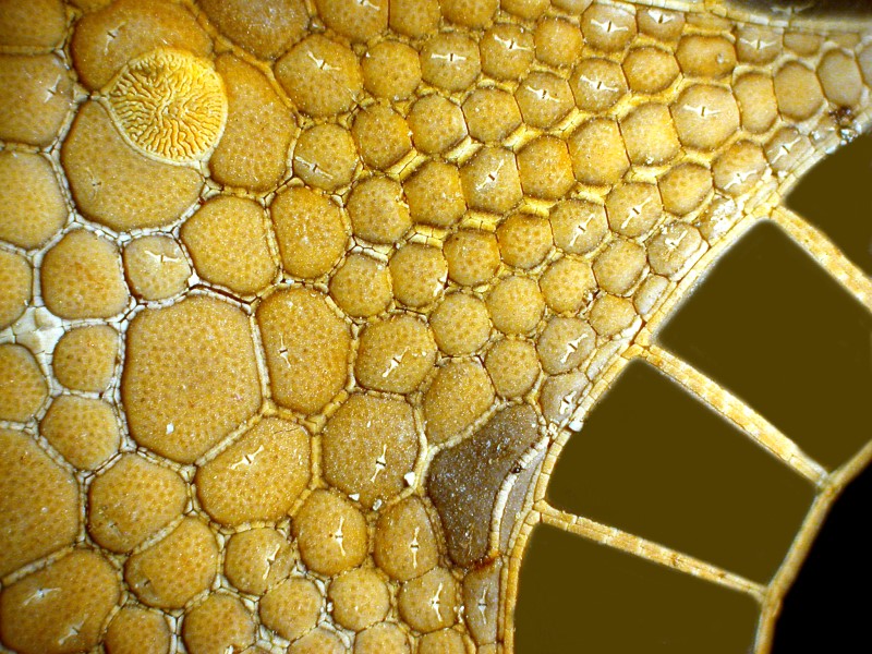
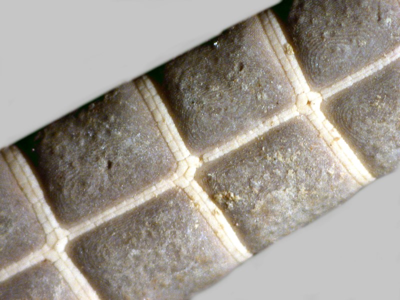
If we look at a section of the arm, we can see both macro- and micro-tiling.
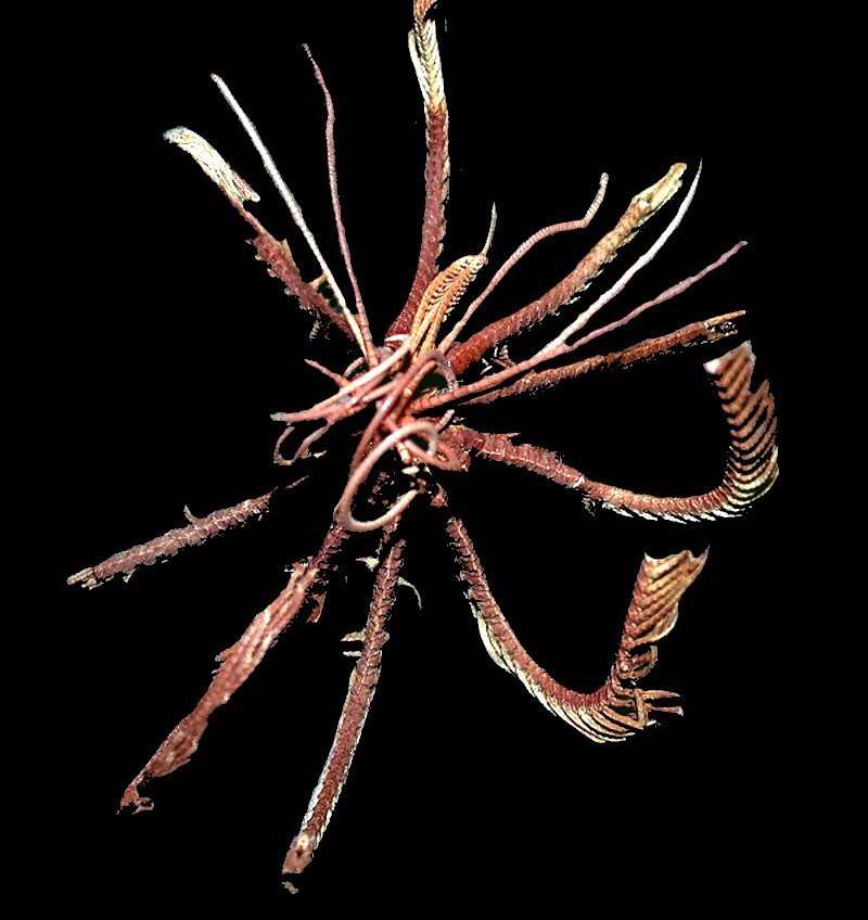
Next, let’s move to a related group, which at a casual, non-critical glance may appear quite similar but, are in fact quite different: the ophiuroids or “brittle stars”, also known as “serpent stars” partly from the derivation of their name in Greek which is descriptive of the motion of the arms. Here is a tiny one clinging to a sea urchin spine.
They are indeed stellate in form and almost always have 5 arms. So, how does one distinguish them from the classical “sea stars”? Fortunately, there are several criteria that allow one to make this determination quite readily: 1) The madreporite in starfish is on the aboral (dorsal) surface whereas in ophiuroids it is located on the oral (ventral) surface–when visible; it is often difficult to detect at all. 2) The oral surface of a starfish has a “channel” running down its length and that is where the tube feet are located, whereas in the ophiuroids, there is a series of plates running down the length of the arms and no channel. The tube feet are reduced to papillae. 3) The connection of the arms to the central disk in starfish is such that they are usually seen as an extension of the disk, whereas in the ophiuroids, the arms are quite distinctly demarcated and it is this jointed character that allows for the fluidity of their movement.
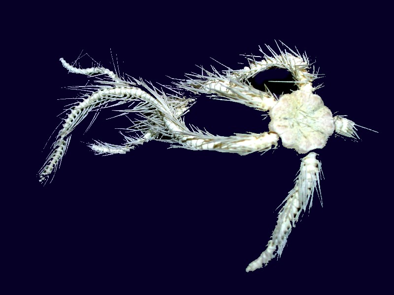
Some species have a “hairy” appearance due to a large number of spines. Below is an example of such a specimen which was damaged on arrival. These creatures are indeed “brittle” and can shed arms when attacked.
However, perhaps the most astonishing thing about these organisms is that at least some species have thousands of eyes! Gordon Hendler, a marine biologist, discovered that Ophiocoma wendtii is sensitive to light. It’s surface is covered with enormous numbers of tiny, calcareous crystalline lenses. In collaboration with a researcher, Johanna Aizenberg of Bell Laboratories, it was discovered that these lenses are superior to what we can produce in that they overcome spherical aberration and birefringence and that their focal point is precisely on bundles of nerves that lie beneath the surface of the lenses.
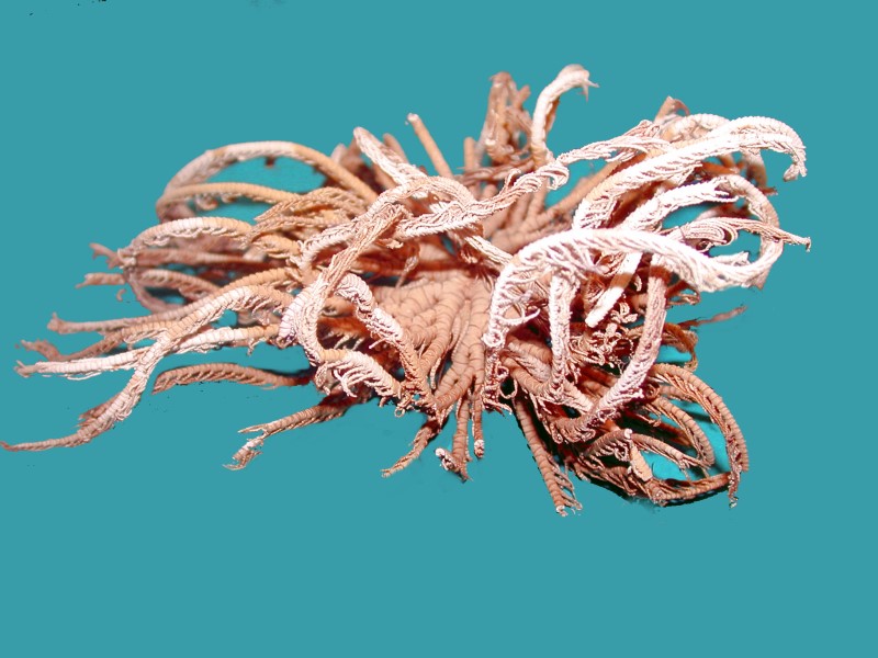
Another group of relatives of the sea stars and brittle stars are the crinoids and comatulids. These are popularly known as the sea lilies and feather stars. Sea lilies have stalks and are anchored to a substrate whereas feather stars have specialized arms that can attach to a substrate and then move to a different location if necessary. Sea lilies are essentially permanently attached. Below are 2 images of small comatulids. As you can see, there is a great deal of branching of the arms.
This gives you a good sampling of echinoderms that are available from the Phillippines and in Part 3 we will look at organisms from other phyla.
All comments to the author Richard Howey are welcomed.
Editor's note: Visit Richard Howey's new website at http://rhowey.googlepages.com/home where he plans to share aspects of his wide interests.
Microscopy UK Front
Page
Micscape
Magazine
Article
Library
Published in the August 2017 edition of Micscape Magazine.
Please report any Web problems or offer general comments to the Micscape Editor .
Micscape is the on-line monthly magazine of the Microscopy UK website at Microscopy-UK .
©
Onview.net Ltd, Microscopy-UK, and all contributors 1995
onwards. All rights reserved.
Main site is at
www.microscopy-uk.org.uk .