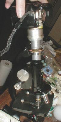
Adapting an Olympus C-2040 digicam to a compound microscope

by David Young, Iowa City, USA


The Olympus C-2040z is difficult to adapt to use with microscopes, so I hope my experiences and results with a homemade adapter proves useful for owners of this and similar cameras in the range. It has already proved to be an ideal and fairly low cost camera for macro photography using simple lens attachments. (See my previous Micscape article).

Design:

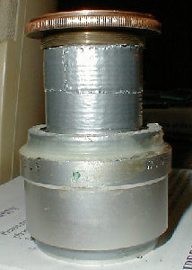
As you can see, it's basically just a tube with a relay lens situated close to the camera's own zoom lens. I don't know the exact power of the relay lens, but it should be as large or preferably a bit larger in diameter than the camera lens. Since the relay lens replaces the microscope eyepiece, it must be a good quality lens, free of scratches etc.


To use:

The eyepiece is removed from the microscope after initially focusing by eye. The adapter is placed over the open end of the microscope drawtube. The sleeve at the end of the adapter tube allows it to slide over the end of the drawtube. (This prevents sideways movement which makes aligning the camera with the 'scope much easier!)

The camera can be aligned using the LCD image as feedback. Lens aperture should be about f2.8 and focus should be set to manual. Use the controls on the microscope to focus the camera. Exposure metering is set to matrix mode with the camera set to aperture priority mode. The optical zoom can be used to make moderate adjustments to magnification.

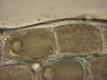
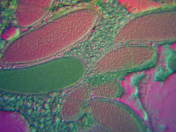
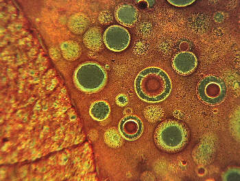
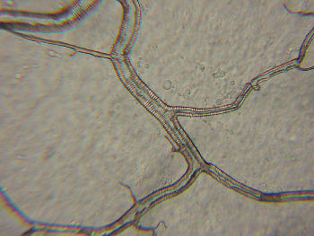
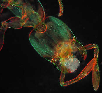
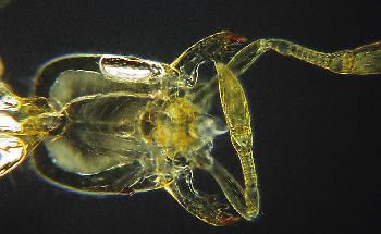
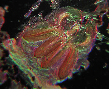
The photos below were taken with the digicam on an AO Spencer microscope.

Comments to the author David Young are welcomed.


Images by the author.

(Editor's note: To fit in the web page, the author's master images have been resized
and sharpened a little to correct softness introduced by resizing.)


Wasp eggs in various stages of development. I dissected the stinger
from a parasitic wasp which included unlaid eggs.   

Wasp eggs in various stages of development. 
Rheinberg illumination

Cross section of the spinal cord of a monkey.

Part of the tracheal system of a meal worm larva. 
Magnified 430x.

The head of a thief ant. A tiny 1/10th inch long ant that is 
common here in Iowa. Rheinberg illumination.

The head of a thief ant.

Part of the venom gland from a formica ant. 
Rheinberg illumination.


Microscopy UK Front Page
Micscape Magazine
Article Library


© Microscopy UK or their contributors.

Published in the December 2001 edition of Micscape Magazine.

Please report any Web problems or offer general comments to the Micscape Editor,
via the contact on current Micscape Index.

Micscape is the on-line monthly magazine of the Microscopy UK web
site at Microscopy-UK

WIDTH=1


© Onview.net Ltd, Microscopy-UK, and all contributors 1995 onwards. All rights reserved. Main site is at www.microscopy-uk.org.uk with full mirror at www.microscopy-uk.net.