|
|
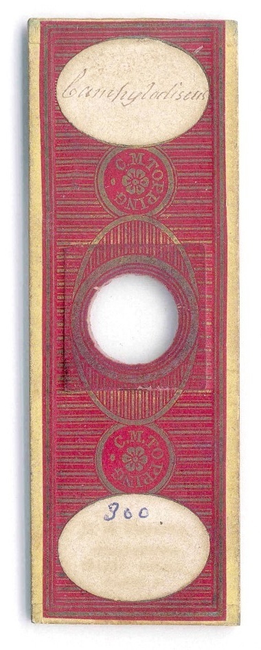
It was sometime between 1851 and 1874, when Charles Morgan Topping prepared the following permanent mount of diatoms. It is a "strewn" mount of Campylodiscus. One of a collection of several Victorian microscope slides I obtained a couple of months ago.
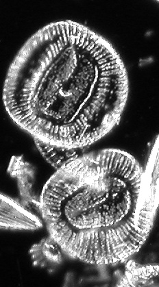
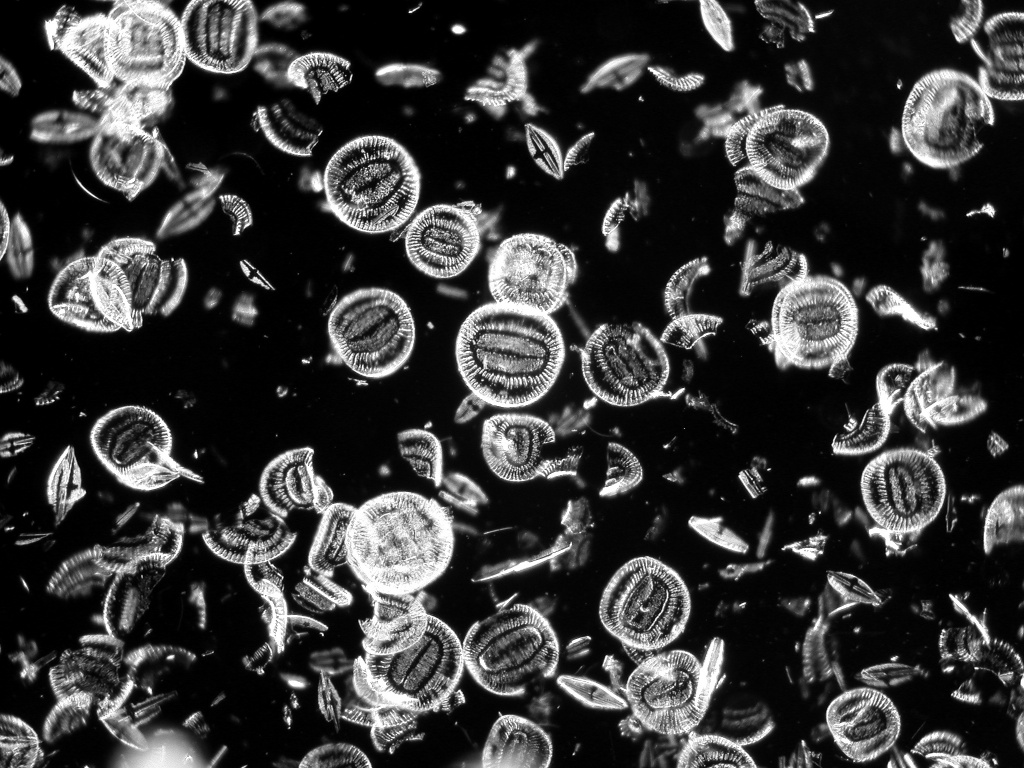
The species Campylodiscus belongs to the major group known as pennate diatoms. These diatoms are bilaterally symmetrical (symmetry about a line). A more thorough classification assigns Campylodiscus to the group Surirelloid. I find that the diatoms of this mount look very similar to one of the diatoms published by F.T. Kutzing in 1844 (see CAS Diatoms Database of the California Academy of Sciences Diatom Collection at http://www.calacademy.org/research/diatoms/).
For the reader not familiar with diatoms, diatoms are golden brown algae (class Bacillariophyceae). Their unicellular material is contained within a highly silicified cell wall called a frustule. Diatom cell walls are often used for permanent microscope mounts (such as for the famous diatom test plates by Johann Dietrich Möller and Klaus Kemp [1]). Diatoms are ideal objects for testing the resolution of a compound microscope [2]. For instance, the spacing between individual striae (lines of puncta) of the diatom Amphipleura pellucida is just around 270 nm.
I took the here presented photomicrograph with a Nikon Coolpix 995 digital camera, which was connected via a trinocular viewing body with a Nikon E200-F compound microscope. It shows a B&W image of diatoms known as Campylodiscus using dark-field illumination (see Fig. 1). One of the most famous mounters in history, Charles Morgan Topping, made this slide (see Fig. 2). He created wonderful mounts of highest quality [3]. The depicted slide was made after 1851 when C.M. Topping started to use gilt-decorated red paper [4]. Unfortunately, I am not able to read the second label on the slide. The print did not pass the test of time. This cannot be said from the mount itself. It aged only slightly over the last 140 years.
The interested
reader should consult the very good introduction into the slide making
of diatom mounts written by Klaus Kemp [5].
Comments to the author, Gregor Overney, are welcomed.
| [1] | Diatom test plates are available from various sources. I recently purchased a diatom test slide from Carolina Biological Supply Company, 2700 York Rd., Burlington, NC 27215, US (see http://www.carolina.com/). |
| [2] | Dave Walker, Giving microscopes a 'workout' using diatom test slides, Micscape Magazine, October (1999); Martin Mach, Test diatoms - what you can expect to see even with modest optics, Micscape Magazine, November (1999). |
| [3] | Brian Bracegirdle, Microscopical Mounts and Mounters, Seacourt Press Limited, Cowley, Oxford, England (1998). |
| [4] | Bob Nuttall, Artifacts as evidence: an early collection of slides prepared by C.M. Topping, Quekett Journal of Microscopy 39, 409 (2002). |
| [5] | Klaus Kemp, Making mounts of butterfly scales and diatoms, Quekett Journal of Microscopy 39, 263 (2002). |
Please
report any Web problems or offer general comments to the
Micscape
Editor,
via
the contact on current Micscape Index.
Micscape
is the on-line monthly magazine of the Microscopy UK web
site
at Microscopy-UK.