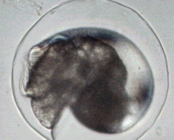
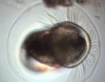
Baby snail in its egg. The picture was taken with polarized light and a x15 objective. Note the cilia in the right hand side picture.
|
|
MARINE SNAILS and PTEROPODS |
By Jean-Marie Cavanihac France |
|
|
|
Everybody knows the terrestrial snails, (some people eat them, but don't be afraid I don't eat them!) But many species of freshwater snails, marine snails and even marine slugs exist too. For an amateur microscopist it's very interesting to study young snails and the eggs. Some marine snails are often found on algae and other snail species create a sort of ball of jelly which have the size of a chicken egg! These balls contains several hundred eggs. Inside the transparent shell you can see all the organs. |
|
|
|
|
|
||
|
Baby snail in its egg. The picture was taken with polarized light and a x15 objective. Note the cilia in the right hand side picture. |
|
||
|
|
It's easy to see the young inside: cilia soon appear and the baby snail begins to move inside the shell. Click HERE to see a video clip (AVI format, 510 kbytes). (Editor's note: If the video doesn't open in your web browser, save to PC and open in an AVI compatible video player). |
|
|
|
|
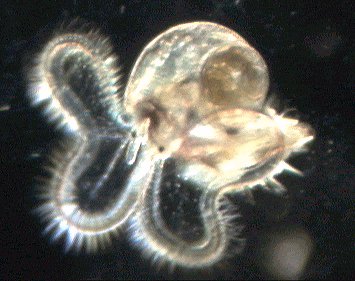
When hatching occurs, the two ciliated crowns allow the baby snail to displace it quickly into open water: it's called the veliger stage. The picture on the left was taken in dark field. |
|
|
|
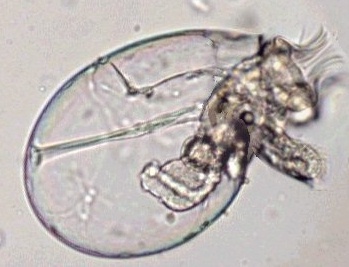
Another specimen taken with a x15 objective. |
|
|
|
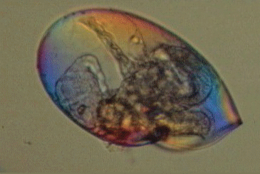
But coming back to the shell: like their terrestrial brothers a sea snail shell is made of calcium salts which have the property to polarize light. Using crossed polarizing filters, (one on the condenser (polariser) and the other on the eyepiece (analyser), you can observe all the rainbow colors in the shell. |
|
You can improve the color density of the effect with a piece of suitable plastic (a cover of a CD case is perfect!) located in the light path between polariser and analyser. When rotating this piece of plastic the colors change as shown in the animation above. It's easy to conserve for some days the little snails especially if you have some algae in your sample. It's a little easier with freshwater species. |
|
|
|
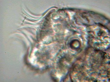
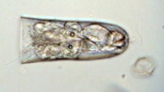
A closer look at a baby which still has a transparent shell and shows some familiar structures: the stomach with vibrating cilia inside, ocular spots and an equilibrium structure called the statolith (see this Micscape article). In the left hand picture taken with a x40 objective the cilia and statholith are clearly seen. |
|
|
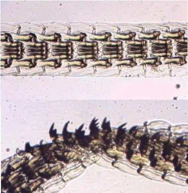
But another organ is very intriguing (but not visible in young specimens). To scrape algae, snails use a sort of rasp containing microscopic teeth. This organ is called a radula (rasp in Latin). Sorry, but I don't have a picture of snail radula but this organ also exists in other marine molluscs like limpets and is very similar. Below you can see the detail of teeth of a limpet radula. The radula can reach more than 2 cm long. |
|
|
|
|
Top: front view
Bottom: side view Teeth are clearly visible. |
|
|
|
|
||
|
|
|
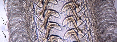
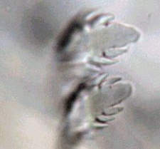
Above is a closer look at the teeth (x15 objective, image size reduced 2 times) and (left hand side) detail of lateral teeth (x40) arrowed in the picture above. |
||
|
|
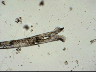
Sometimes in seawater samples, I've found an intriguing subject and some details of its anatomy were close to that of a snail's in appearance but... it was NOT a snail. The shell was not spiral and it was also perfectly linear. My knowledge being limited, I asked the advice of a fellow Micscape contributor, Walter Dioni, (thank you Walter!), who identified my unknown creature: they were pteropods (an old term but I like it because it describes perfectly the animal: in Greek pteros means 'wing' and podos means 'foot'). |
|
|
|
|
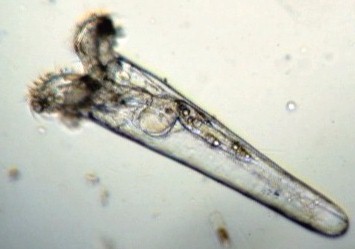
The left hand picture shows a young pteropod with ciliated crown looking like a baby snail. The rounded stomach is well seen. Above the stomach there appears a digestive gland (hepatopancreas) with spherical fat inclusions. |
|
|
The animation left shows how an adult moves who seems to applaud with its feet! The mouth is located at the base of the feet. |
|
|
|
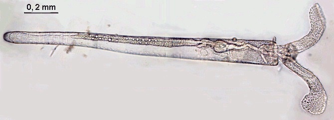
A close view of a younger specimen retracted inside its shell and showing two statoliths.
Below, a picture taken of an adult with a x15 objective and made with 16 individual pictures stitched together! (Picture resized to 1/4 of its real size.) |
|
|
|
|
Nowadays, Pteropods are classified in the families Gymnosomata (naked body) and Thecosomata (with shells). The picture above shows a member of this last family and is probably a Cresseis species. To see beautiful drawing of these creatures see www.mbl.edu/leuckart/L91.GIF …( If you want to see other wonderful drawings of the microscopic world or marine creatures by Rudolph Leuckart go directly to the main page of the site at www.mbl.edu/leuckart.)
|
|
|
||
|
Comments to the author Jean-Marie Cavanihac are welcomed. Microscopy UK Front Page
All drawings and photographs © Jean-Marie Cavanihac 2003 Published in the December 2003 edition of Micscape Magazine. Please report any Web problems or offer general comments to the Micscape Editor, Micscape is the on-line monthly magazine of the Microscopy UK web |