Invasion Of The Body Snatchers
 PAGE 2
{go to start page?}


by Mol Smith Dec. 2013


Our youngsters have never had a better time for being entertained by movies which fictitiously generates futuristic dystopian worlds of vampires and zombies, or mass-extinction through air-born viruses. Often the vampires and zombies are created due to an infection. I thought it might be interesting to consider some of the real invaders of micro-organic forms, and the way they may invade our bodies in exotic ways.
Most people are aware of Malaria, and the vector (carrying, spreading-agent) for that being the mosquito. But what about so many others?

Go Mad On This
Creutzfeldt-Jakob disease


Tonsil biopsy in variant CJD. Prion protein immunostaining. Author: Sbrandner.
Used here under the
Wiki Creative Commons Licence


 Spongiform change in brain in CJD


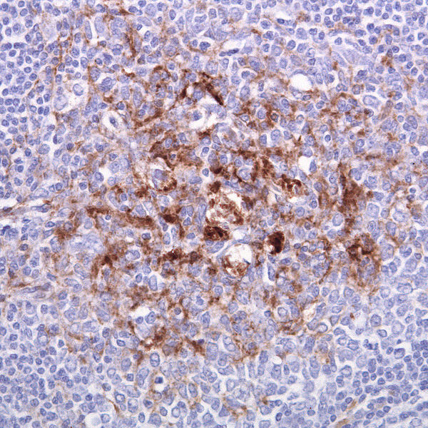
CJD: in its classical form will see you go mad then die in an average time of 4 to 5 months. In it's variant form, 13 - 15 months. It's always fatal and there's no cure.

At times called a human form of mad cow disease (bovine spongiform encephalopathy or BSE) even though classic CJD is not related to BSE, this is something you certainly don't want to get. BSE is believed to be the cause of variant Creutzfeldt–Jakob (vCJD) disease in humans, therefore the two terms are often confused.

 CJD is caused by an infectious agent called prions. Prions are misfolded proteins which replicate by converting their properly folded counterparts, in their host, to the same misfolded structure they possess. The disease leads to rapid neurodegeneration, causing the brain tissue to develop holes and take a more sponge-like texture. (see left).

Most people are only aware of CJD due to the media-scare over one form which causes 'mad cow disease' BSE - (bovine spongiform encephalopathy), and a public awareness that affected beef may have entered the food chain. However, classic CJD is not directly related to BSE.

Prions appear to exist in soil where infected sheep have grazed. They are not living micro-organisms per se. But they have come from animals suffering the prion disorder Scrapie
.

Most cases of prion disease are sporadic; that is, they arise spontaneously for no known reason. Sometimes, although rare, a prion disease is inherited due to a faulty gene. Because the infecting agent is a malformed protein capable of influencing healthy protein in adjacent cells, it can also be acquired by  medical procedures, transfusions, or contaminated food. The sporadic occurring form of the disease affect populations worldwide. The incidence of sporadic CJD is around 1 per million of the population per annum; males and females are equally affected. The incidence of the various acquired prion diseases, however, is more localised to specific groups and populations.

The CJD form associated with humans affected by BSE related vectors, is through contaminated meat. But CJD can also occur in humans through contamination of surgical instruments. The existing sterilisation of surgical instruments will not destroy the prions. Food containing prions (malformed proteins) cannot be made safe by cooking. The protein does not break down at cooking temperatures.

FAQ:
Can I get CJD by contact with another human being?
No.

Can I get CJD if I cook all my meat properly?
Yes. Because CJD is not just caused by contaminated food (meat), and if you eat infected meat, cooking does not destroy the malformed protein - prions which cause the issue.

Medical Perspective
Difficult to diagnose because a biopsy involves invasive procedure into the brain and sample taken might be from an uninfected area. There are no cures or remedies, just preventive health measures to minimise cross-contamination.

About Prions
The protein that prions are made of (PrP) is found throughout the body, even in healthy people and animals. However, PrP found in infectious material has a different structure and is resistant to proteases, the enzymes in the body that can normally break down proteins. The normal form of the protein is called PrPC, while the infectious form is called PrPSc - the ''C'' refers to 'cellular' or 'common' PrP, while the ''Sc'' refers to 'scrapie', a prion disease occurring in sheep. While PrPC is structurally well-defined, PrPSc is certainly polydisperse and defined at a relatively poor level. PrP can be induced to fold into other more-or-less well-defined isoforms in vitro, and their relationship to the form  that are pathogenic in vivo is not yet clear.


   Quick simplified understanding.

Prions do not themselves behave like living components. They are not alive. They consist of a protein form which is structurally folded, which is not how our proteins should be. The prions seem able to influence similar protein in their hosts. They make normal versions of the protein restructure and fold... effectively ending their function in nerve tissue and cells.

And that means brain cells and nerve tissue no longer are healthy.

mol
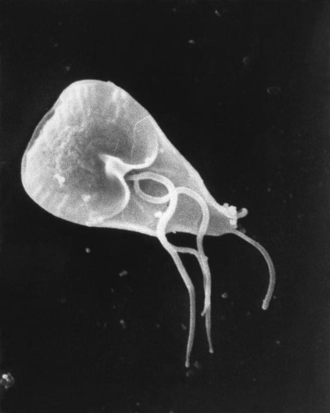
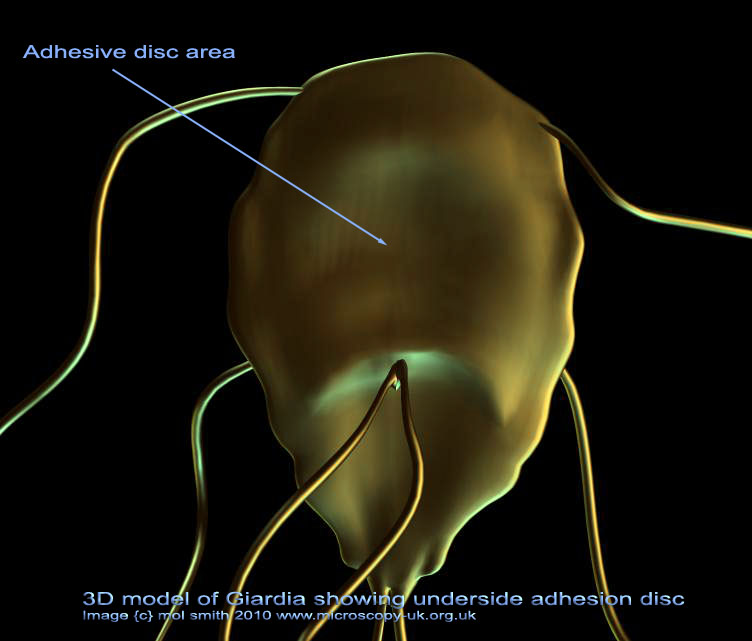
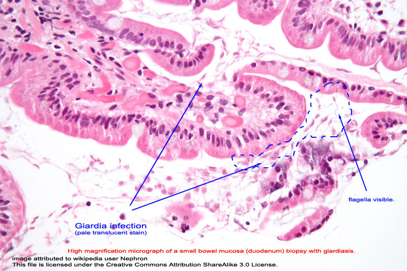

Giardia will make you run

Giardia SEM image - creative commons
                        licence - wiki
Source http://phil.cdc.gov/PHIL_Images/8698/8698_lores.jpg
Author CDC / Janice Carr
Wiki creative commons licence
3d model of giardia protozoan

3D model of Giardia
by mol smith
Read more about Giardia Here


Giardia
And if those first few don't scare you enough to run to the toilet, this one will take you there.
Giardia is flagellated protozoan parasite. If lives inside the intestines of animals, infected with and can can be passed to humans.  Individuals become infected through ingesting or coming into contact with contaminated food, soil, or water, mainly through the presence of waste products from affected animals.

Normally, after a day or two, people infected with this protozoan will get abdominal cramps, violent diarrhea, and will begin vomitting. The infection is untreated can kast for 2 to 6 weeks, and in a few individuals - much longer.  Medication containing tinidazole or metronidazole is used to treat the infection and reduce its sevreity and time suffering.


giardia infection


FAQ

Can I die by being infected?
No. Most people recover naturally. Although infection can last for several months to years with continuing symptoms in some people if not treated. Children with chronic infection may fail to thrive while infected.


This will make you breathless - Pneumonia

pneumonia
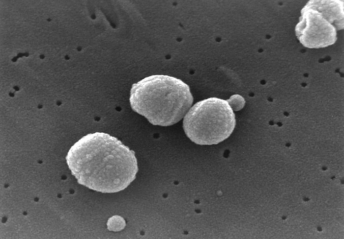
Photo Credit: CDC/Janice Carr
Content Providers(s): CDC/Dr. Richard Facklam
Public Domain via wiki commons licence

The bacterium Streptococcus pneumoniae, a common cause of pneumonia. The disease/infection has been  known to humankind since early Greek times, over 2000 years ago. The symptoms were described by Hippocrates (c. 460 BC – 370 BC)

It was not until the 1800s when Edwin Klebs became the first to observe bacteria in the airways of persons having died of pneumonia in 1875. Initial work identified the two common bacterial causes, Streptococcus pneumoniae and Klebsiella pneumoniae, and was performed by Carl Friedländer and Albert Fränkell in 1882 and 1884, respectively.
 

Pneumonia is not just an infection, it is more known as a condition of the lungs which may be caused by a variety of factors including non-infectious damage through smoking or inhaling toxic dust.

CAP or Community-Acquired Pneumonia is caused by an infecting vector which may be bacteria, a virus, air borne fungi, or parasites such as  Toxoplasma gondii, Strongyloides stercoralis, Ascaris lumbricoides, and Plasmodium malariae.

Half of all cases due to bacteria are caused by Streptococcus pneumoniae. Other commonly isolated bacteria include: Haemophilus influenzae, Chlamydophila pneumoniae, and Mycoplasma pneumoniae.

Poor country populations have high incidents of Pneumonia with high death rates in children. Anti-biotic's can help fight bacteria caused pneumonia but people with existing lung damage and elderly people remain under greater risk of fatal outcomes. Fungal pneumonia is uncommon, but occur more commonly in individuals with weakened immune systems due to AIDS, immunosuppressive drugs, or other medical problems. Fungi causing pneumonia are Histoplasma capsulatum, blastomyces, Cryptococcus neoformans, Pneumocystis jiroveci, and Coccidioides immitis.

Top right:
X-ray of Lung affected by Pneumonia. Note the white (denser) wedge in the right lung, not apparent in the other lung.

  x-ray of lung with pneumonia

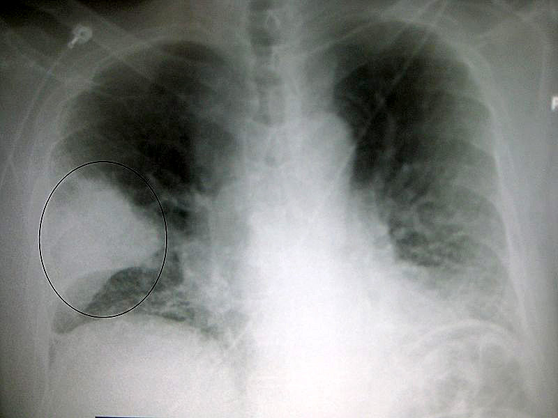
A very prominent pneumonia of the middle lobe of the right lung. Author James Heilman, MD. Used here under the Wiki Creative Commons licence.

CT scan of Pneumonia in lung
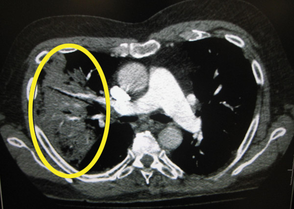
CT of the chest demonstrating right-sided pneumonia ( left side of the image ) by  James Heilman, MD
Creative Commons licence.
Contact the Author using the form below.


previous page        


Microscopy UK Front Page
Micscape Magazine
Article Library


© Microscopy UK or their contributors.

Published in the December 2013 edition of Micscape.

Please report any Web problems or offer general comments to the Micscape Editor .

Micscape is the on-line monthly magazine of the Microscopy UK web site at Microscopy-UK


© Onview.net Ltd, Microscopy-UK, and all contributors 1995 onwards. All rights reserved.
Main site is at
www.microscopy-uk.org.uk.