|
Giardia lamblia - an outline
Giardia was first discovered by Antony
van Leeuwenhoek in 1681 when examining diarrhoeal stools from himself. Although it was not really identified as
a cause for serious infection until quite recently. Debate still continues regarding this parasite. It is a binucleate
flagellated protozoan which can alternate in its life-cycle between an active motile (swimming) trophozoite and
a resistant cyst which is infectious. It is possible to see this parasite in stained sections of the upper portion
of the small intestine of infected people and animals. Leeuwenhoek must have been severely ill, I surmise, because
it is quite difficult to find this parasite in stools due to the fact they have 'sucking discs, 'enabling them
to cling on tightly to the intestinal mucosa! This is when they are in their swimming trophozoite part of their
life-cycle. It is thought they feed on mucous secretions, reproducing in large numbers to form dense problematic
populations, which then interfere with nutrient absorption by the intestinal epithelium.
Infection and disease
Giardia lamblia is responsible for around 30,000 people yearly suffering
waterborne disease causing diarrhea. The protozoan probably causes 200 million infections world-wide per year!
Infection is affected by encysted Giardia being excreted by humans and animals and ending up in 'clean' water supplies.
Many people in the states, exploring the natural environment of remote areas, become infected by drinking from
what appears to them as crystal-clear freshwater. The resultant illness has been coined as 'Beaver Fever' since
it is known that the animal is probably the cause of the encysted Giardia being deposited directly into the water
with its faeces, although deer, sheep, and rodents contribute too.
The Giardia cysts survive for months in cold water and may be present in any water system especially naturally
occurring ponds, storm water storage systems, as well as clean-looking mountain streams. As they are resistant
to traditional water treatments such as chlorination and ozonolysis, they may also occur in city reservoirs. Problems
with infection may also also occur in baby or child-care day centres through direct or indirect contact with contaminated
child faeces if hygiene practice control is poor.
Viewing Giardia with
a light microscope
The protozoa can be seen
with high magnifications under a light microscope, although they more often appear as elongated crescents such
as in this stained section below Fig.1
(please see copyright
information on resources page). I have marked an area here with a dotted
line so you can see a cluster protozoa.
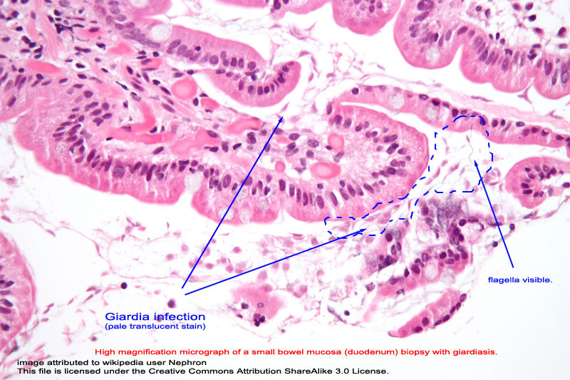
Fig. 1
A scanning electron microscope can provide us with a much better view of this entity so that we can understand
more about its ability to 'cling' to the wall of the intestinal lumen. So let's take a closer look here...
|