If an earthworm is drawn over the tips of the fingers, a roughness will be felt although the surface appears smooth and glistening. This roughness is due to the presence of bristles called chaetae or setae.
If a typical segment is examined with a lens it will be seen that at the side, and also ventrally, are spots where the chaetae protrude, in fact these can often be seen with the naked eye. Each spot will reveal a pair of chaetae. There are, therefore, on each segment eight chaetae in all, arranged in four pairs, two pairs on each side. Chaetae are present on every segment, except the first and last, and are embedded in chaetigerous sacs, which are ingrowths of the epidermis, each chaeta being secreted by a single cell at the base of the sac.
The chaetae are made of chitin, a nitrogenous polysaccharide, which is hardened and darkened by impregnation with insoluble proteins known as scleroproteins.
Chaetae are involved in the locomotion of the worm and this can be illustrated by allowing a worm to move over a piece of rough paper and then a sheet of glass.
On the paper, progression will be seen to be effected by a succession of waves of contraction and elongation along the body, assisted by the protrusion and retraction of the chaetae. The scraping of the chaetae can be heard as they get a grip of the roughness of the surface of the paper. When placed on the glass, the difficulty the worm experiences in movement will be obvious.
In forward movement, the anterior part of the body elongates and the chaetae in this part are at the same time retracted within the body wall, so as to offer no impediment to the gliding movement.
Having extended the anterior part to its fullest extent, with a consequent reduction in girth, the anterior chaetae are protruded and, by their curved nature, get a firm anchorage on the substratum. With this hold the contraction of longitudinal muscles, within the body wall, draws the body forward, the chaetae on the moving portion being withdrawn. In turn the middle and hind portions of the body behave similarly and progression is accomplished. Protractor and retractor muscles are used for protruding and withdrawing the chaetae, the protractor muscles being associated with the circular muscles in the body wall and the retractor muscles with the inner surface of the longitudinal muscle layer.
A microscopical preparation of the chaetae is a very simple procedure and is best carried out at the end of an earthworm dissection, when the alimentary canal will have been removed.
About 1.25 cm. of the full width of the body wall is cut off, this ensures the ventro-lateral portions are included. Place the material in a test tube and add a small quantity of 5% potassium hydroxide, no more than about half an inch of liquid in the tube. (See safety footnote).
Warm over a very small flame, agitating the liquid until it boils. Continue boiling until the material has apparently disappeared.
Fill the tube with cold water and after allowing a few seconds for the released chaetae to settle at the bottom of the tube, decant off the greater part of the liquid. This decanting should be done into another test tube, in case the chaetae have not fully settled.
Place the thumb over the mouth of the test tube and tip it up sharply and then holding the inverted test tube over a watch glass, release the thumb slowly and allow the liquid to escape into the watch glass.
When examined under the low power of the microscope, several isolated chaetae should be seen. If no chaetae can be seen, then insufficient time was not allowed for them to sink in the test tube and the decanting operation should be repeated with the liquid, which was poured off into the second test tube.
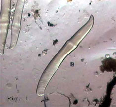
The ordinary chaetae are elongated bright yellow objects, rather fusiform in shape with the thickest portion in the middle. (Fig. 1)
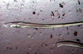
If the clitellum was included in the portion of the body wall treated, some of the specialised, so called genital chaetae may be seen. These are longer and more slender than the ordinary chaetae and these penetrate the body of the other worm during the copulatory process, helping to hold the pair together. (Fig. 2)
Comments to the author Mike Morgan are welcomed.
Related links
The author's earlier article on 'Earthworm dissection' provides some guidelines and references to learn more.
Figure 1: The ordinary chaetae. (Low power objective). Figure 2: The specialised genital chaetae. (Low power objective).
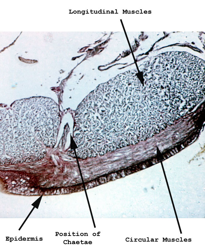
Figure 3: Part of a transverse section of the earthworm. This shows the position of the chaetae and the circular and longitudinal muscles.
(High power objective).
Safety footnote: Potassium hydroxide solution is a caustic alkali and suitable skin and eye protection should be worn. Burns and scarring can occur if spilt on the skin and possibly blindness if it gets in the eyes. Be very careful warming alkalis; anti-bumping granules are recommended and direct vessels away from you. Emergency eyewash facilities in addition to washing facilities are recommended. Workers should become familiar with the hazards from appropriate Material Safety Data Sheets (e.g see this link) and take all recommended precautions. An appropriate working area with correct equipment should be used. The procedure should not be carried out by youngsters.Disclaimer: This article is offered in good faith by the author. Neither the author, Microscopy-UK, Micscape, Onview.net nor any of its administrators or contributors assumes any responsibility for injury to persons or property damage incurred by using the chemicals and procedures described.