Micscape Article: Earthworm Dissection
by Mike Morgan 1996
 Introduction
Dissection of the earthworm involves fine dissection and requires steadiness of hand and
precision. For a general dissection, the worm is immersed in 50% alcohol when, after 10
-15 minutes it will die in an extended condition with muscles relaxed, enabling an easier
dissection to be performed. For the preparation of the contents of the seminal vesicles,
the worm should be killed with chloroform and the specimen should not be covered with
water. Dissection of the worm is performed in a wax-bottomed dish and small pins are used
to display the dissection.
Technique
An initial incision can be made with a razor blade or scalpel and this cut followed up by
a pair of sharp pointed scissors. The body wall is pierced with the lower blade of the
scissors, well behind the clitellum, in the mid-dorsal line. A pin is placed through the
first segment and another through the tail of the worm, so that the worm is fully
extended. The pins are inserted at a sloping angle, to ease the strain, along the length
of the dissection. When pinned the dissection should be covered completely with water. The
dissection will show the alimentary canal, the position of the seminal vesicles, and
pseudo hearts. The alimentary canal may be lifted with forceps and careful cutting of the
septa will enable this to be pinned aside. This will reveal the ventral nerve cord and the
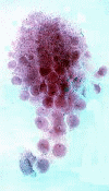
circum-pharyngeal ring. The ovaries will be found in segment 13 of the worm. Using fine
forceps the ovary and septum, to which it is attached, is removed (see figure on left
above).
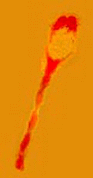 Using a needle, the nerve ring can be
loosened from the pharynx, which is then pulled through the ring and cut off. The pharynx
should go back through the nerve ring. The nerve cord (shown in the figure on the right)
is then cut off, about one inch behind the nerve ring, and removed. Both the ovary and
nerve cord can be stained with borax carmine and mounted in Canada Balsam.
Using a needle, the nerve ring can be
loosened from the pharynx, which is then pulled through the ring and cut off. The pharynx
should go back through the nerve ring. The nerve cord (shown in the figure on the right)
is then cut off, about one inch behind the nerve ring, and removed. Both the ovary and
nerve cord can be stained with borax carmine and mounted in Canada Balsam.
A preparation of the seminal vesicles is made by cutting them off and teasing them in a watch glass, with physiological saline. A small drop of the milky fluid is placed on a cover slip and dried in warm air (e.g.. above a spirit-lamp). The smear is fixed in absolute alcohol, dried and stained in Ehrlich's haematoxylin, before mounting in Canada balsam.
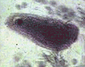 This will show the stages of developing
spermatozoa and, in most preparations, the protozoan parasite of the earthworm,
Monocystis sp., will be seen in various stages of its life-history. The image on the
left shows the trophozoite of Monocystis.
This will show the stages of developing
spermatozoa and, in most preparations, the protozoan parasite of the earthworm,
Monocystis sp., will be seen in various stages of its life-history. The image on the
left shows the trophozoite of Monocystis.
Useful aids to this dissection
1) Dissection Guides V.--Invertebrates by H.G.Q. Rowett
2) Biology Staining Schedules for First Year Students by R.R. Fowell
Comments to the author Mike Morgan.
Also see Micscape article 'Monocystis'.
Also see the Micscape article 'Earthworm dissection: Chaetae'. This describes the preparation of the chaetae (stiff hairs) of the worm for microscopy.
Microscopy UK Front Page
Micscape Magazine
Article Library
© Microscopy UK or their contributors.
Please report any Web problems or offer
general comments to the Micscape
Editor,
via the contact on current Micscape Index.
Micscape is the on-line monthly magazine
of the Microscopy UK web
site at Microscopy-UK