Monocystis
by
Mike Morgan, UK
|
(Underlined words are defined in
the glossary)
Several species of Monocystis are parasitic in the seminal
vesicles of the earthworm. They belong to the class Sporozoa
and are placed in the order Gregarinida.
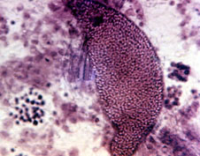 The adults, or
mature trophozoites, are commonly to be found within the
seminal vesicles of the worm. There is a thick pellicle
beneath which, in the plasmagel, are longitudinal myonemes.
In the granular plasmasol there are paramylum
granules and an ovoid nucleus with a prominent nucleolus.
Locomotion of the parasite is characterised by contraction of the
myonemes during wriggling; it is known as gregarine motion. (Fig.
1 right).
The adults, or
mature trophozoites, are commonly to be found within the
seminal vesicles of the worm. There is a thick pellicle
beneath which, in the plasmagel, are longitudinal myonemes.
In the granular plasmasol there are paramylum
granules and an ovoid nucleus with a prominent nucleolus.
Locomotion of the parasite is characterised by contraction of the
myonemes during wriggling; it is known as gregarine motion. (Fig.
1 right).
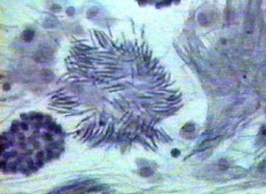 The parasite
feeds on the cytoplasm of a sperm morula by extruding
enzymes and absorbing the digested products through the pellicle.
It will frequently move to another morula and consume the
cytoplasm, before it is fully-grown. Often numerous sperm tails
adhere to the pellicle, giving the Monocystis a ciliated
appearance. (Fig. 2 left).
The parasite
feeds on the cytoplasm of a sperm morula by extruding
enzymes and absorbing the digested products through the pellicle.
It will frequently move to another morula and consume the
cytoplasm, before it is fully-grown. Often numerous sperm tails
adhere to the pellicle, giving the Monocystis a ciliated
appearance. (Fig. 2 left).
Excretion and respiratory activities are carried out by
diffusion.
When two mature adults come together they secrete a common
wall and form a conjugative cyst. Within the cyst mitotic and
meiotic divisions occur. There is no further development until
another earthworm, which first entails the liberation of the
cysts into the soil, swallows the cysts. This latter is
accomplished by birds, or other animals, eating the earthworm.
The cysts are not digestible and are voided with the faeces.
Another worm now swallows the cyst, the cyst coat is digested and
the freed sporozoites migrate to the seminal vesicles by
boring through the gut wall into the coelom of the earthworm.
Cysts may live in the soil for a considerable period.
The majority of earthworms are infected by the parasite. In
fact, in many years, I have not found the absence of the parasite
in the seminal vesicles of any worm examined. Quite often there
is a heavy infestation. Despite this, the presence of the
parasite seems to cause little inconvenience to the worm. Sperm
are produced in such quantity that the destruction of quite large
amounts has little effect on the reproductive capacity of the
worm.
Smear Preparation of the Contents of the Seminal
Vesicles.
 For this
preparation the worm should be killed with chloroform and opened
dry. A mid-dorsal incision is made, about segments 9 - 15. This
will clearly show the white seminal vesicles. These are cut off
and placed in a watch-glass. The material is covered, with about
five times its bulk, with 0.75% saline, and is then teased
thoroughly with needles, to release the contents of the seminal
vesicles. A drop of the milky fluid obtained, is placed on a
cover-glass, dried by warming and fixed in alcohol. This is then
stained with Ehrlich's haematoxylin and again dried by warming.
The cover-glass is placed on a microscope slide, in the centre of
which a drop of Canada Balsam has been placed, and is then
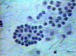
examined microscopically. Apart from the presence of the
parasite, all the developing stages of the spermatozoa (the
morula, resembling a blackberry) will be seen. (Fig. 3 above
right).
For this
preparation the worm should be killed with chloroform and opened
dry. A mid-dorsal incision is made, about segments 9 - 15. This
will clearly show the white seminal vesicles. These are cut off
and placed in a watch-glass. The material is covered, with about
five times its bulk, with 0.75% saline, and is then teased
thoroughly with needles, to release the contents of the seminal
vesicles. A drop of the milky fluid obtained, is placed on a
cover-glass, dried by warming and fixed in alcohol. This is then
stained with Ehrlich's haematoxylin and again dried by warming.
The cover-glass is placed on a microscope slide, in the centre of
which a drop of Canada Balsam has been placed, and is then
examined microscopically. Apart from the presence of the
parasite, all the developing stages of the spermatozoa (the
morula, resembling a blackberry) will be seen. (Fig. 3 above
right).
Useful reference for staining technique:
Biology Staining Schedules by R.R. Fowell. H.K. Lewis
& Co. Ltd.(London)
Another useful reference is Dissection Of
The Earthworm. Whitehouse & Grove. University Tutorial
Press Ltd.
Comments and feedback to the author Mike Morgan are welcomed.
Also see Micscape article 'Worm dissection'.
Glossary
Morula: solid round mass of cells, resulting from the division of
an egg.
Myoneme: a muscle fibre.
Paramylum: stored carbohydrate, resembling starch.
Pellicle: relatively stiff, thin surface layer covering the cell.
Plasmagel: jelly-like state of the outer cytoplasm.
Plasmasol: the fluid inner cytoplasm.
Seminal Vesicle: the organ that stores sperm in invertebrates,
such as the earthworm.
Sporozoite: the minute spore, developed by Sporozoans, which
infect the
host animal.
Trophozoite: a growing stage in the life cycle
of some Sporozoan parasites, when they are absorbing nutrients
from the host.
Return to article.
Safety notice and disclaimer:
dissections and the use of chemical reagents should only be
undertaken by experienced personnel using the appropriate
precautions. No reponsibility is accepted by On.View Ltd.,
Microscopy UK, Micscape or it's contributors for any damage to
property or injury to persons undertaking such work.
© Microscopy UK or their
contributors.
Published in March 1999 Micscape
Magazine.
Please report any Web problems
or offer general comments to the Micscape Editor,
via the contact on current Micscape Index.
Micscape is the on-line monthly
magazine of the Microscopy UK web
site at Microscopy-UK
WIDTH=1
© Onview.net Ltd, Microscopy-UK, and all contributors 1995 onwards. All rights
reserved. Main site is at www.microscopy-uk.org.uk with full mirror at www.microscopy-uk.net.