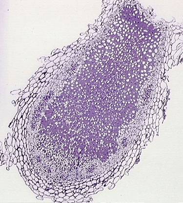
In the previous article we looked at mycorrhizal symbiosis among plants and fungi. Another form of symbiosis can be found between nitrogen-fixing soil bacteria called rhizobia and plant roots. This is correctly called rhizobial symbiosis; hosts most frequently found are leguminous plants like beans (soya beans, broad beans and others). This type of symbiosis is readily observable since the nodules formed are numerous and usually are 1 to 3 mm in size. The following 3 figures illustrate such tiny but visible nodules on the roots of broad beans.
 |
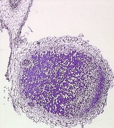 |
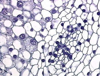
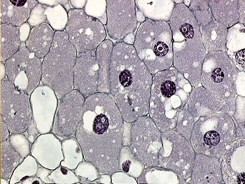
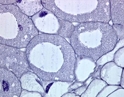
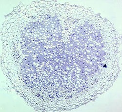
Figs 1, 2 (above) and 3 (below) illustrate root nodules of 1 to 3 mm in size which were sampled from growing broad beans. After cleaning and fixation in formalin, they were processed for light microscopy. The outer part of the nodules shows large, empty-looking but in reality not empty, plant cells (2 micron thick sections; Crystal violet stain).
 |
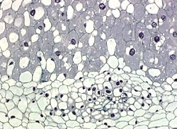 |
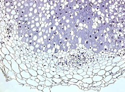
Figs 4, 5. These large cells are heavily infiltrated by Rhizobium bacteria and may sometimes show large cytoplasmic vacuoles as well.
Sections illustrated in Fig. 7 ang Fig. 9 are stained solely with Hematoxylin (a universally recognized, rather specific, stain for nucleic acid and nuclei) and Fig. 8 and Fig. 10 with crystal violet.
Fig. 7. This image was obtained by using a 40x objective.
Fig. 8 This image and Fig. 9 and 10 below were obtained using a 40x objective and an intermediary projection magnifier of 1.6x. Cells filled with bacteria in figures 8-10 are between 100 to 200 µm in size.
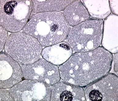
Fig. 9
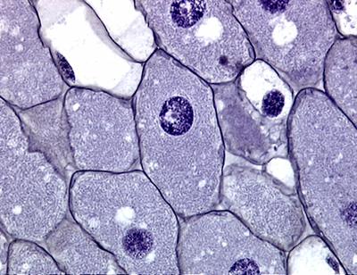
Fig. 10
From this short introduction it becomes clear that these Rhizobium bacteria live mainly inside the cells (intracellular symbiosis*). They are able to alter metabolic and molecular processes in the cells and cause cellular enlargement. The nodules formed by this bacterial proliferation exist alongside and within the growing root system of the host plants. These tumorous forms are certainly benign and mutually useful structures, conducting most of the N2-fixation for the host plants. Rhizobial nodules are the result of a long-standing and selective evolution, a good example of living together with mutual benefits.
* Please note that not only are some kinds of bacteria intracellular, but all viruses live and replicate inside living cells. They are then rightly called an 'obligate, intracellular parasite'.
Additional information about microbiological features: these rod-shaped, nitrogen-fixing bacteria belong to the Rhizobium, one of five genera of the family Rhizobiaceae. All members are Gram-negative bacteria and most are flagellate. Dissemination of rhizobial bacteria in soil is world-wide. Beans can easily be grown in small plots of soil; microscopic examination and possibly experiments with rhizobial nodules can readily be done since they are handy, a few millimeter in diameter and provide attractive cellular patterns of plant cytology. Smears on glass slides may be obtained from the cut surface of such rhizobial nodules and can be stained with any available solution including Giemsa, toluidine blue, methylene blue, light green, hematoxylin, acridine orange, crystal violet, Gram, and very many others.
Hint : The interaction between plant roots and rhizobial bacteria is definitely a good model for biology, microscopy and symbiosis studies in the classroom.
Some of the numerous, interesting Internet links chosen for you by the author:
Rhizobium Research Laboratory of the University of Minnesota gives brief results and good illustrations.
The Rhizobium-Legume Symbiosis explains various aspects of this kind of symbiosis.
A large number of Rhizobium species are cited with regard to their historical features.
Encyclopaedia Britannica gives a good description of rhizobial association.