Summary : The term 'symbiosis' indicates the general concept of living together which has a wide range of applicability in biology. We find symbiosis at both the macro- and microscopical size. As a process, it brings about mutual benefit to the host and symbiont. Mycorrhizas are examples of a very important cooperation between growing plant roots and the so-called mycorrhizal fungi, whereas rhizobial nodules are examples of an equally important cooperation between plant roots and nitrogen-fixing bacteria. Not surprisingly, both mycorrhizas and rhizobia can be found around the tiny roots below the soil surface. Careful exposure of a plant's root system will suffice to reveal such macroscopical symbiotic structures to the naked eye.
Encyclopaedia Britannica, defines symbiosis as: any of several living arrangements between members of two different species, including mutualism, commensalism, and parasitism. So, both beneficial and harmful associations are included by definition in the term 'symbiosis'.
Mycorrhizas are beneficial associations between the growing root system of a plant, which is the host, and a mycorrhizal fungus as a symbiont. In such a mycorrhizal association, the host and the symbiont belong not only to different species but to different Kingdoms, as well.
Why should members of two different species live together in a very close relationship? What kind of benefit comes about for both parties? How do hosts find their particular symbionts or how does a symbiont associate itself with a specific host? These and many other questions with respect to symbiosis were posed in the past and are still the subject of extensive studies. As the title suggests, mycorrhizal associations will only be explained briefly in this article.
At the very beginning of life, and certainly during a long period thereafter, Earth was an empty planet. Emerging living organisms could therefore live alone and spread without much constraint or strong competition. There was relatively little divergence of life forms; characterized by their organizational simplicity at that time. Then, one of the fundamental processes of biology, called endosymbiosis, took place. As defined by Dr. Lynn Margulis, endosymbiosis deals with the symbiotic relationship of early prokaryotes, which are cells without nuclei, and their development into the ancestors of modern eukaryotic cells during the evolution of life.
Following on from the prokaryotes, cells containing nuclei—eukaryotes—began to co-exist, spread and differentiate. Eukaryotes then acquired characteristics which led to the development of the so-called colonial organisms. Soon, they also developed the ability to differentiate into various cell types, forming tissues and organisms starting from stem cells. This briefly summarises what was happened during the last 3.6 billion years and how philogeny, the family tree of life, diversified.
The divergence of living beings continued as successive extinctions, changing environmental conditions and ecosystems helped to fill all possible niches on Earth. An organism's existence became a struggle to overcome environmental constraints and heavy competition. A very useful invention and an effective strategy to remain alive and reproduce was found by living together, a symbiosis. It is obvious that symbiosis is more than just living in a community.
Most symbiotic associations are mutually beneficial, but sometimes may be harmful, parasitic, and usually leading to the death of one of the partners. The chances of remaining strong and alive are certainly higher in a symbiotic life form.
In a previous article, a typical example of symbiosis such as that observed in lichens was presented. Heinrich Anton de Bary was the first to show (in 1866) that lichens consist of a fungus and an alga in intimate association; he coined the term symbiosis in 1879. In a comparable fashion, plants and fungi can associate to form mycorrhizas. As is well known, plant cells contain chlorophyll whereas fungal cells, by definition, explicitly lack chlorophyll. The former are able to photosynthesize using CO2 and H2O in the presence of light. But fungi are NON-photosynthetic. They are heterotrophs and have to find the basic food stuff they depend on. This is a costly, energy consuming process. In a mycorrhizal association, fungi take most of their food from the roots in which the symbiont (fungi) may infiltrate either superficially or deeply.
What kind of benefit can plants expect from fungi to compensate for their generous supply of food to them? The answer may be found in the functional properties of the symbionts. The mycorrhizal fungi are very successful at absorbing and accumulating minerals, especially phosphorus (P), from either organic or inorganic sources. When the availability of P in soil is low, the plants easily become P-deficient. The net effect of such a condition is a sensible decrease of the growth rate. Thus, the ability of mycorrhizal fungi to accumulate minerals is a pay-off to the host struggling with nutrient stress, especially in P-deficiency. Also zinc and copper can readily be translocated by fungi to the plants.
Mycorrhizal fungi develop and replicate by such manners typical to this Kingdom. They may belong to the Basidiomycetes, Ascomycetes or Zygomycetes. They all have characteristically filamentous structures and may produce visible fruiting bodies both below and on the soil surface. Amanita muscaria (the fly amanite), Scleroderma citrinum, Laccaria laccata, Boletus edulis, Cantherellus cibarius are just a few of the widely known fruiting bodies of mycorrhizal fungi.
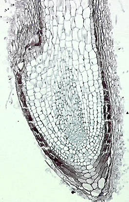
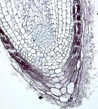
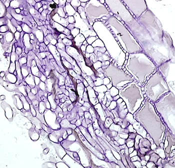
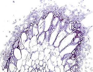
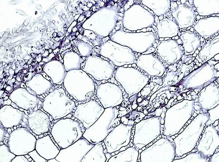
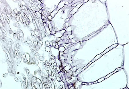
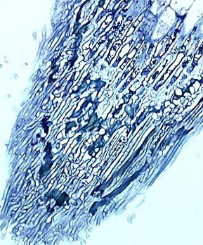
The figures in groups shown below are obtained from the same material and even of the same microscopical fields. Note that the images are displayed in increasing order of magnification. Information on the (ultra)structural features of filamentous fungi and also of cells & tissues in general, can be found by following the highlighted links. From a microscopists point of view, seven types of mycorrhizal associations have been described. Of these various forms, microscopical features of ectomycorrhizas are illustrated below as an introduction to this large topic. Remember that in ectomycorrhizas we find a visible fungal network on the surface of the growing roots of many plants like firs and pines. In contrast, VAMs are invisible to us since they are microscopical and intracellular structures. VAMs are found, for example, inside the cells of tiny roots of grasses and clovers.
The samples used for microscopic study and photomicrography are at most 1 mm in diameter.
|
|
 |
|

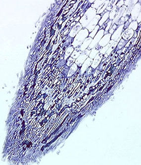
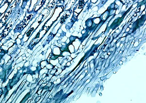
Figures 1-4. The distal part of a young, growing plant root with ectomycorrhizal development. In these images, we observe that filamentous fungus covering the surface envelops the root as a mantle and superficially infiltrates between the cells of the root.