| The
Condenser
None too technical thoughts concerning the Brightfield Condenser By Paul James, UK
|
| The
Condenser
None too technical thoughts concerning the Brightfield Condenser By Paul James, UK
|
When the very first experiments were in progress concerning microscope design some two or more centuries ago, it soon became apparent that the specimen needed to be much brighter than normally required for direct vision. The reason was simply that as magnifying power rises the image intensity falls inversely.Fortunately the remedy was exceedingly simple and must have been employed in the earlier days of microscopy. The solution was to use a small short focus bi- convex lens or concave mirror to concentrate and therefore multiply the light intensity upon the subject using light shone from below the specimen.
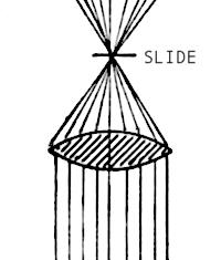 |
| Single element
light concentrator
or "condenser" |
In one clean swoop this substage lens, we now call the condenser provided the light levels needed for higher magnifications.The term "Condenser" evolved naturally to encapsulate the essence of what was happening when more light was focussed on the specimen.......Light Concentrator might have been used as an alternative.
Angular illumination and resolution
In the earliest days of microscopy any improvements to image quality were brought about by a process of trial and error, or often as not through serendipity..........by simply stumbling on a new inventive step? Achromatism also improved image quality by the early 19C.
It must have been increasingly obvious to microscopists in the evolution of the science that if the condensing lens was of short focus, the image of the specimen was not only a little brighter but more clearly defined. Later, a variable stop aperture, we now call the iris (so called because of the similarity to our eye's iris) was contrived to allow convenient variation of light intensity.
The reasons behind the apparent increase in resolution were not clearly understood until Prof. Abbe in conjunction with Carl Zeiss revealed the true nature of the property of light in operation in the realms of microscopy.
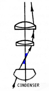 |
The essence of this revelation is that the maximum resolution for a given objective is determined by the angle of the cone of light falling onto the specimen. The wider the cone, the greater the resolution obtained, subject to the limitations of the objective in use. The diagram above shows a typical medium power objective and condenser lens with one ray of the extreme edge of the cone furnished by the condenser. The angle between the axis and this ray determines the maximum resolution possible in the image. It is expressed as NUMERICAL APERTURE...................NA for short. It is calculated simply by looking up the SINE of HALF the full CONE angle as shown in blue above. Thus the sine of half the cone angle of 40 degrees brings a NA value of 0.64. This is the same as the angle of cone accepted by a x40 objective, so that its full cone would be twice this or 80 degrees. Condensers produce maximum light cones of up to about 1.3 NA in certain circumstances.Subsequently the design and manufacture of condensers were taken as seriously as those of the objective and eyepiece, for the rather obvious reason that the proper illumination of the specimen with a suitably wide cone of light was essential if the highest quality imagery and resolution was to be realised from the instrument. A single element bi-convex lens cannot provide a wide enough cone of colour free light to satisfy the aperture angles required by the higher power objectives. Multi-element combinations in condensers were therefore necessary.
Condenser types
Of the many variations of brightfield designs manufactured over the years, 2 basic types evolved and remain in use today. They are essentially the same in principle because they furnish a bright converging cone of light onto the specimen from below the stage The first is known as a 'dry' condenser, and the second is known as an oil immersion condenser.
The essential difference between the 2 forms is that the 'dry' condenser has a limited aperture of 1.0 NA in theory ( Most achieve about 0.95 NA ), and the oil immersion types can exceed this and form light cones of up to approximately 1.2-1.3 NA when oiled to the undersurface of the slide.
Each design by different makers will have slightly different focal lengths and so the image of the light source produced will vary in size also. This is very important and should be a consideration when deciding to obtain any condenser, since this image should fully cover the field of view of the lowest power objective in use............hopefully.
Abbe (Dry and Oil immersion)
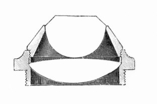 |
| Cross section of an example of an old
Abbe design 1.2 NA. |
This is a very old design and has been known under various names, but is generally now called the Abbe. It is the simplest, cheapest and most commonly used condenser of highish NA. Two elements of glass bring about the convergence of light. But being a two element design means that its achromatic corrections are restricted, and it fails to provide a 'solid' cone of light of even illumination because of its lack of spherical corrections. It is however capable of adequate illumination of most specimens and powers up to and including x40 objectives. Numerical apertures of between about 0.9 to 1.3 are catered for, and some can function well enough to be used with oil immersion objectives when oiled to the undersurface of the slide.The aplanat is a variation of the simple Abbe, but usually of 3 element design which furnishes a more geometrically perfect cone of light, though its chromatic aberrations are similar to the Abbe's. This is altogether a moderately cheap design to manufacture, yet is capable of being used for most applications of microscopy. The Russian 'Lomo' Aplanat 1.4 NA is a good example of this form, and possesses the great advantage being capable of oblique movements in its mount.
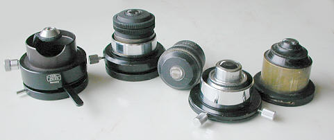
Flip-up Abbe Forms
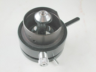 |
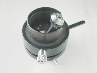 |
This Carl Zeiss Abbe condenser shown above has a very convenient method of altering the field coverage and aperture simultaneously when using low and higher powers in quick succession. The knurled control knob is twisted to move top element aside the optical axis revealing the larger bottom element which has a wider field and lower aperture (0.6 NA) for objectives between x10 - x20. When in place the top element provides a maximum of 1.3 NA. for oil immersion x100 objectives when oiled to a slide, or for up to x40 objectives when used dry. There are some variations of this type of condenser, and they are made precisely for rather obvious reasons. It is an extremely practical and successful condenser.Achromats (Dry Max 1.0 NA)
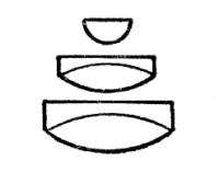 |
| A 'dry' 5 element form |
These designs are usually of several elements, and though similar in essence to their Abbe/aplanat counterparts, have higher colour and spherical corrections. Thus they yield finer colour free cones and being more geometrically perfect, the cone is able to fill the objective more uniformly with colour free light. They are almost as well corrected as objectives...........They are quite expensive and are desirable in use because they impart little or no colour to the illuminating cone. Numerical apertures up to 1.0 NA are available, and thus they are to be used in an unoiled or dry state. Therefore they can illuminate fully all achromats, fluorites/apochromat objectives up to and including the x 40 powers.
Achromats (Oil Immersion Types Max 1.4 NA)
 |
| Typical complex design |
This 'oil immersion' design is very highly corrected and resembles the oil immersion objective's internal optics to a large extent. It is therefore capable of providing a 'solid' and very precise colour free cone of light upon the specimen. When oiled to the undersurface of the slide, its maximum aperture of about 1.3 NA can be fully realised, thus fully utilising the oil immersion x 100 objective's maximum resolution.They are understandably expensive, and are only necessary when the very highest quality imaging is required at the highest powers in bacteriology and pathology etc. The field is understandably small, needing only to serve the requirements of the x100 oil immersion objectives *, and its physical size therefore is also reduced.
* They are capable of being used dry for lower powers such as the x40 objectives. Anyone considering the purchase of such a condenser should understand that it is specifically designed for the higher powers and its ability to provide a well illuminated field which can be used for even lower power objectives such as the x20 and x10 varies between makers.
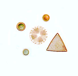 |
| Diatoms were taken using a Vickers
achromatic oil immersion 1.25 NA condenser.......used as an objective ! Demonstrating its build quality. |
Some Points1) A condenser's full aperture capability is only realised when it is focussed on the specimen. Lowering it effectively reduces its aperture and also of course its field width too.
2) The condenser's iris diaphragm controls its effective aperture when focussed, and should not be used primarily to reduce light intensity. Though it is understandable that many do so because their illumination's intensity cannot be easily controlled. (See author's earlier article on diffuse lighting.)
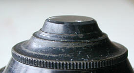
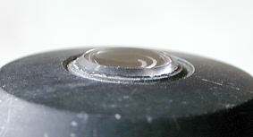
3) One particular fact which is I think misunderstood by those starting microscopy, is that when a condenser is used 'dry' (no oil contact to the undersurface of the slide) its maximum theoretical effective NA can be no more than 1.0, and in reality slightly less. Thus a condenser which is marked '1.25 NA' can only provide this wide cone of light when it is oiled to the undersurface of the slide. But a condenser of 1.0 NA or less cannot furnish a wider cone even if oiled to the slide. Usually those condensers whose aperture exceeds 1.0 NA have top elements which have slightly protruding top optics, to facilitate intimate glass/oil/glass contact.
 |
 |
| Old Abbe design with very proud top.
Also user removable top element. So it's max 1.2 NA can be reduced with wider field coverage. |
Vickers Achromatic condenser showing
top element sufficiently proud (1.25 NA) |
4) The field coverage of some condensers even when in their low power configuration is still too small, but this can be broadened effectively by using ground glass. (See author's earlier article on diffuse lighting.)
ConclusionsFor much amateur work in microscopy, the Abbe/aplanatic condenser is quite adequate. The achromatic versions produce cleaner, more faithful imaging of the illumination's light source, but the differences depend much on the quality of the condensers used. The simple fact as I have found is that the most profound effects on the quality of low to medium power brightfield imaging, assuming proper substage adjustments, depend very much on the quality of the objective /eyepiece combination, and less so on the condenser type. (The iris, if used incorrectly by closing too far down, is counter productive as it will ruin the image from the very best objectives and condenser combinations)
As power and resolution increases, as with the x40 apochromats and achromatic x100 oil objectives, so does the need to very accurately focus and precisely centre the condenser, but here the differences between an achromat and aplanat condenser, and even some Abbes will be subtle, and difficult to notice in some circumstances. Without doubt it is far better to be able to focus and accurately centre an Abbe/aplanatic condenser on your 'scope than to possess a more sophisticated achromat and be unable to centre it properly.
The " Law of diminishing returns " applies here, because the superiority of the more costly condensers brings about only very marginal increases in image quality, and so the less costly Abbe or aplanat condenser in a centerable mount will serve the amateur microscopist extremely well for most visual and photographic applications. The swing or flip up top element form is probably the most effective and convenient condenser of all.
| All Comments to the author Paul James are welcomed. |
Please report any Web problems
or offer general comments to the
Micscape
Editor,
via the contact on current
Micscape Index.
Micscape is the on-line monthly
magazine of the Microscopy UK web
site at Microscopy-UK
WIDTH=1