|
by Rene van Wezel, UK |
Figure 1: Silica skeleton of Dictyocha fibula from acid cleaned material. Brightfield. Size 35 µm.
Everybody looking at acid-cleaned diatom material from the sea will come across a silica skeleton from this group of organisms now and then. It forms the outer scale of an amoeboid organism with chloroplasts and a flagellum.
They live in the upper water layers in all the seas and oceans, generally in low abundance. Dividing once every 1-2 days, they live (mostly?) of the sun, as feeding on detritus or maybe on the occasional bacterium has never been reported.
Although the skeleton is bound to give them protection against predators, it can be shed easily. They can live on happily ever after as a blob of protoplasm with a nucleus - or more than one if they fancy growing as a multi-nucleated organism.
Sexual reproduction has never been observed. In fact, nobody knows how many species there actually are. There may be only one species living nowadays.
The taxonomy of the genus is based on the shape of the silica skeleton, with its Latin name derived from dictyoor net-form, meaning the holes in the skeleton. However, clonal cultures in the lab can form very differently shaped skeletons from the naked amoeboid stage. But when a scale is present, replication of the skeleton is fairly consistent.
|
|
|
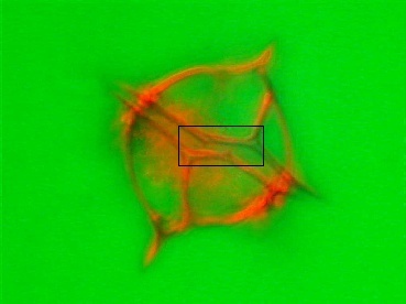
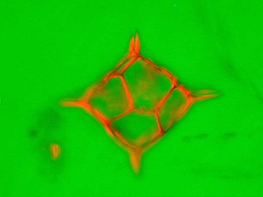
Figures 2/3: Double skeletons during
asexual division. Rheinberg contrast.
Click
each image to view animated sequential focus sequence in a new window.
'Image Cycler' script written by D Keith Higgs. One of the
free JavaScripts provided by
The
JavaScript Source.
Coming across the silicoflagellate skeletons locked together in asexual reproduction is actually quite a rare sight. In acid cleaned material for diatom analysis, the silicoflagellate skeletons will be separated, just like the two parts of a diatom will fall apart by the aggressive cleaning. Seen from the top (figure 2/3) the shape is like the skeletons found normally, but the side view (figure 4) shows clearly how the skeletons are locked together.
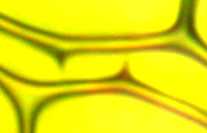
Figure 4: The connection points that hold the double skeletons together. Brightfield.
The skeletons are filled with some fluffy material and a flagellum couldn't be detected. It is possible that the critter already decomposed, or the Lugol-fixative used couldn't preserve the organism very well, see also figure 5.
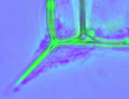
Figure 5: Protoplasma feet (pseudopodia) sticking out under the spines. Colour phase contrast.
Even though silicoflagellates occur around the world, biologists do regard this group as insignificant living fossils, more of interest to the geologist than for understanding their role within the oceanic ecosystem.
Their uncertain make-up certainly also didn't help to make them better known. Sometimes they were classified as an animal, sometimes as a plant. When ultrastructural data became available the critter was finally put down close to the Chrysophyceae or Yellow-green algae (which is, not surprisingly, quite a heterogenous group).
Technicalities:
The sample was derived from Station 2737 (ca. 10 km from Mizen Head, SW coast of Ireland) during a cruise on board the Celtic Voyager, 18-31 August 1998.
Vertical net hauls were preserved with Lugol's iodine solution and permanent mounts were made in glycerine jelly, or acid cleaned for diatom analysis.
I am indebted to the crew of the Celtic Voyager and the Marine Microbiology section of the Martin Ryan Institute in Galway, Ireland, where I had the opportunity to study the plankton flora around Ireland.
Also thanks to David Mann for his opinion about possible feeding habits of silicoflagellates.

Figure 6
All comments to the author Rene van Wezel are welcomed.
Please report any Web problems or offer general comments to the Micscape Editor.
Micscape is the
on-line monthly magazine of the Microscopy UK
web
site at
Microscopy-UK