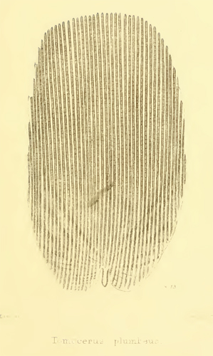
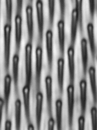
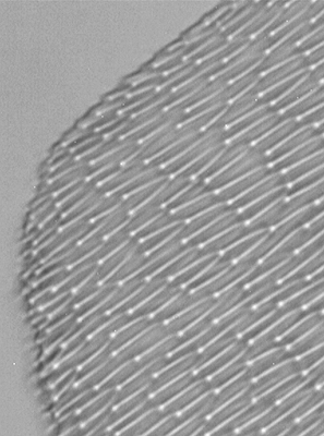
Left and above: Plate X and caption from E M Nelson's paper
'On the Podura Scale' in 1907 (4). Click image left for larger version.
(Screen capture from the free Acrobat pdf format of the Journal volume at www.archive.org)
Exploring classic insect test objects for the microscope: II - Scales of 'Podura' (springtail)
by David Walker, UK
(In Explorer browser, setting 60% reduction in 'Print Preview' with 'Show Pages Individually', retains layout on A4 pages.)
Series Introduction: Using certain subjects as tests for microscope optics has a long history (refs. 1-3, 18, 23, 25, 30, 34). Some species of diatoms are well documented as tests and still very popular with enthusiasts, but using other subjects including animal hairs and various insect parts are also long established.
A selection of test objects in the author's slide collection will be shared in this occasional series in hope they are of interest. As others have noted, given the potential variability of a subject between mounts and the sometimes vague specification of the species, it's arguable if insect parts can be regarded as reliable test objects. But it's both great fun and instructive to inspect these slides while consulting the old books and to repeat the studies they recommended. The enthusiast nowadays can also try techniques developed since the Victorians such as phase and interference contrast if available.
"It is strange, but true, that while the Podura scale has been the most looked-at object of any, it still remains the microscopist's enigma, and this, moreover, in spite of the fact that its structure is by no means so very minute for a microscopical object."
The eminent microscopist E M Nelson in the introduction to his 1907 paper 'On the Podura Scale' (4).
Scales of 'Podura' - some historical background
The potential interest in this slide to the modern enthusiast lies only partly in viewing the slide itself; equally fascinating is the extensive literature written on this subject and the lively debate it has prompted. It was one of the earliest to be described as a test for microscope objectives (Nelson notes that it was by Wollaston in 1828, refs. 4,6) and its use overlapped a key period in microscope objective development (5). 'Podura' are springtails (Collembola), the scales can occur on various body parts of these small hexapods, but the scales are not present in all genera (21). The species with the 'test scales' is now recognised to be Lepidocyrtus curvicollis Bourlet 1839 (5) which has scales on body, legs, head and antennae (35).
In the scales' long history as a test subject they have been useful in a number of roles; in the earliest work, for assessing the ability of newly developed achromatic objectives to resolve the scale markings and for assessing chromatic and spherical aberration (5). Richard Beck in an 1862 paper (9) showed a sequence of drawings of the scale markings' appearance with both poor and well corrected / set-up objectives. Conrad Beck remarked in 1938 (11):
"It forms the most sensitive test for tube length that has yet been met with".
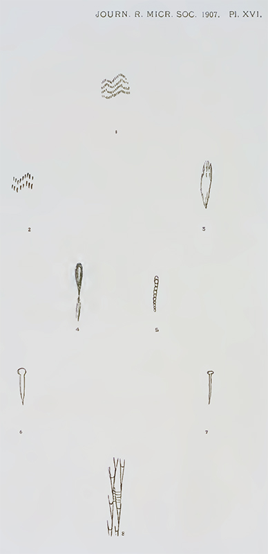
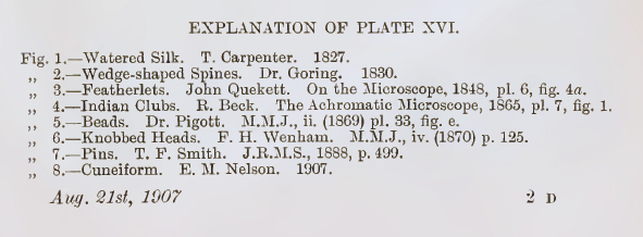
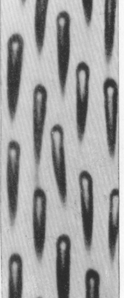
As various aspects of microscope development matured (objective optics, substage condenser and its role, light and lighting techniques), there was a wealth of published reports describing how the now readily resolved markings appeared and each worker's interpretations. Thankfully, the eminent microscopist E M Nelson eloquently summarised and assessed many of these often conflicting studies in his 1907 paper "On the Podura scale" (4), as well as offering his own observations. The variety of interpretations of the markings were shown in Plate XVI accompanying the paper and is shared below; note the varied descriptions in the caption! Some workers regarded the markings to be 'apertures' or 'depressions' rather than discrete raised structures, but Nelson presented observations that suggested that this was not the case.
|
|
Left and above: Plate X and caption from E M Nelson's paper (Screen capture from the free Acrobat pdf format of the Journal volume at www.archive.org)
|
Piggott's interpretation in 1869 of the markings as 'beads' (Fig. 5 in Plate XVI above), in particular provoked much debate (4) and was discussed in an article by Michels entitled 'The Microscope and Its Misinterpretations' in 1875 (24).
By the early 20th century, the accepted appearance of the scale markings by a number of workers using good optics and a well set up microscope seems to be that typically shown by Spitta's turn of the century excellent photomicrographs (7), reproduced later in this article; the markings were described by Spitta as 'notes of exclamation', Conrad Beck later describes them as 'quills' (11). The ability of a given class of optics to show this type of structure well was discussed (8) and the effect of different lighting techniques on their appearance e.g. crossed polars (4), off-axis oblique and incident lighting (9).
However, Nelson in 1907 (4) from his own careful studies remarked:
"An examination of a Podura scale in a critical manner with modern apochromatic objectives, illuminated by a full axial cone from an achromatic oil immersion condenser, shows that the exclamation marks have V-shaped heads, causing them to assume the appearance of the cuneiform marks on the Assyrian inscriptions. Both the pin and knobbed heads are due to some obliquity in the illumination, especially in a direction from root to tip."
The 'cuneiform' shape is shown in the plate from Nelson's paper above (Figure 8). N E Brown writing in 'Watson's Microscope Record' in 1931 (26), cited Nelson's work and also believed from Brown's own studies that the correct presentation of the markings to be the cuneiform shape (see below). Both Nelson and Brown also described and illustrated transverse lines seen on the scales; Nelson describing them as 'minute horizontal filaments of great tenuity' and Brown as 'fine oblique veinlets'. Both agreed that skilful manipulation of the microscope was required to see these. Later TEM studies by Baker and McCrae (5), see below, show evidence of 'irregularly arranged transverse marks' across the scale although the TEM studies don't seem to show the longitudinal markings themselves in the form shown.
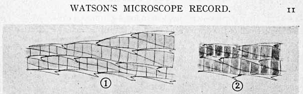
The appearance of the Lepidocyrtus curvicollis scales as described by N E Brown in 1931 (26), probably with a 1/12th objective NA1.3 which Brown mentions in text; compare with Nelson's Figure 8 in earlier plate above. Crop of Plate sourced from the complete 'Watson's Microscope Record, Issues 1-47, 1924-1939' on CD, compiled by Steve Gill (Little Imp Publications) and shared with his kind permission. This invaluable resource is available from Savona Books and excellent value.
Nelson in 1907 believed that the scales remained of value as test objects and later remarked in same paper (4):
"As a test for dry lenses, from N.A. 0.3 upwards, the Podura scale is still unrivalled. Differences between objectives can be detected by a Podura scale, which no other test will reveal."
Despite the intensive scrutiny of the scales over a long time period, including studies by many of the leading microscopists of the 19th / early 20th century, in Nelson's introduction to his 1907 paper he commented (4):
"It is strange, but true, that while the Podura scale has been the most looked-at object of any, it still remains the microscopist's enigma, and this, moreover, in spite of the fact that its structure is by no means so very minute for a microscopical object."
Edmund Spitta skilfully presented the scale markings in his 'Photo-microscopy' book, 1899 but remarked (7):
"What these markings are due to is a matter of great dispute, some thinking them depressions, whilst others regard them as hair-like appendages."
Even as late as 1938, Conrad Beck in his book 'The Microscope' commented on the uncertainties of the true scale structure (11):
"It has a structure which gives the appearance of a series of regularly arranged quills with a black margin and white centre—what that structure actually is has not been determined."
In his 1907 paper (4) Nelson after assessing the work of others, offered his own thoughts on the real structure:
"In conclusion, it has been seen that these exclamation marks are neither featherlets, nor beads, nor perforations ; so much for what they are not : but what are they ? They are probably elevations which adhere closely to the scale for their entire length."
Moore in 1883 came to similar conclusions, as stated in a report of his work (12):
"Dr. A. Y. Moore agrees that the markings on Podura scales are caused by spines. These are attached to the scales, not at the small end only, but by nearly the whole of their under surface. If they were attached at the end, it is highly probable, he thinks, that they would become broken or bent from their normal position by a slight rub, whereas scales are found which have been scratched and the spines still remain, but in an injured condition."
Later electron microscopy studies, summarised below, would show that both Moore and Nelson had correctly interpreted the true nature of the scale markings, despite the EM revealing that their true structure showed little resemblance to the optical microscope's interpretation.
Scale variability and availability
Natural objects inevitably have limitations as a consistent test subject. Andrew Pritchard, a noted maker of microscope optics, microscopist and writer, remarked on this in his splendid book 'The Microscopic Cabinet' published in 1832 (25). He devotes a chapter to 'Test Objects' where he describes his own careful observations including of the test scales 'Podura plumbea' and are illustrated in the book's Plate 12.
Regarding the 'Podura' test scales, Dallinger in 'The Microscope and its Revelations', 1901 remarked (10):
"Its scales are of different sizes and of different degrees of strength of marking (fig. 728, A, B) and are therefore by no means of uniform value as tests."
Joseph Beck showed splendid drawings of typical 'test' scales and 'ordinary' scales in the Appendix to Lubbock's monograph (17, 21), shown below.
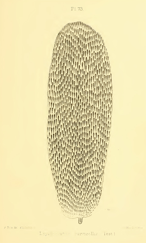
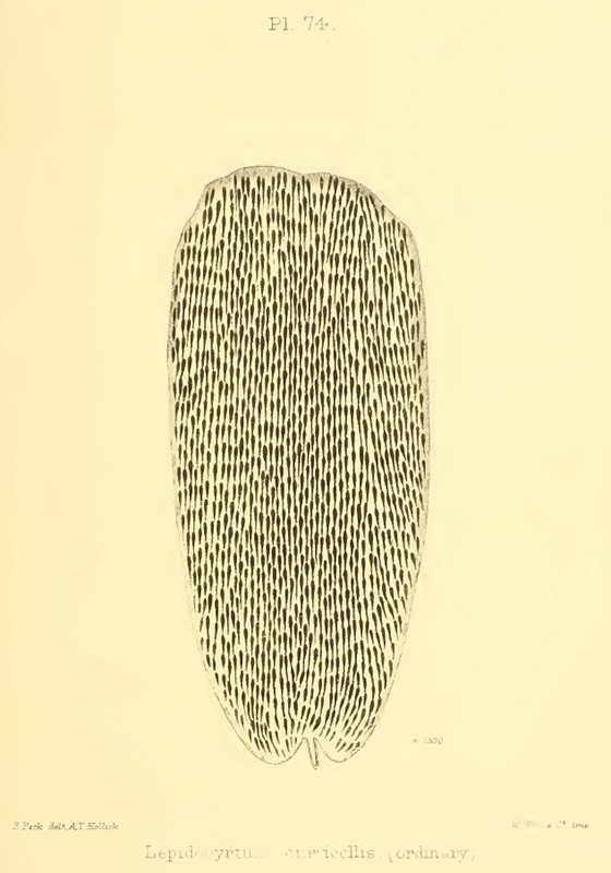
Plate 73 and 74 'Lepidocyrtus curvicollis' scales from Joseph
Beck's, 'Essay on the scales of the Collembola and Thysanura',
an Appendix to Lubbock's 'Monograph on the Collembola and
Thysanura'. The drawings are by Richard Beck. Plate 73 show Beck's 'Test' scales, Plate 74 show what he defined as 'Ordinary' scales.
(Screen capture from the free Acrobat pdf format of the Appendix included in the Monograph at www.archive.org).
In the Appendix article accompanying these plates, Beck remarks (17):
"I am unable to explain the reason for the difference in the size and appearance of the scales. They are taken from insects, entomologically speaking, the same, and yet I have never found the two sorts of scales on the same insects, and have never found the insect with the large scale in the same habitat as the one having the smaller and more common form."
Spitta discussed the limitations as a test object as well as its roles for assessing different categories of objectives. He also notes in 1909 (8) that:
"Most of these specimens are of old date, and consequently mounted with rather thick covers."
... and remarks on the tube length corrections that were required to correct for this.
Nelson also remarked on availability (4):
"It is to be regretted that good test scales have become so rare and difficult to obtain. Poduras are quite common, and are moreover, easily bred."
Spitta's and Nelson's comments may suggest that recently prepared slides weren't widely available by the early 20th century and possibly reflected the demise of its widespread use as a test object. Although given the springtail species of interest was 'quite common' as Nelson notes and still regarded by him as a valuable test object (4), it's not clear to the present author the reason for the apparent lack of well prepared slides of Podura scales at that time (comments welcomed). The variability of the scales within samples of the same species, noted by Joseph Beck above, may have thrown doubt on the scale's reliability as a test subject.
Classic test subjects such as diatom frustules continued to be discussed as tests further into the 20th century, e.g. by Needham in 1958 (13). But to date have only read one report of optical studies more recent than 1938 (C. Beck, 11) of Lepidocyrtus curvicollis scales to elucidate their structure, e.g. by using modern techniques such as phase or interference contrast (see Notes 1 and 2). Definitive confirmation of the Podura scales' actual structure may have remained elusive until the widespread availability of the electron microscope.
Note 1: Baker and McCrae in 1966 (5) reported their positive phase contrast studies of dry scales as a complement to their electron microscopy studies.
Note 2: The Cumulative Index of 'The Microscope' journal published by the McCrone Research Institute lists five articles on the Podura scale published between 1937 and 1958, but copies of this journal at the British Library are currently not accessible.
Update 16/02/2011. Four of the five 'The Microscope' journal references to 'Podura' have now been accessed (35-39). A short article by Grover (36) summarises some common British springtail species and their features to help readers identify the correct test species L. curvicollis.
An intriguing photomicrograph shared in a letter to 'The Microscope' by a Mr. G Freestone (Coventry) claims to show the 'Podura' scale mounted on edge to show what he describes as the 'fox's brush effect'. Disappointingly, no information on how this scale was prepared and photographed is given (a later Journal note does ask for details). Frans Janssen, a Collembola taxonomist, has inspected the image and remarks (28):
"In my opinion the author was confused. He shows a macrochaeta, not an edge mounted scale. We know now from Baker and McCrae [TEM studies, see below] that the micro 'hairs' are only present on the outer side of the scales. The picture shows 'spines/hairs' on both 'sides' of the 'scale'. Macrochaeta of entomobryids are said to be ciliate multilaterally. In other words they are furnished with small 'hairs' on all sides. In my opinion this is what the picture shows. Thus it is not showing a section of a scale."
Note on nomenclature
There was a variation of nomenclature in 19th century reports of the springtail species whose scales became adopted as a test object. The potential reasons of the naming variation coupled with conflicting observations was remarked on by Richard Beck (9, 29). In ref. 29, he notes:
"The author of the present paper considered these differences of opinion to have arisen from various sources, but more especially from the fact that none of the writers quoted here identified the scales they have described with any particular species, and the only attempt that any of them has made to figure the insect, occurs in the 'Micrographic Dictionary ;' it is not, however, an original drawing, and can only be recognised as a species which has no scales whatever upon it."
Frans Janssen notes that the species illustrated in the 'Micrographic Dictionary' is the hairy but scaleless Orchesella villosa (28).
The situation wasn't helped by editions of the popular 'Micrographic Dictionary' including the following statement in its 'PODURA' entry (27), present author's underlining.
"The body is covered with scales (PL. 1, Fig. 12), which are used as test objects. Those of P. plumbea, the so-called common springtail, are usually recommended ; but we believe that the most common Podura is not this species. This is, however, a matter of little importance, because the scales of several species, belonging to even different genera, are exactly similar, both in form and markings."
This entry was included in editions as late as the fourth edition of 1886, i.e. well after the publication in 1873 of Lubbock's monograph on the Collembola (21). This monograph included Joseph's Beck's Appendix with his brother Richard's splendid drawings of the scale markings from many different species (17). These clearly show the variety of forms that the scale markings can take.
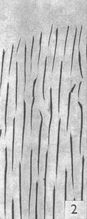
Some of the earliest descriptions (1829+, ref. 6) and many later to circa 1860, referred to the species 'Podura plumbea', others as to just the scales of 'Podura'*. In 1862 Richard Beck published a paper entitled 'On the SCALES of LEPIDOCYRTUS ____? hitherto termed PODURA SCALES, and their value as TESTS, for the MICROSCOPE' (9). Later the test scale species was referred to by many workers as Lepidocyrtus curvicollis (19, 17), more correctly by Baker and McCrae in 1967 as Lepidocyrtus curvicollis Bourlet (5) although some continued to just refer to them as scales of 'Podura' into the 20th century (4, 8). Lubbock in the 1873 edition of his 'Monograph on the Collembola and Thysanura' (21) has species entries for Tomocerus plumbeus (Podura plumbea) and Lepidocyrtus curvicollis and they remain distinct species today and have very different scales (see below).
(*The term 'Podura', before the work of Lubbock in the latter half of the 18th century had a higher taxonomic role, today it is a genus (21, 27).)
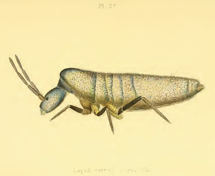
Plate 25 'Lepidocyrtus curvicollis' from Sir John Lubbock's magnificent 1873 edition of his 'Monograph on the Collembola and
Thysanura' (21). The position of the head with respect to thorax is a distinctive feature of this species which is found in dry cellars (22b). The scales in this species are irridescent (22b). The plates in the monograph are credited to a Mr. Hollick who Lubbock notes was deaf and dumb.
The 'UK Collembola taxonomy and ecology' website hosted by Roehampton University has a page devoted to the species with photos and distribution and which notes it can be up to 3 mm in length.
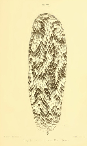
Plates 73 and 68 from Joseph Beck's Appendix to Lubbock's monograph showing the test scale of Lepidocyrtus curvicollis (left) and 'Podura teres plumbea Linn.' ('Tomocerus plumbeus, Linn' in Lubbock's 1873 Monograph). The scales are very different.
(Screen capture from the free Acrobat pdf format of the Lubbock monograph at www.archive.org.)
Frans Janssens, Dept. of Biology, University of Antwerp and co-editor of the www.collembola.org website has devoted a considerable amount of his time to reviewing the complex published literature on the 'Podura' test scales to assess the potential causes of the 'naming confusion' from a modern taxonomic standpoint (28). These findings will be published in due course on the 'Collembola' website.
Transmission electron microscopy studies: 'Podura' scales' true structure elucidated
Baker and McCrae in 1965 (5) and 1967 (14) used transmission electron microscopy to study the scales' 'ultrastructure'. The author's stated:
"The present investigation was undertaken partly to clear a protracted dispute, partly to help in the interpretation of what is seen when excessively small objects are viewed by light microscopy, and partly as a contribution to insect morphology."
In the first paper they studied untreated freshly collected scales of "Lepidocyrtus curvicollis Bourlet" using a tilting stage in the TEM for three-dimensional views. (The first production scanning EM was introduced in 1965, ref. 16). As a complement to their electron microscopy studies, they studied and remarked on the optical appearance of the dry mounted scales in brightfield and phase but didn't show images. Their comparison of the scales as seen by TEM at the same scale as Spitta's reproduced classic optical image (left and middle image below) is a striking illustration that the optical microscope presents an interpretation of the true structure, even when the discrete markings are not difficult to resolve.
They remarked that their optical microscopy studies gave the typical markings to have a length in the range "over 3 µ to more than 5 µ", and "the longer marks are a little less than 1 µ wide at their furthest point". However, their TEM studies showed the markings to be only 0.125 µ at the widest point. This width is well under the limit of resolution of the optical microscope and as the author's noted, the optical microscope is essentially showing an interference figure of the thin line markings.
Left: Photomicrograph of Podura scales scanned by the present author from Spitta's 1909 'Microscopy' book (8) to retain quality. This image mirrors Baker and McCrae's own Figure 1 where they scanned the same photo; as they did, the image is presented at the same relative scale as their Figure 2. It would have been interesting to see a side by side comparison of their TEM image with an optical microscopy image
of their own scales; the authors note that the scale marking density of their sample under TEM is higher than that of Spitta's.
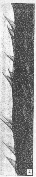
Second from left: Figure 2 from Baker and McCrae's paper (5). Their caption reads: "Part of a scale of L. curvicollis as seen by [transmission] electron microscopy. Reduced to x3000, for comparison with Fig. 1."
Third from left: Figure 4 from Baker and McCrae's paper (5). Their caption reads: "The folded edge of a scale of L. curvicollis, to show the projecting parts of the spines. x18,000".
(Note the magnifications quoted apply to respective author's original printed work, not reader's screen mags which will vary.)
Right: Present author's image of own 'Lepidocyrtus' slide (see below), Zeiss 100X NA1.3 Neofluar objective, phase, green interference filter, image resized to approx. same scale. The marking density is closer to Spitta's for the present author's slide than Baker and McCrae's TEM image.
Credits and Acknowledgements: Figures 2 and 4 above reprinted from the Journal of Ultrastructure Research, 15, J R Baker and J M McCrae, 'The Ultrastructure of the “Podura”
Scale (Lepidocyrtus curvicollis, Insecta, Collembola), As
Revealed by Whole Mounts', 516-521, 1966, with permission from Elsevier. The Journal of Ultrastructure Research, later the Journal of Ultrastructure and Molecular Structure Research, is now the Journal of Structural Biology.
(The publishers, via the Copyright Clearance Center, kindly allowed up to two figures to be used from each of their papers, (refs. 5 above and 14 below) without incurring copyright fees. Note that Baker and McCrae's original image quality of the figures presented are potentially limited by the Xerox scan of the paper.)
In the 'Results' section of their paper (5), Baker and McCrae present TEM evidence to support their observations:
"Further study by electron microscopy reveals that each mark is in fact a slightly raised ridge situated on one side of the scale, and continued as a projecting spine at the end pointing toward the free end of the scale".
and later, they remark :
"It will be noticed that the underside of the projecting mark, except the extremity, is attached to the surface of the scale by a delicate membrane of low electron density."
It is recalled from the earlier Historical Background in this present article, that this is essentially the conclusion Moore came to regards the markings in 1883 (11) and later by Nelson (4). Baker and McCrae duly credited and cited Moore's first correct observation of the true structure of the scale markings in their 'Discussion'.
In the second paper, they reported their studies of transverse sections through both the scales and insect which revealed a further insight into the scale structure and the relationship between the scales and the cuticle. They also acknowledged that prior work had come to light; Parisia and Lanzavecchia in 1957 (15) were the first to publish an electron micrograph of the scales of Lepidocyrtus curvicollis.
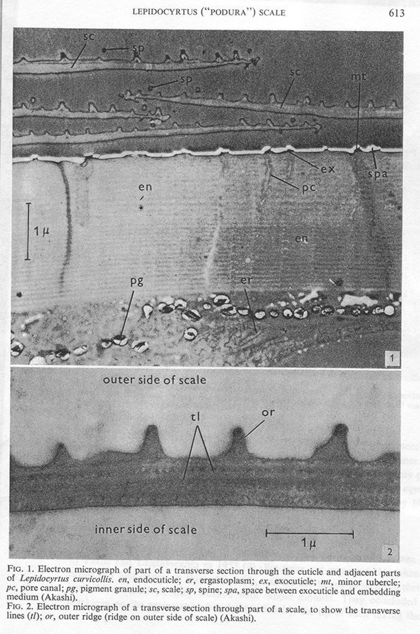
Figures 1 and 2 from Baker and McCrae's paper (14). (Figures 3 and 4 in their paper, not shown, presented "Electron micrograph of transverse sections through the edges of two scales.")
Credits and Acknowledgements: Figures 1 and 2 above reprinted from the Journal of Ultrastructure Research, 19, J R Baker and J M McCrae, 'A Further Study of the Lepidocyrtus
(“Podura”) Scale (Insecta, Collembola)', 611-615, 1967, with permission from Elsevier. The Journal of Ultrastructure Research, later the Journal of Ultrastructure and Molecular Structure Research, is now the Journal of Structural Biology.
Some of the key illustrations and observations from Baker and McCrae's papers are shared above, but urge readers interested in the 'Podura' scales (L. curvicollis) as test object to obtain copies of the papers, they are a fascinating read, including their 'scene setting' historical overview. Bradbury includes a short discussion of Baker's and McCrae's work in his coverage of the 'Podura' test scales in his splendid 2002 review of 'Test Objects' (29).
The present author would be very interested to hear of any more recent EM studies of the Lepidocyrtus curvicollis scales' ultrastructure e.g. by scanning electron microscopy.
 Studying an example of 'Podura' scales
Given that the TEM work of Baker and McCrae shows that the width of scale markings at the widest point are 0.125 µm and are well below the resolution of the optical microscope, the present day enthusiast may well ask whether they are 'chasing their own tail' studying what, as those authors remark, is an interference figure. Using the modern optical microscope to elucidate the true structure is clearly unproductive, but does still offer an opportunity to retrace the footsteps of the earlier workers studies of a fascinating test slide with an extensive and complex history.
The scope of the present author's hobby level studies certainly doesn't aspire to be any more than a 'retracing the footsteps' type study and exploring how this particular example of the scales appears on a Zeiss Photomicroscope III with a range of techniques, in particular to see how 'modern' techniques such as phase, interference contrast, near UV and autofluorescence interpret the markings even though they are below the resolution limit; while hopefully keeping a wary eye out for the limitations and potential pitfalls of studying a demanding dry mounted test subject.
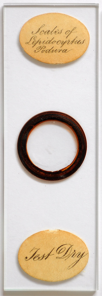
The author had one microscope slide of this subject labelled 'Scales of Lepidocyrtus Podura' and 'Test Dry' (shown right), the mounter is not stated but the oval label style and italic script typeface is similar to that of Watson / Wheeler. Dry mounts seemed to be the popular method of studying the scales (5). To date the author hasn't found any references to work using alternative mountants—as they were used for other test objects of the time such as diatom frustules. The true structure of the scale markings was proving elusive to determine and there was a possibility of natural oils on the scales (and mountants?) masking structural detail (see below).
For examples of how prepared microscope slides of the 'Podura' test scale typically varied both in presentation and labelling throughout the 19th and 20th century: Howard Lynk has a splendid series of early test slides labelled 'Andrew Pritchard' including 'Podura' scales on his superb 'Cabinet of Curiosities' website ('Pre 1840' section). The address on the slide '263 The Strand' likely dates it to the 1830s (33), so a very early example. Bracegirdle illustrates examples in his definitive book 'Microscopical Mounts and Mounters' (32) and also in his fascinating paper on the test slides personally owned by W B Carpenter (31).
On the author's slide the coverslip thickness was unknown. Focussing from the top to undersurface of coverslip gave ca. 0.15 mm for slip thickness using a LOMO Biolam's known fine focus increments. Using a potentially sensitive objective, a LOMO 40X NA0.95 dry apo with coverslip thickness correction collar, the optimum image of scales attached to the underside of coverslip
required a collar correction of ca. 0.15 rather than the default 0.17; scales unattached to slip at lower focus plane also were optimally seen at ca. 0.15.
By using published affects of tolerance of objective NA to slip thickness and required tube length corrections (13a, 22), for the objectives the author used for imagery i.e a max NA of 0.75 dry and using less sensitive immersion objectives for high power work, the maximum tube length correction required was estimated to be 20 mm, 12 mm was already built in to author's Zeiss microscope for projection work. Trials using additional tube length correction didn't noticeably affect the imagery with
objectives used. All scales imaged below were those at or near the same focus plane as the underside of slip to minimise affect of dry mount, with monochrome light used at higher powers to minimise any chromatic aberration. The main affect of the dry mount noticed was a limitation on using the full aperture of immersion objectives and an inability to use either annular oblique or darkfield with immersion objectives.
On completing the article the author acquired two additional examples shown below. Any relevant comparisons may be shared at a later date.
|
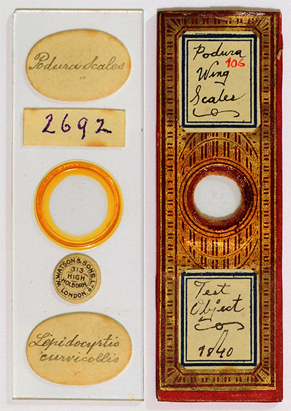
Right: Two 3 x 1 inch slides sourced at completion of the article and not further discussed. Both appear to be dry mounts as is typical for this test slide. 'Podura Scales. Lepidocyrtus curvicollis'. The form of the maker's label and address dates it to 1908 - 1957 (33a). This is labelled with the full correct species name of the test scale. (Genus first reported ca. 1862 by Richard Beck (9), full species name ca. 1868 e.g. by McIntire (19)). ****** 'Podura Wing Scales'. 'Test Object 1840.' 'Wing Scales' is incorrect, springtails are wingless. Slides dated this early are uncommon if dated correctly either at the time of making or later. To the author the style maybe somewhat later (comments welcomed). The slides dated 1840 in Bracegirdle's book made by Topping use a simple papering (32c). If dated correctly, this is only one year after the accepted wide introduction of the 3 x 1 inch slide as standard (32b). A similar pattern of gold tooled red papering is shown by Bracegirdle (32a). Update August 2011: Also see later Micscape article in the August 2011 issue. Notes on a selection of old 'Podura' microscope slides, including two by G A Clout - presents examples of this popular test subject and how the slide variants reflect aspects of its history of use. |
|
Note on equipment: A Zeiss Photomicroscope III was used with a Nikon D5000 consumer digital SLR remotely controlled with Nikon Control Pro 2, focus where possible on screen with Live View. Projection eyepiece Zeiss 10x Kpl W on short collar. 'HFW' in an image captions is the horizontal field width.
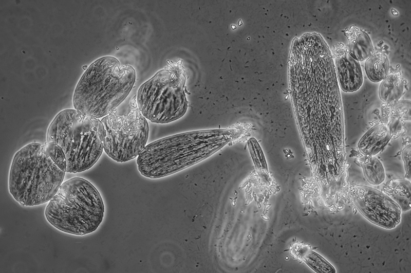
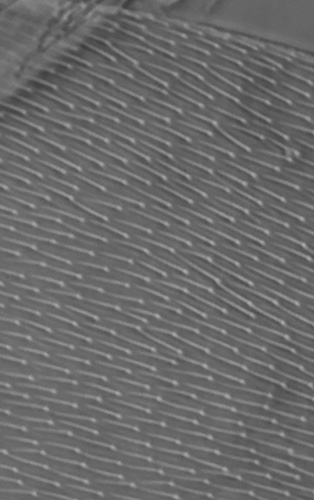
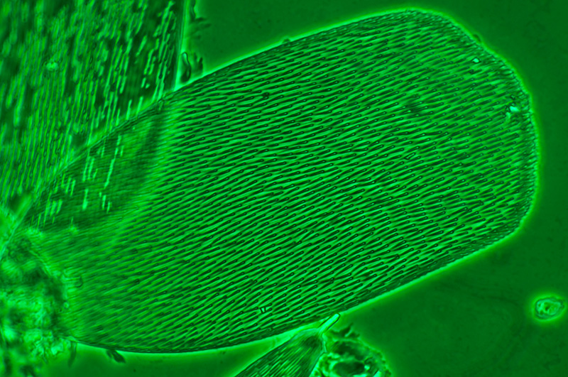
Overview of scale types: It is uncertain whether the slide scales were sourced from one springtail specimen but there is clearly a wide variation in scale morphology and size from this slide example shown below. Pritchard illustrated a variety of scale forms, including a large disc shaped scale similar to that shown below, in his 'Microscopic Cabinet' 1832 (25). With the 16x NA0.35 objective, the scale markings are unresolved and take the appearance of what has been described by Nelson and others (4) as 'watered silk'.
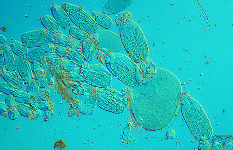
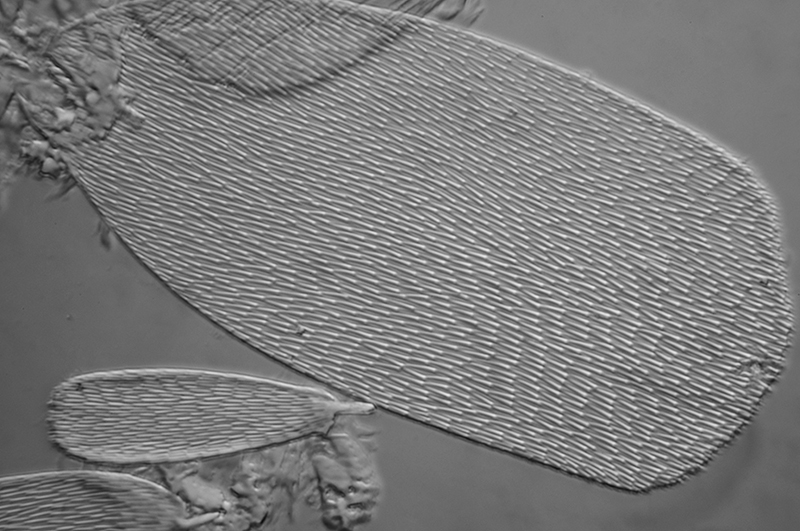
Nomarski DIC with lambda (560 nm tint plate; the author prefers this wavelength to the 530 nm red tint). Zeiss 16x NA0.35 objective.
The scales are in two distinct focus planes, one essentially touching the underside of slide and the other on the slide surface. Stack of two images to combine these two focus planes using CombineZM software. HFW = 510 microns. Scale length in image varies from ca. 36 to 141 microns.
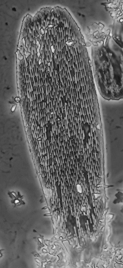
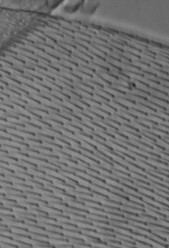
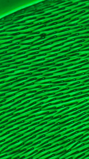
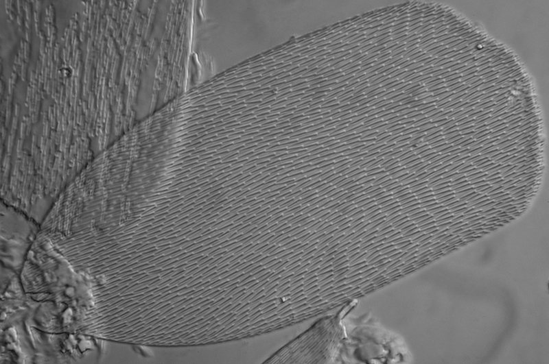
Phase Zeiss 25X NA0.60 objective. At this numerical aperture the scale markings are readily resolved (detail from lefthand image, right).
Scales in a lower plane are not stacked in this image causing the glare spots. HFW (left image) = 322 microns. Scale on right is 127 x 38 microns.
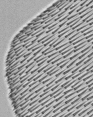
Brightfield at high power: Out of digital camera images in brightfield are of low contrast but with a well corrected objective such as the Zeiss planapo used, there is no evidence of chromatic aberration in the main scale below, for which this scale formed a useful test for 19th century workers.
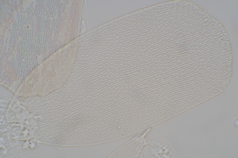
Brightfield. Zeiss 100x NA1.30 planapo objective, aperture ca. 2/3rds (the max this dry mount allowed). Electronic flash.
White focus point shown; use mouse rollover to see the darker lower focus point where the markings are more wedge shaped.
HFW = 82 microns. Scale is 81 x 39 microns.
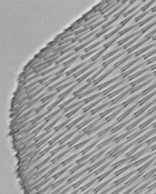
Off-axis Oblique: This technique was superior to brightfield for showing the markings at higher contrast. The end of the scales showed as a whiter mark.
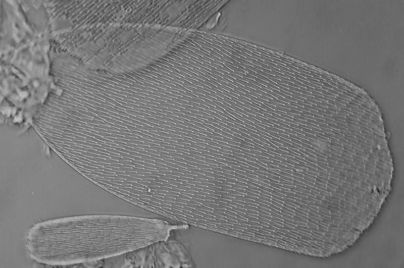
Oblique using 25% off-centre brightfield port in phase condenser. Oblique axis parallel to scale' long axis. Zeiss 100x NA1.3 Neofluar objective. Electronic flash, green interference filter. Scale size as above.
Focus point showing the white and black appearance: left - detail from image above; right - lower focus plane.
The feintest touch of the fine focus creates a slightly different view as shown above, reminiscent of the 'black and white' focus points of some diatom test objects. Richard Beck in 1862 (9) commented on and illustrated this effect for a well corrected objective, but he distinguished this effect from the different views seen of the scale markings in or slightly beyond the focus for a poorly corrected objective.
The present author, despite repeated studies under various conditions and tube length checks, hasn't been able to reproduce with the slide possessed the cuneiform appearance and cross veining which both Brown and Nelson report (see earlier).
Near UV: To see how the scale markings were interpreted in brightfield at shorter wavelengths, a Knight Optical interference filter 400 nm (35 nm bandwidth at 50% transmittance) was placed on the field iris of the Photomicroscope. The typical results are shown below.
Near UV, 400 nm with brightfield, Zeiss 10X NA1.3 planapo objective with Zeiss achromatic-aplanatic NA1.4 condenser. The images left to right show lower, 'mid-point' and upper focus level of the markings. These images are just the slightest touch of the fine focus apart. The scale used is that for the other techniques shown above. Camera - Opticstar PL-130M 1.3 Mpixel monochrome C mount camera with no eyepiece but tube length correction. Tonal balance stretched in images. (The IR/IV/visible sensitive camera is sold for star guiding on an astronomical telescope.)
Phase at high power: The markings were clearly seen with phase contrast from medium to higher powers and exhibited the exclamation mark type appearance; typical scale example shown below. The central clear feature of the scale markings seen in brightfield was discussed by some workers and noted as evidence of a well corrected objective; this appearance was similar in this slide example under phase. Baker and McCrae in 1966 (5) noted that the markings of their dry mounted scales under positive phase were 'bright throughout, apart from a narrow black outline', although were not illustrated so unable to compare.
By phase, a typical marking was 3 - 3.5 microns long and ca. 0.5 um wide, i.e. about half the width reported by Baker and McCrae for their scales under the optical microscope (5).
Phase, Zeiss 100X NA1.3 Neofluar, green interference filter. Detail from same image shown right. HFW = 82 microns. Scale is 81 x 39 microns.
Crossed polars: Nelson in 1907 (4) repeated an older observation that the scale markings 'de-polarise light' and reached a brightness maximum at one orientation between crossed polars; he used this observation as evidence that the markings were raised structures and not depressions. The present author's dry mounted scales showed the same properties with an example shown below.
The markings, notably the brighter end showed a forked / double image which was not a vibration artifact as used electronic flash and was also seen visually. This may be an artifact of some type, or the appearance of the raised separated end of the structure under crossed polars, as now known from TEM studies.
Crossed polars with scale markings orientated to maximum brightness, Zeiss 100x NA1.3 Neofluar objective (detail from same image shown right). Green interference filter. Tonal balance stretched to darken background; extinction is not perfect at magnification used.
Nomarski interference contrast: Images of the scales with a 100x objective were taken with the scale aligned both parallel to and at right angles to the prism shear axis and typical examples are shown below. As for oblique, the technique shows the markings as somewhat more linear and thinner features and less like the 'exclamation marks' but didn't seem to reveal more true structural information than other brightfield techniques. Baker and McCrae's electron microscopy studies (5) showed that lower frequency transverse structures at right angles to the markings were present; similar features were remarked on illustrated by Nelson in 1907 (4, also Fig. 8 in Nelson's Plate XVI shown at beginning of present article). Neither DIC nor phase revealed evidence for these structures on the author's slide example.
The scale markings are seen to extend beyond the upper edge of the scale, a feature which was used by some past workers, (or in folded over scales, 4) as evidence that the markings were not depressions.
Nomarski DIC, scales at right angles to prism shear axis. Zeiss 100X NA1.25 planachromatic objective (the design recommended by Zeiss to properly match the no. III DIC prism). HFW = 82 microns. Scale size is 81 x 39 microns.
Nomarski DIC, scales set parallel to prism shear axis. Zeiss 100X NA1.25 planachromatic objective. As the scale markings are birefringent, they are bright in this setting.
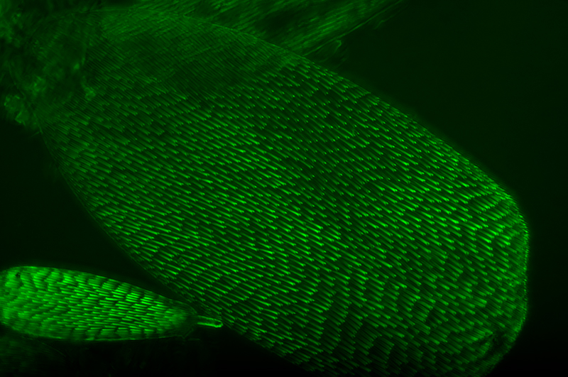
Autofluorescence?*: As the scales were mounted dry they would not be prone to the mount masking any autofluorescence (from past experience, many old Canada Balsam mounts are particularly prone to this; see 'Forays into Fluorescence 2'). There was weak autofluorescence of the dry mounted scales with the standard blue excitation on the Zeiss IIIRS epi head used with the Photomicroscope III, but required a high aperture objective Zeiss 63X NA1.4 planapo to capture enough light from a 100W quartz halogen lamp to photograph. (A planapo may not be ideal either for weak autofluorescence!)
*With such weak autofluorescence, efficient high NA light gathering objective and long camera exposures, reflected incident light imagery from any residual light leakage from the always less than perfect excitation and barrier filters cannot be ruled out. But repeating the study with a high intensity 450 nm blue LED gave a similar result, suggesting the autofluorescence is genune.
The cause of the abraded look of many Podura scales, seen in this and other techniques, was much debated in the past; some believed it to be the partial loss of the upper surface of the scale, others as the effect of natural oil on the scales touching the slip and masking the structure (4). Baker and McCrae's TEM study (14) seemed to show no clear evidence of distinct separable layers, so the oil is more likely.
The 'peg-like' structure top left of image below is the attachment point for the scale.
An aspect of this image puzzles the author, perhaps scholars of optical theory can comment. In autofluorescence the subject is self luminous, yet the markings, although somewhat slimmer seem similar to the transmitted image appearance; from TEM studies the markings seen by transmitted optical microscope studies are now known to be an interference figure of a ca. 0.125 µm feature. Would the self luminous image also show interference and/or be predicted to look similar?
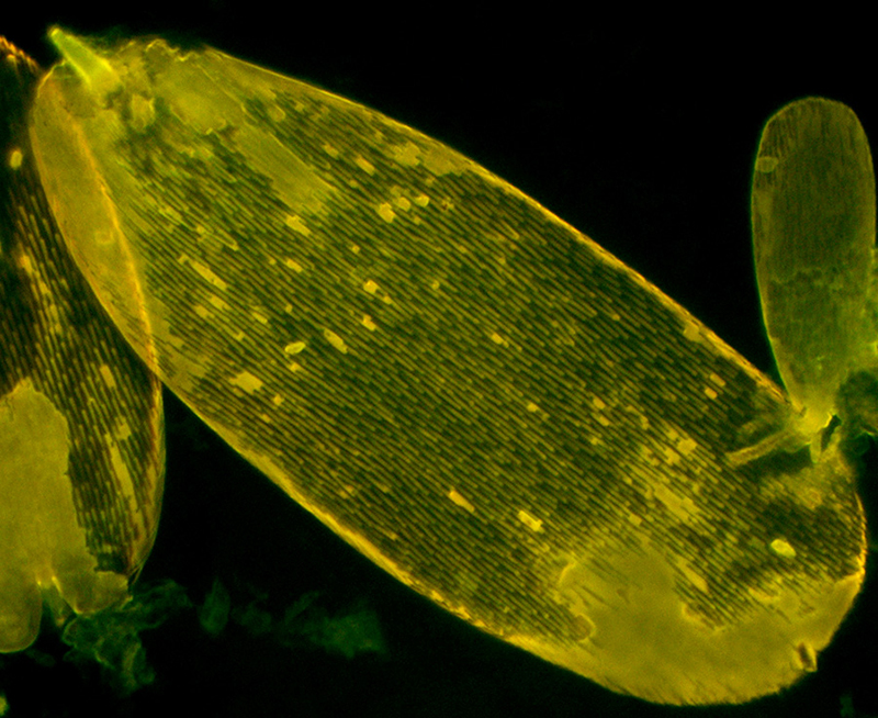
Autofluorescence, 'Zeiss IIIRS blue'* excitation from 100W quartz halogen lamp. Zeiss 63X NA1.4 planapo. Exposure 10 secs, ISO 400, tonal balance adjusted to darken background. The scale markings appear to have a somewhat more linear look than the 'exclamation marks' of transmitted brightfield. (*From Zeiss IIIRS specs this is: Excitation filter - BG12, 'FT 510 chromatic splitter, barrier filter 50'.) HFW = 133 microns. Scale is 99 x 39 microns.
Incident brightfield: Richard Beck was one of the 19th century workers to report incident light studies of the scales (9), he studied uncovered dry scales using an 1/8th objective (ca. 80X on an old 10 inch tube) with side lighting and illustrated the effect of orientating the light along the axis of the scale and at right angles.
The author doesn't have an ideal high mag incident light setup for either axial or off axis lighting. But tried it out of interest, using a Zeiss epi head and immersion objectives corrected for coverslips. For mounts with coverslips, glare is very prominent but can be suppressed to some extent with crossed polars. A tolerable albeit low contrast image was obtained shown below.
Incident light shows some promise; there is evidence that of the techniques tried, the markings are arguably least like the 'exclamation marks' of typical transmitted studies, but are taking on a longer, thinner, linear form perhaps more akin to their now known true structure as shown by electron microscopy. It would be interesting to repeat this with a better epi set-up, i.e. scales mounted without coverslip and dedicated epi objectives. Epi DIC at high power would also be interesting.
Incident axial brightfield with green interference filter, crossed polars and 100W quartz halogen lamp. Zeiss 100X NA1.3 Neofluar. Tonal balance stretched to reduce glare. The scale was that used for other techniques for comparison. HFW = 82 microns.
The pale halo effect was also seen visually and may be an artifact of the less than ideal epi imagery.
Again using incident axial brightfield, on another example of a Podura scale, size 76 x 29 microns, using a deep blue Lee filter and 100W quartz halogen lamp. Zeiss 63X NA1.4 planapo. Tonal balance adjusted. The scale markings again have a more linear look than the 'exclamation marks' of transmitted brightfield, perhaps more so than for fluorescent. HFW = 133 microns.
Final comment: As remarked earlier, because of their potential variability, such subjects can't really be regarded as objective tests for microscope optics. However, because this type of test subject is well documented, studying them while consulting the work of the early writers is very worthwhile; you almost feel to be in the virtual company of these eminent microscopists while repeating the studies which they so eloquently describe. Although as the comparison of the scale markings under both optical and electron microscopes by Baker and McCrae so strikingly shows, the optical microscopist needs to remain constantly wary of making any definitive remarks of true fine structure of the subjects they study!
The author would be very interested to hear from readers who possess well labelled, perhaps dated examples of the 'Podura' test scale. Also how the scales appear with a ca. 40x objective of the microscope, to confirm if the test slides were Lepidocyrtus curvicollis or other species, e.g. the species 'Podura plumbea'; as illustrated above, the scales of the two species are very different. This may help gain some insight into how far back and how extensive the misnaming of the test scales was in the 19th century.
The author David Walker is an amateur microscopy enthusiast not a historian, so would particularly welcome any comments / corrections on the attempt at summarising aspects of the very extensive and at times contradictory history of the 'Podura' scales as test object, or on any aspects of the author's own slide imagery.
Acknowledgements:
Many thanks to Brian Stevenson for
kindly sourcing and providing a copy of ref. 5 and 14.
Thank you to Steve Gill for permission to show part of plate from the CD of 'Watson's Microscope Record'.
Jutta Schickore kindly emailed a copy of her recent extensive paper 'Test Objects for Microscopes', ref. 34.
Thank you also to Howard Lynk for studying his early examples of Podura scales, including a slide by Pritchard and for sharing the images. These images have greatly aided Frans Janssen in his forthcoming taxonomic review of the early workers' studies.
Howard Lynk has compiled the splendid resource 'A Cabinet of Curiosities' at www.victorianmicroscopeslides.com
A particular thanks to Frans Janssens, Dept. of Biology, University of Antwerp and co-editor of the www.collembola.org website for his patience and time in providing a valuable insight into the complexities of springtail systematics from the 19th century to the present and also suggestions on the present author's article. Any errors, including in the brief comments on taxonomy in the above article are however solely the present author's. As remarked earlier, Frans is carrying out a modern taxonomic survey of the complex literature and which will reported in due course on the 'Collembola' website (Publications section).
Web resources
'UK Collembola taxonomy and ecology' - hosted by Roehampton University, London. Has a page for Lepidocyrtus curvicollis providing notes on the species and illustrating typical specimens and UK distribution map.
www.collembola.org - an extensive resource with extensively illustrated section devoted to all aspects of the Lepidocyrtinae.
Postal Microscopical Society (UK) Springtail Group - a splendid resource encouraging microscopists of all skill levels to study this relatively understudied group.
References: Many of the older journals and books are available on www.archive.org.
1. C R Goring, 'On Achromatic Microscopes, with a description of certain Objects for trying their Defining and Penetrating Power', Quarterly Journal of Science, Literature and Art, 1827, 23, 410-434. (Link is to Google Books full download.)
2. W B Carpenter, 'The Microscope and its Revelations', London, 1857, second edition. Section 108, 'Test-Objects', 183-189.
3. Cheese mites and other delicacies: the introduction of test objects into microscopy, Endeavour, Volume 27, Issue 3, September 2003, 134-138, Jutta Schickore. (Link is to an Abstract and full access to subscribers.)
4. E M Nelson, 'On the Podura Scale', J. Royal Microscopical Society, 1907, 393-404 and Plate X.
5. J R Baker and J McCrae, 'The Ultrastructure of the “Podura” Scale (Lepidocyrtus curvicollis, Insecta, Collembola), As Revealed by Whole Mounts', J. Ultrastructure Research, 1966, 15, 515-521.
6. W H Wollaston, 'A description of a microscopic doublet', Philosophical Transactions, 1829, 12. Cited by Nelson in ref. 4.
7. E J Spitta, 'Photo-Micrography', 1899, London, 139-140. Figs. 11 and 12 in Plate IV. (Figs. also reproduced in his later 'Microscopy' books.)
8. E J Spitta, 'Microscopy, the construction, theory and use of the microscope', first edition, 1909, London.
9. R Beck, 'On the SCALES of LEPIDOCYRTUS ____? hitherto termed PODURA SCALES, and their value as TESTS, for the MICROSCOPE', Trans. Microscopical Soc, 1862, Vol. X, New Series, 83-88 and Plate X.
10. W B Carpenter and W H Dallinger, 'The Microscope and its Revelations', 1901, vol. 2, 979.
11. C Beck, The Microscope', 1938, London, 235.
12. A Y Moore, 'Markings on Podura Scales', J. Royal Microscopical Society, 1883, 3 (2), 501.*
13. G H Needham, 'The Practical Use of THE MICROSCOPE', Illinois, 1958, 253-256.
13a. Ibid. p. 233.
14. J R Baker and J M McCrae, 'A Further Study of the Lepidocyrtus (“Podura”) Scale (Insecta, Collembola)', J. Ultrastructure Research, 1967, 19, 611-615.
15. V Parisia and G Lanzavecchia, Rend. Ist. Lombardo Sci. Lettere, 1957, B, 92, 197. (Ref. 9 from ref. 14.)
16. Wikipedia, Scanning Electron Microscope entry.
17. J Beck, 'Essay on the scales of the Collembola and Thysanura', Appendix to ref. 21.
18. J W Griffith and A Henfrey, 'The Micrographic Dictionary', third edition, 1875, p.773 - 776, ('TEST-OBJECTS' entry). (Link is to www.archive.org full download of the 1856 first edition.
19. S J Mcintire, 'On Podurae', Quekett Journal of Microscopy, 1868, 1, 73-76. Commenting on the species in his earlier paper ref. 20.
20. S J McIntire, 'Podurae', Hardwicke's Science Gossip, 1867, March 1st, 53-59.
21. Sir John Lubbock, 'Monograph on the Collembola and Thysanura', 1873, London, Ray Society. Ref. 21a, p. 55. Beautifully scanned and free to download in various formats from www.archive.org.
22. R P Loveland, 'Photomicrography, A comprehensive Treatise', 1970, New York, volume 1, p.67.
23. A Pritchard, 'An Account of Test Objects for Microscopes', The London and Edinburgh Philosophical Magazine, 1833, 3rd series, vol. II, 335-342 and Plate III.
24. J Michels, 'The Microscope and its Misinterpretations', Popular Science Monthly, 1875, vol. 7, June. (Available on 'Wikisource'.)
25. A Pritchard and C R Goring, 'The Microscopic Cabinet ...', 1832, London.
26. N E Brown, 'Some Interesting Test Objects', Watson's Microscope Record, 1931, 24, 10-14. (A splendid DVD of the complete series of this publication by 'Little Imp Publications' with an Introduction by Brian Bracegirdle is available from Savona Books, UK.)
27. J W Griffith and A Henfrey, 'The Micrographic Dictionary', third edition, 1875, p.612 - 613, ('PODURA' entry).
28. Frans Janssen, co-editor of www.collembola.org, personal communications.
29. R Beck, 'On the SCALES of some SPECIES of THYSANOURA, and more especially the value as TEST OBJECTS of those SCALES hitherto considered as belonging to PODURA PLUMBEA.' Quarterly Journal of Microscopical Science, 1862, Vol. II (New Series), 122 - 125.
30. S Bradbury, 'Test Objects', Quekett Journal of Microscopy', 2002, 39, 215 - 224.
31. B Bracegirdle, 'W B Carpenter's Personal Test Slides', Quekett Journal of Microscopy, 2006, 40, 333 - 342.
32. B Bracegirdle, 'Microscopical Mounts and Mounters', Quekett Microscopical Club, 1998. Podura scale slides shown include: Plate 1, slide N. Plate 4, slide N by W M Bale, Australia. Plate 5, slide J by R & J Beck, Plate 9, slide J, by G A Clout, UK.
32a. Ibid. Figure C, plate 54, p.212. 32b. Ibid. 'SLIDES, SIZES OF' entry, p. 86. 32c. Ibid. Figures A and B, Plate 36, p. 76.
33. B Bracegirdle, 'Notes on Modern Microscope Manufacturers', Quekett Microscopical Club, 1996, London., 'PRITCHARD A.' entry, p. 59.
33a. Ibid. 'WATSON. W' entry, p. 78.
34. J Schickore, 'Test objects for microscopes', History of Science, 2009, 117 - 145.
35. S P Hopkin, 'A key to the Collembola (Springtails) of Britain and Ireland', Field Studies Council (UK) AIDGAP Series, 2007, p. 111.
35. Anon, 'Postal Microscopical Society' section, The Microscope, 1937, no. 1, p.42. Short collecting note. Commented on in ref. 36.
36. J J Grover, 'Notes on the Podura', The Microscope, 1937, no. 1, 95 - 96, and Plate 13.
37. D B Spence, 'From My Notebook', 'Podura Scales' entry, 1947, no. 6, p.267. Short note suggesting 'micrurgical manipulators and the electron microscope' for studies.
38. G Freestone, 'Letters and Notes from Readers, Podura Scale Markings', 1948, no. 6, p.326.
39. D B Spence, 'From My Notebook', 'Podura Scales Marking' entry, The Microscope, 1948, no. 7, p.50. Queries method of taking photomicrograph shown in ref. 38.
Revision history:
Published February 13th 2011.
February 16th - 'The Microscope' Journal refs. added and notes. Additional links added.
Published in the February 2011 edition of Micscape.
Please report any Web problems or offer general comments to the Micscape Editor .
Micscape is the on-line monthly magazine of the Microscopy UK web site at Microscopy-UK
© Onview.net
Ltd, Microscopy-UK, and all contributors 1995 onwards. All rights
reserved.
Main site is at www.microscopy-uk.org.uk
with full mirror at
www.microscopy-uk.net
.