|
Mysteries Surrounding Dried Stuff: Part 2 A Battered Chirodota Sea Cucumber From the Philippines by Richard L. Howey, Wyoming, USA |
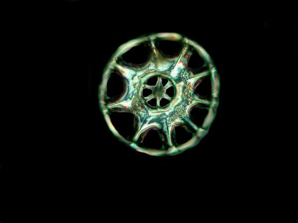
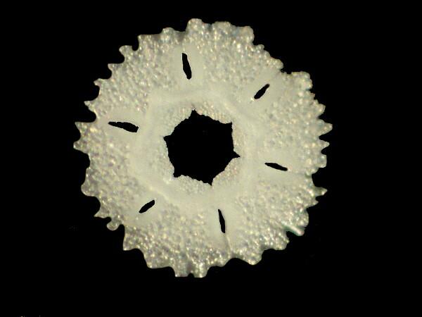
Chirodota, you will remember, has those wonderful wheel spicules embedded in its skin.
You will recall that in our last exciting episode, I had finally decided to rehydrate this specimen which process produced what, at first, looked like an undifferentiated, slimy mess–something I have encountered not infrequently over the years. So, with micro-forceps in one hand and a micro-dissecting needle in the other, I began my probing and poking.
A Brief Excursus For Those Interested In Micro-Tools
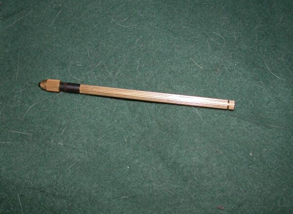
I recently found some small, single-ended pin vises at Findingking.com
I had a packet of Minuten Naedln (Carolina Biological lists them as Minutien Pins in a packet of 500 for $23.95 plus shipping, as of this writing–Oct. 24, 2010) which will make a lot of micro-dissecting needles, so you might want to get a couple of fellow enthusiasts to share the cost with you.
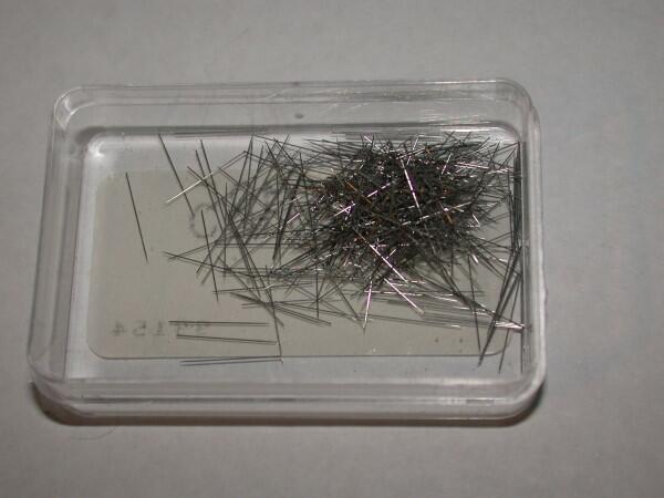
These are very small, extremely sharp, and not the sort of thing you want to get imbedded in a thumb or finger. They are stiff, brittle, and ideal for very delicate probing. They are a bit tricky to position in the pin vise and I always handle them with forceps and am very careful to avoid a spill. Trying to pick these little monsters up would be a nightmare. When the needles are finally positioned in the vises, it is very difficult to see them with the naked eye and so the ones that I haven’t yet fitted up with needles, I keep in a separate drawer.
Back to Chirodota. As I poked around, still removing bits of toilet tissue from the sea cucumber tissue, I found numbers of wheel spicules distributed closely together in very small areas. This made me think that Hamlin might very well have been right about a sort of process which bundled the spicules within a highly transparent membrane. These wheels have 9 open spaces between the spokes on the outer rim and 6 spaces in the central disk.
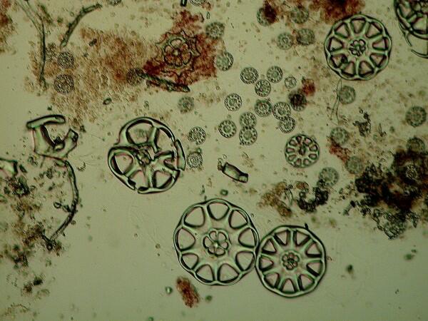
And, imagine my pleasure to find them in glorious abundance. However, as I looked at the tissue (sea cucumber, not toilet) around them, I noticed areas surrounding the spicules which showed tiny glints, so it was Clorox time. I cut a very small section, put it on a slide with a crop of the bleach and watched while it quickly dissolved the organic material. I then used a micro-pipet to remove as much of the bleach as possible without disturbing the calcareous material, added a drop of distilled water, a cover glass and put it under the compound microscope using Nomarski DIC. What I expected was some tiny platelets of calcareous crystal “grains”; instead this is what I found.
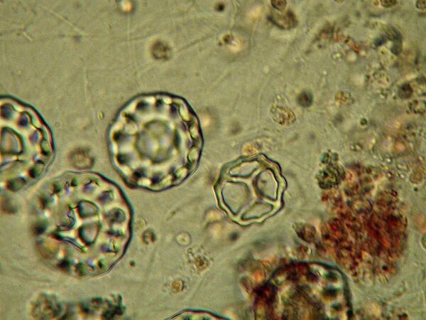
Even tinier wheel spicules! However, whereas the larger wheels have a 9/6 arrangement of spaces, the small ones have a 12/4 arrangement as you can see from the image above.
The larger ones occur in considerable numbers, but the numbers of the smaller ones are simply staggering. One thing I am fairly sure of is that these are not simply “baby” spicules waiting to grow up. I was delighted to find such a morphological oddity in a single species. Someone out there very likely knows the genus and species of this amazing little creature; all I can say is that I’m fairly certain it’s a chirodotid. While trying to find more information on this fascinating group, I consulted Libbie Hyman’s superb reference Echinodermata in her 6 volume series on invertebrates. At one point she comments that during the 18th and 19th Centuries there was intensive interest and investigation of sea cucumbers and that in the 20th Century, such research has been largely relegated to a small group of taxonomists. Talk about the decline and fall of civilization!
I continue to go back to this specimen. I have preserved all but a few small pieces in 70% alcohol since it was beginning to develop that distinctive effluvium which belongs neither to roses nor lilacs–in short, it was starting to really stink. Before I did this, I treated one more tiny “fresh” sample from an area that looked like it had something oddly different and differently odd about it. Clorox treatment, rinse, move to fresh slide in distilled water, add cover glass, and examine. I was expecting some “C’-shaped spicules which show up fairly commonly in holothuroids (that’s a snob word for sea cucumbers), but, no. What I found instead were irregular rod-shaped structures some of which you can see below.
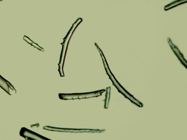
The rods are fairly birefringent, the plates and wheel spicules less so, as you can see from the image below.
This is in large part a result of the way the micro-crystals get deposited along an axis in a particular structure.
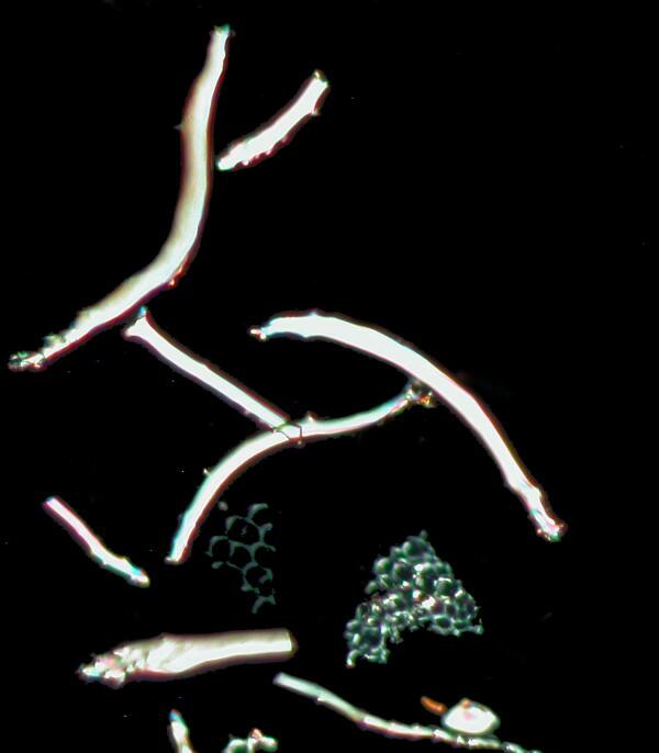
In addition, there were a few, fenestrated, thorny rods which were indeed embedded in the tissue but still, nonetheless, could have been accidental deposits from another organism around which some of the sea cucumber’s tissue grew. Three possible contenders are 1) small, immature spines from a developing regular echinoid (regular sea urchin), 2) spines from a spatangoid echinoid (an irregular sea urchin or 3) spines from an ophiuroid (brittle star). It could be any of these because of the strongly similar fashion in which such spines develop. However, sea cucumbers are also echinoderms, so it’s not inconceivable that a holothuroid might produce these structures, but it seems to me rather unlikely.
What is clear is that we have at least 4 different types of spicules in this single specimen that do indeed belong to it and typically, sea cucumbers have a ring of calcareous structures just below the tentacles which form a ring. Now, if in the mashed remnants of this specimen, I could find some things that remotely look like tentacles, then perhaps I could find this ring, but so far I haven’t been able to locate any promising hints, but I haven’t given up and eventually I will likely completely dismantle the remains, so I will keep an eye out for anything that might fit the role of this support ring.
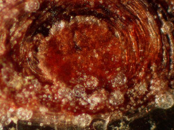
Furthermore, it is just possible that there is a calcareous internal madreporite which is a sort of filter to keep debris out of the water vascular system but, In Chirodota I have no idea what such a structure would look like. In starfish, madreporites are off-center on the dorsal surface and look like a bit of embedded coral and so are easily recognizable. Here is an example from a small starfish.
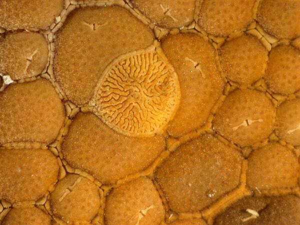
I doubt that one can be found in this mangled specimen, but I did manage to isolate the tips of several tube feet (podia) and I was curious about them because some echinoderm podia have supporting, embedded skeletal plates which allow them a good basis for increasing or decreasing suction when they are active. Here is a lovely example from the tube foot of a sea urchin and although, I managed to get an image of this one where the plates are still connected, you can nonetheless see that the disk consists of 6 plates in a circular arrangement.
In the Chirodota tube foot which I subjected to the bleach treatment, I was surprised to discover all 4 types of spicules. This is unusual; the dominant type is the slender , slightly curved rods and from what little I could see as the tissue dissolved–remember all of those blasted little bubbles that are produced as the bleach attacks the tissue–it appears that they are laid out in a roughly circular pattern in the tissue without being directly connected to one another. The larger wheel spicules are distinct, but not abundant, and finally, there are a few bits of fenestrated plates. These are somewhat of a puzzle, because it’s not clear to me whether these are just fragments of some larger support or connecting structure or not. In any case, these podia are exceptionally intriguing and, in their dried state, present yet one more curiosity to be examined. I mentioned before the odd bright, orange-red pigment associated with part of this specimen and also quite distinctly with the podia.
Pigments play an extraordinary variety of roles in the biological world–a subject for a whole series of essays–and they often present very difficult challenges to us when we try to figure out their function. In this instance, I can only report the presence of some orangish-red, powdery residue, since I have neither the expertise nor the equipment to do a micro-chemical analysis.
All of this rambling is by way of telling you, that even when you encounter some dried-up, mangled specimen that, at first glance, seems utterly uninteresting and not worthy of your attention–think again, look again. There are amazing mysteries sometimes in things that seem very drab and ordinary.
All comments to the author Richard Howey are welcomed.
Editor's note: Visit Richard Howey's new website at http://rhowey.googlepages.com/home where he plans to share aspects of his wide interests.
Microscopy UK Front
Page
Micscape
Magazine
Article
Library
Please report any Web problems or offer general comments to the Micscape Editor .
Micscape is the on-line monthly magazine of the Microscopy UK website at Microscopy-UK .