
|
An imaging conundrum in diatoms. Frithjof A.S. Sterrenburg Heiloo, The Netherlands |
Last month, I mentioned that there are several diatom species that have been mistaken for the classic test object Gyrosigma balticum because diatomists had simply overlooked a characteristic structure, which I then described as the “apical micropore crescent” in 1991. No special optical instrumentation like DIC is needed to make this visible, any good oil immersion objective and slightly oblique illumination will do, so the apical micropore crescent could have been observed and described long ago.
In the January 2013 Micscape article, I showed two different shapes of this structure in the “true” Gyrosigma balticum (where there is a tiny crescent) and a “non-balticum” (sporting a very long crescent running along the entire apex of the valve). In the present article, I’ll show an entirely different form of this structure in another “non-balticum” species.
This diatom was found in a sample from New Zealand by my good friend Stuart R. Stidolph and superficially, it looks much like Gyrosigma balticum (Fig. 1).
When you take a closer look (Fig. 2) at the apex of the valve, however, you’ll notice a marked difference: instead of a single tiny continuous crescent, there are two clearly separated “half-crescents”.
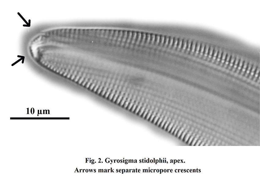
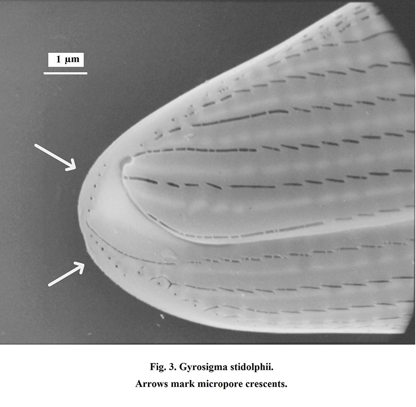
This evidently called for confirmation in the scanning-electron microscope (SEM) and Fig. 3 shows the ultrastructural aspect: two segments of 5-6 tiny pores, which are about 0.25 to 0.3 µm apart. The actual size of these micropores is fantastically small, about 1/30th to 1/40th of a micron, or 30 to 25 nanometres! Because of this specific difference in ultrastructure, together with differences in the shape of the raphe fissures, I described this “non-balticum” as the new species Gy. stidolphii.
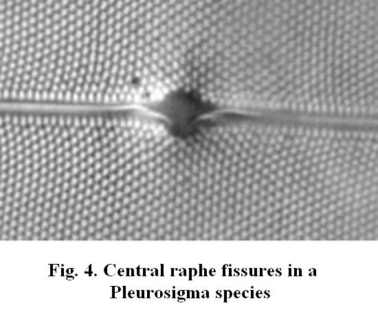
The ultramicroscopic size of these micropores raises a question, however: do not physical optics teach us that the light-microscope (LM) cannot resolve details that are smaller than about half the wavelength used, say somewhat less than 0.25 µm? So how can we see these micropores that are about ten times smaller? And if we look at other diatom structures in the SEM, we continue to wonder how such incredibly fine detail is at all visible in the humble light-microscope. Figure 4 shows the LM image (slightly oblique illumination) of the raphe fissures in a Pleurosigma species.
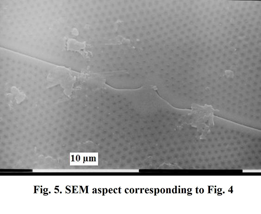
The SEM image of Fig. 5 again shows that these fissures are far narrower than the resolution limit of the LM; in this case the fissures are about 1/20th of a micron wide, or 50 nanometres. And yet they are not only visible, their shape is even faithfully represented in Figure 4!
So how to explain this imaging conundrum? The crux is that the limit of about 0.25 µm refers to resolution: the LM cannot separate two points that are closer together than circa 0.25 µm. That is visible in Fig. 2: some of the micropores are clearly separately visible, others are so close together that their images tend to fuse. But structures smaller than this resolution limit can still yield a faithful image, as these examples may have demonstrated.
The moral: do not underestimate the LM, it’s much more trustworthy than the scare stories about artefacts suggest. Artefacts are actually rare unless you are guilty of serious mismanagement like excessive “stopping down” of the condenser.
Published in the February 2013 edition of Micscape.
Please report any Web problems or offer general comments to the Micscape Editor .
Micscape is the on-line monthly magazine of the Microscopy UK web site at Microscopy-UK
©
Onview.net Ltd, Microscopy-UK, and all contributors 1995
onwards. All rights reserved.
Main site is at
www.microscopy-uk.org.uk.