|
DIATOMS AND MICROSCOPE OPTICS – SOME THOUGHTS. Frithjof A.S. Sterrenburg Heiloo, The Netherlands |
Just before last Christmas, Dave Walker sent me a pdf of a few pages from Murphy and Davidson, “Fundamentals of Light Microscopy and Electronic Imaging”, ed. 2, asking whether the examples of diatoms shown had been identified correctly. As I read these, I became aware that although diatoms are still favoured test objects in microscopy, microscopists have not generally recognized advances in diatomology over the past quarter century or so. Also, these pages contained some statements related to microscopy itself that were questionable.
Diatom identification
On page 112, there is a low-power image of a diatom test plate with 8 forms. The following corrections in the nomenclature are necessary:
Navicula lyra: it has long been known that the genus “Navicula”, even after the cleaning-up by Cleve in 1894, was a sort of garbage-can containing diatoms of very different structural types. One of the first major corrections was to remove Navicula lyra and a whole group of lyrate diatoms to the new genus Lyrella (Karayeva 1978). The correct name for over 30 years has thus been Lyrella lyra.
Stauroneis phenocenteron: read Stauroneis phoenicenteron.
Surirella gemma: this diatom does not have the structure corresponding to the type of the genus, Surirella striatula, and has therefore been assigned to the genus Petrodictyon (D.G. Mann, 1990). The correct name has thus been Petrodictyon gemma since more than 20 years.
Frustulia rhomboides: see www.microscopy-uk.org.uk/mag/artjul02/fsdiatom.html, in which I described how Jahn and Lange-Bertalot discovered a major mistake of identity, not just a nomenclatural error. The correct name has been Frustulia saxonica since then.
On page 188, Fig. 10.10 two images are shown that are claimed to represent Gyrosigma balticum. Even a modest familiarity with diatoms would show that this is not the case, and although the image on page 112 does not show a G. balticum (see further), a quick comparison would at any rate have prevented an identification of this Pinnularia as a Gyrosigma.
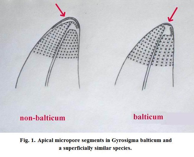
There is a true misidentification (not a nomenclatural, but a taxonomic, error) in the test slide shown on page 112. I mention this misidentification as a matter of interest, not as a critical remark, to illustrate that diatom taxonomy is a specialism. The purported Gyrosigma balticum is not that species. In 1995 (see reference) I showed that there are diatoms that superficially resemble balticum, but are actually totally different. Two LM drawings of the apical structure of such a "non-balticum" and a true balticum are shown in Fig. 1 here, from my notebook. These structural differences were subsequently confirmed in the scanning electron microscope (SEM).
Although the image on page 112 of the book discussed is taken at low power, it is still clear that the apices of the supposed balticum have large hyaline (black in the darkfield image) areas, indicating a "non-balticum". There are at least 4 such "non-balticums", all different. The difference in apical structure (crescents of tiny foramina, arrowed in my drawings) is clearly visible in LM with a good oil immersion of even a century ago with oblique lighting (at the correct azimuth!). People simply did not look...
The “non-balticum” shown is still suitable as a test object, because its line- and pore-spacing are fully comparable to those of G. balticum.
Diatoms as test objects
On page 112 it is said that diatoms are “commonly used to calibrate the magnification of microscopes”. This is not the case, diatoms are not suitable for calibrating the magnification as the line- or dot-spacing are not “constant for a species” as claimed in the book discussed, but can often vary by more than 10%. Diatoms are commonly used to test the quality of the objectives - and even more so, the expertise of the microscopist.
On page 113 it is said that in the absence of a diatom test plate, commercial diatomaceous earths and scouring powder can be used for an exercise in examining the resolution of a microscope. However, such industrial diatomaceous earths are (always/mostly?) from fossil deposits (e.g. Lompoc) and will certainly not contain the usual test diatoms such as Pleurosigma angulatum.
In point 7 on page 113 states “examine the diatom Pleurosigma (P. angulatum is meant here, but there are scores of totally different Pleurosigma species) or Diatoma….”. I wonder how Diatoma did wriggle in: the usual test slides do not contain Diatoma species, which are not suitable as a test object as pore spacing in this genus is typically 50-70 in 10 microns! Also in point 7: oiling the condenser would not make any difference in the resolution of P. angulatum as its structure can already be resolved with a good 45/0.65 lens, Diatoma species would still be beyond resolution.
General microscopy
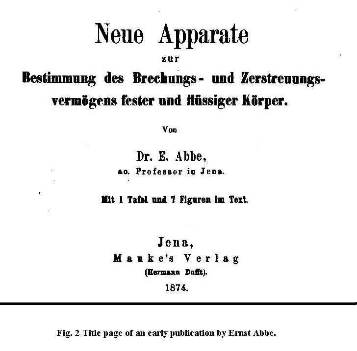
For inexplicable reasons, Ernst Abbe is called "Abbé" in the text, suggesting that he was either of French extraction or perhaps even a clergyman. This orthography would also lead to incorrect pronunciation of his name, the version "Abbé" would be pronounced as "Abbay", with emphasis on the last syllable. The correct German pronunciation is "Abbuh", with emphasis on the first syllable. A book dealing with microscopy should at least give the doyen of microscopy his proper name and a quick search on the internet would have helped, see Fig. 2 here.
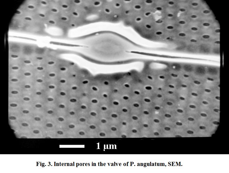
On page 104 the diameter of the "pores" in the valve of Pleurosigma angulatum is said to be 0.4 microns. This statement must be based on an error no microscopist should make: interpreting an optical diffraction effect as representative of the true dimensions of the structure imaged. My Figure 3 here shows the internal pores in the valve of Pleurosigma angulatum in SEM, which have an actual diameter of circa 0.2 µm.
In point 4 on page 114, the instruction is given to examine Pleurosigma angulatum with a “dry 100x objective”. Such objectives have indeed been produced (the ancient Zeiss F) but they are rare and a contemporary microscopist is very unlikely to possess one. If the instruction means that the reader should use an oil immersion without oil that would seriously interfere with the state of optical correction of the objective.
For just four pages, I find this a worrying harvest. Next month I will discuss an interesting imaging conundrum encountered in diatom studies.
Reference:
Sterrenburg, F.A.S. (1995). Studies on the genera Gyrosigma and Pleurosigma (Bacillariophyceae). Gyrosigma balticum (Ehrenberg) Rabenhorst, G. pensacolae sp. n. and simulacrum species. Botanica marina, 38, 401-408.
Published in the January 2013 edition of Micscape.
Please report any Web problems or offer general comments to the Micscape Editor .
Micscape is the on-line monthly magazine of the Microscopy UK web site at Microscopy-UK
©
Onview.net Ltd, Microscopy-UK, and all contributors 1995
onwards. All rights reserved.
Main site is at
www.microscopy-uk.org.uk.