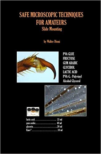
INTRODUCTION
In
a research histology laboratory, or a pathology laboratory, mounting is
the last procedure in the series that ends with a permanent histological
preparation on the table, well after the 1) fixing, 2) paraffin embedding,
3) sectioning, 4) staining, 5) dehydrating and 6) clearing operations.
Leaving
aside some exceptions, amateurs rarely engage in research that needs long
and complicated histological techniques. Most of their work is made on
live material. Though at times, many of them may need or want to preserve
some materials for future study, or to make a comparative collection of
samples, or to see cytological detail such as a cell nucleus.
After
some preliminary treatments they would like to mount the objects or organisms
as semi-permanent or even permanent preparations.
Normally
they manipulate whole organisms or parts dissected from them and many times
microscopists mount their critters without staining. Some subjects can
even be mounted without any previous manipulation at all, especially if
they are dry objects. In this series on microscopic techniques, this makes
the study of mounting media easier and useful, reversing what would be
the usual presentation schedule.
Canada
balsam.- The standard mountant for histology,
and also for taxonomy, be it zoological or botanical, is Canada Balsam,
a now scarce and very expensive natural resin. This is prepared by collecting
the resin exuded by Abies balsamica
(the balsam fir) and diluting it in solvents (many of which are now considered
toxic e.g. xylene).
From
experience to date, Canada Balsam mounted preparations last over a century.
As
Canada Balsam does not mix with water, mounting in it implies the use of
a sequence of dehydration, starting with low grade alcohols, followed by
high grade alcohols, absolute alcohol, mixed clearing
agents plus alcohol, clearing
agents, clearing
agents mixed with xylene,
pure xylene,
and balsam dissolved in xylene,
toluene
or benzene could
be used instead of xylene.
But
all three solvents are equally toxic and dangerous. (Text in red
implies toxic substances.) The development of some synthetic media as substitutes
for balsam dont solve the problem, they are proprietary trade marks, equally
expensive, that need the same steps, and use the same (toxic) solvents.
There are less toxic and less dangerous proprietary substitutes but they
are expensive.
Alternatives.-
Amateurs normally desire an easier way to have their critters mounted and
normally dont need a '100 years proof' mounting media. They need easy
to find, easy to use, non toxic, inexpensive, and reasonably lasting media
(perhaps months, perhaps some years) that assures a clear view with good
contrast of the morphological traits he (or she) is searching for.
There
exist several of these mounting media. A few of them are resinous, several
are aqueous. But normally only one is extensively cited in an amateurs
bibliography. It is glycerin jelly, a very useful option, but not the only
or most easy to use. I will review those media, selecting only the non-toxic
ones, and providing formulae and notes for their application. My formulae
in many cases are not the original, nor even the classic formulae. I have
resorted to easy to find ingredients, many of domestic use, raised here
to laboratory rank.
Refractive
Index.- Any time I know the value, I give
the Refractive Index (RI)
of the proposed mountant. Refractive index is important because it governs
the contrast between the detail you are searching for and the background,
and also the transparency of the observed sample against the bright field
of the microscope. A media with a higher index imparts more transparency.
The mounting media must always have an RI higher than the mounted sample.
Some aqueous media have an index of about 1.41 (very pure water has an
index of 1.33) but Canada Balsam has an RI of 1.524, very near that of
the glass of slides and coverslips. Naphrax which is used as a specific
mountant for diatoms, whose frustules are made of a material similar
to glass, has a very high RI of more than 1.65. There is now even
a synthetic media that reaches a really high 1.70+ RI.
Natural
media or synthetic ones of RI = 1.5 or more are used routinely in histological
mounts of tissues, previously stained and cleared. Most objects (including
micro-crustacea, or arthropods) look good when recently mounted in them,
but show additional undesirable clearing as time passes by, and many important
details, such as setae in cladocera, or copepods or acarii, could become
invisible. For these materials the modest RI of the friendly aqueous media
are actually a better choice.
Selected
mounting media.- We shall review pure
water, glycerin, sugars (karo and fructose), gum arabic, gelatin
, and PVA. I
disregard Damar,
a very economic alternative to balsam, generally used as a xylene solution,
because it is not soluble in any easy to obtain, or safe, solvent. Together
the selected products provide a selection of aqueous media, two of them
liquids (antiseptic water and glycerol) the others solids, and also one
easy to use synthetic resinous medium, NPM
(Nail Polish Mountant) that I proposed in
a previous article in Micscape Magazine (see references).
Standard
subject - To review the practical solidifying
mountants and to provide some comparative images to judge its behavior,
it's best to start with an easy to mount standard subject. And to use a
dry one, that needs no fixative, nor any stain to be applied before mounting.
I selected fly wings
as my, more or less easy to find, standard objects. Of course for some
special mountant media I needed and added some alternative test objects.
Equipment.-
Most
of the cited equipment is obvious. But throughout the article I speak about
capsules.
In professional papers the descriptions would be most probably watch
glasses, Syracuse glasses or cavity blocks. These are useful pieces
of equipment. If you have them, or could buy them. dont hesitate, they
are the best choice.
But
if you dont have watch glasses make a visit to your old relatives. They
surely have consumed some medicine tablets sealed in those dimpled plastic
sheets. If they take care to not push up the tablet, thereby ruining
your prospective laboratory equipment, they can provide you with an assortment
of sizes of concave plastic recesses (capsules) which are very useful for
your laboratory work. Select the largest for the actual purpose. I have
concave circular capsules of 10, 12, 15 and 17 mm in diameter, and also
some useful ones of 8 x 22 mm.
Here
in Durango I have identified one almost ideal large plastic capsule: the
caps of one brand of ice-cream cones are plastic cupules of 5.5 cm in diameter.
I must make a big sacrifice to acquire one dozen of them, but you understand
that science is a priority for me.
Of
course dont use them with powerful solvents such as xylene, or acetone.
If your materials allows you the use of a white opaque background (indeed
it is an advantage in the process of staining) you have recourse to the
plates with several concavities, or to the little individual dishes that
watercolour artists use to mix their colors.
In
addition you may need droppers, tweezers or forceps, fine pointed brushes,
wire loops, mounted needles, fine pointed scissors, and a small scalpel.
I've searched for but havent found a substitute for test tubes. There
are very useful. If you can, you must buy a dozen or so of 1 cm of internal
diameter and a length of no more of 8 to 9 cm. Richard Howey has made a
sound review of the laboratory materials an amateur can use more often.
Please read his articles.
(See the references.)
NOTES
AND FORMULARY LIQUID MEDIA
AW.-
ANTISEPTIC WATER
(Defined
here as water containing some diluted fixative, i.e. the water containing
the fixed sample.)
This
technique has much to do with the usual method of temporary water mounts
(wet mounts) you use when studying water samples, microscopic algae and
organisms you have just collected in your favorite pond. Except you dont
use pond water and live organisms. This is not of course a long lasting
mounting medium, but it is useful when you don't want to have your critters
drying out, after a long session of microscopic work.
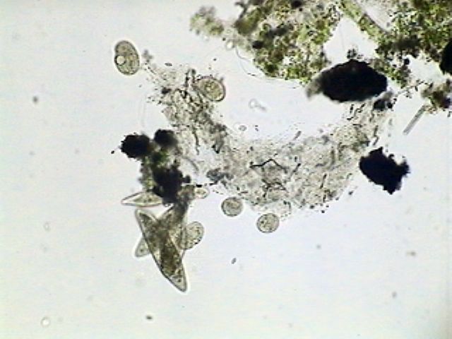
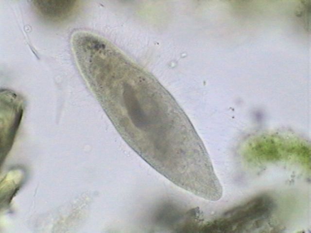
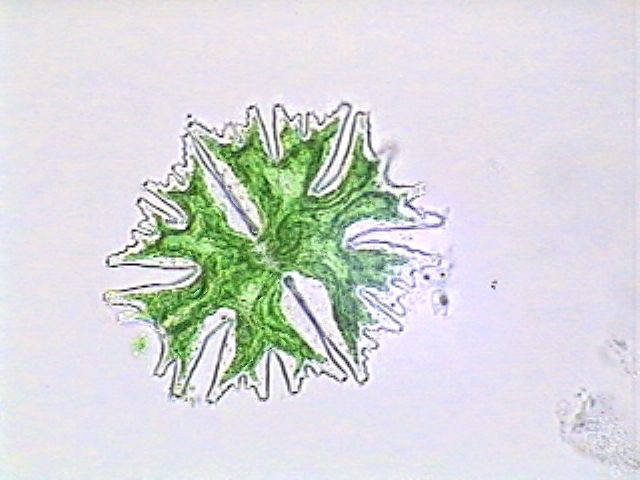
 |
 |
|
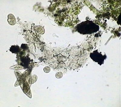
A collection of vegetable
debris with Paramecium and what is probably a Tetrahymena.
|
Paramecium alive.
x40. Rheinberg oblique illumination.
|
Many
times we return from our field trip with some different samples with one
characteristic in common: they are a collection of concentrated planktonic
microorganisms, or a handful of detritus, some times very fine, from many
possible origins, with probably thousands of interesting
.but hidden organisms.
Suppose
you have a plankton sample. You mount your drop between slide and coverslip,
and start your search. You find many interesting subjects that you want
to measure, or to draw or photograph. In a few your sample may be crushed,
or lost. You must continuously add water with a fine pointed pipette to
the coverslip border
or, better yet, you can seal the water medium to stop
evaporation.
Do
not absorb the excess water with absorbent paper, this can remove just
the critter you are most interested in. Let the preparation evaporate just
to the point in which there is no more water outside the coverslip. Now
with a fine brush, or the tool I describe afterwards, put one little drop
of sealant in each of the four corners. Give the sealant opportunity to
set, and continue sealing all the borders.
If
you have not fixed your critters, with time they will asphyxiate and
disintegrate. It would be a good precaution to take two samples. You take
home one of them alive and treat it with all the precautions Richard Howey
has explained in his recent Micscape article.
You
can fix the other using one of the recommended traditional formulae, that,
before the new trend for safety were mostly composed of formalin, glutaraldehyde,
mercury chloride and other chemicals . They are now reported to be toxic,
and not recommended for amateurs.
One
useful, effective and safe fixative, that I have designed to fix protozoa,
rotifers and the like, has a mild action, and which even preserves the
green color of algal plastids for a while, is:
GALA 20
GALA 60 60
(professional
formula)
GALA
20 is less prone to distort delicate organisms such as protozoa. For most
of the other microinvertebrate groups use GALA 60.
Preparing
the formula.- Dont be deceived by the low
concentration of the active substances in the formula. It works. Put 100
ml of water in a suitable flask. Mark the level accurately. Empty the flask.
With a 20 ml hypodermic syringe withdraw 1 ml of lactic acid, 5 ml of glycerol,
10 ml of vinegar (that is: 9.5 ml of water and 0.5 ml of acetic acid),
and some water. Agitate to mix. Put the mixture in the flask. Syringe out
some water, agitate to wash the remnants of the lacto-acetic mixture. Pour
into the flask. Repeat one or two additional times. Add the alcohol (21
ml at 96% = 20.2 ml absolute alcohol and 0.8 ml water more or less).
Now replenish the flask with water to the 100 ml mark, and stir until the
solution is homogeneous. Alternatively, if you are in the rich group
of amateur microscopists, use your measuring pipettes and graduated cylinders,
to prepare your formula.
Label
your flask as GALA 20 fixative (because it is composed of Glycerol,
Alcohol
20%, Lactic acid
and
Acetic acid)
or GALA 60 if you have used the higher alcoholic formula.
Fixing
the sample.- To fix your plankton sample (or
any aqueous sample with suspended organisms of the same order of sizes)
you must add to it 1/10th of its volume, of this solution. (1 drop to 9
drops, or 1 ml to 9 ml, etc) and agitate well to mix immediately. If you
want to fix larger animals (some micro-arthropods, hydracarina, some anesthetized
worms, arthropod larvae etc.) put them directly in the concentrated fixative
(2, 3 or more volumes of fixative by 1 volume of biomass).
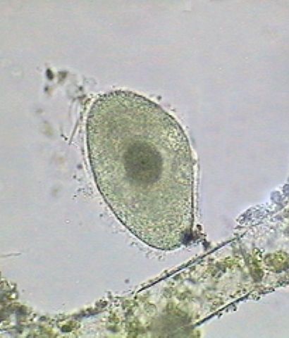
|
|
|
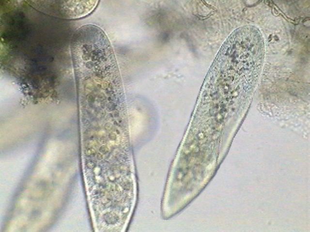
Paramecium fixed
and mounted in GALA 60. As I did not used any clearing agent it is somewhat
opaque. Nevertheless the exploded trichocysts and the macronucleus are
visible.
|
Monostyla lunaris.-
a rotifer fixed and mounted in GALA 60
|
Searching
your sample.- Of course you can search your
materials as is, or apply some color to better differentiate them from
the detritus, or to identify some organelles. One beautiful and useful
dye is Rose Bengal,
but as for all the good reagents of old times it is forbidden now. It is
toxic. In future articles on staining I hope to review some safe (but inferior)
substitutes.
To
make your preparation, to aid with the evaporation problem, and to give
the subjects a minimal clearing that mimics the live appearance of many
micro-invertebrates, mix one drop of fixed sample with a drop of glycerin,
or even lactoglycerol (see formula below). Allow to stand for one minute,
cover and start your observations. If the materials promise a long working
session proceed to seal. You can search by this technique thecamebae,
gastrotrichs, rotatoria, nematodes, microalgae
and the like. Some specimens, especially some of the protozoa,
microoligochaeta and other soft bodied organisms
dont support this drastic procedure and become dehydrated and wrinkled.
With these materials you'd better proceed to mount in glycerol or lactoglycerol
(see formula below) with all the precautions I explain later.
Sealant
media.- For sealants you can use solid vaseline,
paraffin wax or beeswax, Valap (see below), or nail polish. Your choice
depends on how long you intend to store your preparation. The selection
order is also the order of duration of the different media. A liquid preparation
sealed well with nail polish could last some months. The other media need
to be applied melted, they remain more or less soft, and can be easily
ruined, except Valap, that adheres well to glass, but is also easy to remove
if you want to recover your slide, and actually is the other media to be
recommended. This is its formula:
Paraffin
1 part in weight
Vaseline
1 part in weight
Lanolin
1 part in weight
Mix,
melt at a low heat and pour in a shallow profile can and cover. Allow to
solidify and melt the quantity you need with the following sealing tool.
One
sealing tool: you can melt the solid sealing
media and use a small fine pointed brush to apply it, but it is very difficult
to apply a neat coat to all four sides of your coverslip. An old economic
sealing tool can be of help here.
Take
a metallic wire, 2.5 mm in diameter, bend the tip at a right angle, to
make the bent portion the same length as the side of the cover slip you
use most often, and if you wish, make a loop at the other end as an aid
to manipulate the tool.
Now,
with a spirit lamp, or a cigarette lighter, apply some heat to the sealing
wire. Touch the surface of the solid sealing medium. It melts. With the
tip of your tool, touch every angle to fix the cover with a little drop
of medium. Now apply the heated side of the wire to the medium and to each
border. The molten medium spreads evenly along the sides and you have a
very good seal all around. It takes only a few trials to become an expert.
Use this tool with Vaseline, the waxes and Valap.
Comments:
These simple, temporary mounting methods, are probably the ones which you
would use most often, hopefully with much success. They are useful with
hydracarina, thecamebae, nematoda, loricate rotifers, most of the Gastrotricha,
Chlorophycea (which retains its green color for a long while), euglenoidina
(many of them showing his flagellae), desmids and Cyanophycea.
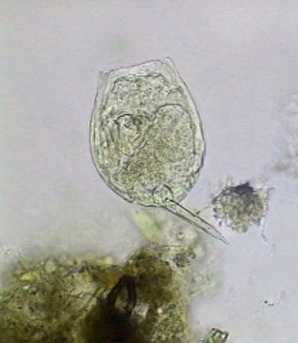
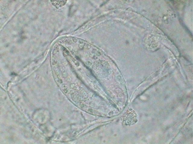
|
|
A desmid retaining the
color of its chloroplasts after two weeks fixed and mounted in GALA 60.
|
The
entomostracans (cladocera and copepods) have calcium in their exoskeleton
that could be dissolved by this highly acidic fixative. So you better fix
and preserve them with 70% alcohol. If you fixed them with GALA, because
they were mixed with other critters, select them after a few hours, and
store in 70% alcohol.
Ciliates
can be well preserved, showing their nucleii (macronucleii at least), and
cilia. Those that have trichocysts, as Paramecium do, show them
exploded most of the time.
Only
for comparatively long lasting collections, morphological detailed studies,
and demonstration purposes, or when your materials need clearing, as in
many arthropods for example, you must resort to the more dense, permanent
and more difficult to use mounting media.
 |
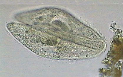 |
|
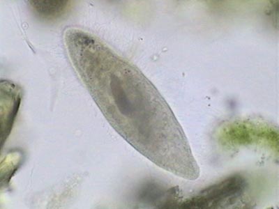
Left picture:
Probably a Tetrahymena. Right picture: A couple of conjugating
Paramecium. Both fixed and mounted in GALA 60. The pictures
increase the general opacity and the contrast of the nuclei. They are less
contrasty, but easily visible in the mounted materials, without using dyes
or clearing agents.
|
A
note on mounting samples in aqueous media:
Quite frequently, water evaporation applies a high pressure to the subjects
with the risk of delicate subjects being crushed. To avoid this you must
resort to the inclusion of some supports for the coverslip. Those supports
must be thin enough to permit a high resolution objective to be used, but
thick enough to give room for the observed subjects.
Use
at will pieces of thin coverslip, paper, plastics, hairs or textile fibers.
Many thin adhesive tapes can be cut into thin strips and neatly adhered
beforehand to the slide.
Think
before you have resort to these methods, because you cant revert the situation.
If you think that it is possible that you may change your mind and want
to slightly squeeze an organism, it is best that you use, instead of any
of the fixed height materials, tiny balls of very soft wax, or drops of
solid vaseline. These allow you to put a more or less controlled pressure
on the coverslip, to stop the displacement of an organism or to reveal
more clearly its internal organization.
Labeling.-
Even with these not so permanent preparations you must label your slides.
At least write the name of the mounted subjects,
the source of the materials, any treatment applied, the mounting media,
and the date. Add your name if you want.
Even
if the slides are to last for only some weeks or a couple of months, when
you make a revision you may have forgotten those details, and be sorry
you did not label them. You can make the labels on your computer. But you
must write the information with a good indelible black drawing ink
or perhaps
you could finish with an illegible label caused by accidentally wetting
it.
MOUNTING
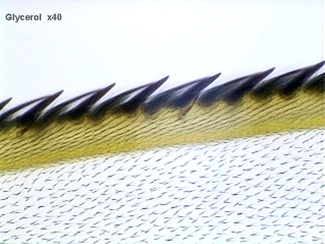
IN PURE LIQUID GLYCEROL
PG.-
PURE GLYCEROL
Glycerol,
also known as glycerin, is a common product, cheap, and easily acquired
in any drugstore. It's a very hygroscopic alcohol, with a weak syrupy consistency
and when anhydrous has an RI = 1.46.
Buy well sealed small bottles of glycerol and open it only for the time
needed to take out the drops you use.
It
is most probable that glycerol was used, as a temporary mounting medium,
from the very beginning of microscopic techniques. It is most easy to use.
Put a drop of glycerol on a slide. Include the test object (our fly wing),
removing it from water, or alcohol, cover
and go to the microscope.
Many
microscopists seduced by this simplicity, the very high compatibility of
glycerol with many solvents, and the added fact that it is a mild clearing
agent, that imparts a fair transparency to the small biological materials
it impregnated, tried to make it the mounting media for their permanent
preparations.
The
straightforward method is to seal the glycerol mount.
It is not at all easy, but it is possible.
When
you want to turn your glycerin mounted slide into a more or less permanent
preparation, put the slide on the table over one or two sheets of paper.
Cover it with one sheet of absorbent paper (thick kitchen towels work,
or even toilet paper) and slide the edge of your hand from end to end of
the preparation, taking care not to exercise an excessive pressure. This
squeezes the excess of glycerol from under the coverslip into the towel.
|
|
|
Stoma on the epidermis
of the underside of a leaf of Tradescantia virginica. Fixed in AFA
(alcohol, formalin, acetic acid). Mounted in glycerin. No stains applied.
|
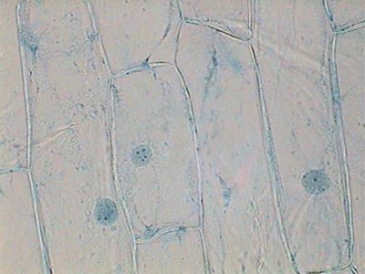
The well known epidermis
of an onion. Fixed in AFA. The staining method will be explained in a future
article. Mounted in glycerin.
|
Uncover
the slide very carefully, wrap your finger in some thin non-absorbent plastic
sheet, support one side of the coverslip, and with extreme care wipe
with a dry cloth all the oily remnants you can. Now you need to seal the
coverslip.
First
of all put one drop of nail polish on each of the four corners, and allow
to dry completely (not less than half an hour). Now support your coverslip,
moisten a cloth with a little alcohol and wipe very carefully all the contours
of the coverslip. Any rough movement and you'll ruin all your work. Use
a dry cloth to finish. Both the slide and cover slip must be dry, with
no glycerol present at all.
Now
seal all four sides with a nail polish layer that overlaps more or less
1.5 mm of the cover, and on the slide. Or use your sealing tool and Valap.
Particularly take care to also cover the four corners you fixed earlier.
Allow to dry completely. Wipe with alcohol, and apply another, very carefully
applied, sealant layer of NP. Normally Valap wont need a second layer.
Make
a check next day. Glycerol is highly hygroscopic and absorbs water greedily.
If there is a minimal opening in your sealant there'll be a glycerol-water
mixture flowing out. Dry, wash with alcohol, seal the opening. Make a daily
check for one or two weeks. When you are satisfied there are no more leaks,
finish the seal with automotive or hobby paints.
As
the mounting media is a fluid, you must file these preparations in flat
horizontal trays, otherwise the subjects can be displaced. Carefully label
them of course.
As
to the permanency of these glycerol preparations, it's worth pointing out
that J.G. Baer, a French helminthologist, reviewed in 1931 the taxonomic
characters of Temnocephala mexicana, Vayssiere, 1899 from the type
slide mounted in glycerol, and filed in the collections of the Museum dHistoire
Naturelle, at Paris, France for 31 years. (T. mexicana is a platyhelminth
that lives on some crabs.)
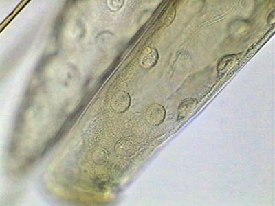
 |
 |
|
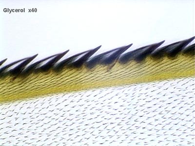
This is the first picture
of the test object. Fly wing collected and preserved in alcohol
70%, and mounted in glycerin.
|
Head of the larvae of
a Sarcophaga fly. Fixed in 70% alcohol, it was gradually passed
through increasing concentrations of glycerin and mounted in 100% glycerin.
|
Difficult
materials.- As I said, glycerol is hygroscopic.
It acquires water from all substances that contain free water. When a soft
biological object is surrounded with glycerol, water will pass from the
object to the glycerol, and is replaced by it. The problem is that glycerol
diffuses to the biological materials at a slower rate than when water goes
out. The results of this imbalance is the biological materials are shrunk
and distorted. To make a useful mount of this kind of subjects we must
resort to two useful tricks.
1)
In a series of capsules put a series of increasing concentrations of glycerin,
starting with the 10% side of the scale (20, 50, 75 could be the other
steps). Put your materials in the first one and leave enough time. (Of
course you must guess, and try some times, until you found the correct
one for your actual material. Twenty or thirty minutes are wise initial
guesses.) Using a suitable spatula or a little brush or a wire loop, or
even an eyedropper, transfer the materials with care from one capsule to
the next, leaving them for the same time in any one. Perhaps after more
or less two hours you are done. Transfer the subject in the drop onto the
slide, as was explained before.
2)
Often the first alternative is good enough. but Seinhorst
(1959) working with difficult nematodes, designed
a technique to gradually replace the fluids with glycerol without disrupting
the organ structure of the worms. By the same nature the method is excellent
for any delicate material.
The
materials, first collected in water, were then transferred to a capsule
with a 1% solution of glycerol in 20% alcohol (more or less).
alcohol
96
21 ml
water
78
ml
glycerol.
.....1
ml
Now
take a wide mouthed flask, with a screw cap, not very tall. One of those
short and wide containers that hold creams for the skin or hair fixatives,
are good. Put in the bottom a cap from another smaller diameter flask,
as a platform to support the capsule. Pour in 96% ethanol almost to the
height of the platform, and put the capsule with your materials on the
platform. Spread some solid vaseline on the rim of the flask. Screw on
the cap of the interchange chamber tightly and set aside for 12 to 24
hours in a warm place. Its better if you apply some heat, say 35-40ºC.
In the interim all water in the capsule was to be replaced by alcohol.
Now
fill the capsule with a solution of 5% glycerol in 96% ethanol, and put
it in a partly closed container. In 3 or 4 hours at 40º almost
all ethanol has evaporated, and your subjects must be in almost pure glycerol.
Normally you can now proceed to mount in pure glycerol.
3)
One special case is the mounting of mixed microscopic microinvertebrates,
which because of their minute size cannot be manipulated individually,
or in sorted uniform groups, and must be mounted in some liquid media.
(The samples we spoke about in the antiseptic water section). The following,
although not easy to apply, is a useful protocol.
a.
Fix and preserve the materials in a suitable fixative (GALA is a good
alternative). Allow at least 6 hours for a good fixation.
b.
Have a look at the sample to see if they are the critters you are searching
for.
If there is a need for some staining (await a future article) this is the
moment to
apply it.
c.
Take a sample (say 19 drops). Add 1 drops of glycerin mixing very well
after
the addition. You now have a solution with 5% glycerin. Set aside for 15
minutes.
d.
Add a new drop of glycerin, mixing well.The concentration is now near 10%.
e.
With a syringe and a fine hypodermic needle remove half of the supernatant
liquid. If you work with care you now have 10 drops with more or less 9
drops of
sample water and 1 drop of glycerin. (10% glyc.) Add 2 drops of glycerin
and mix well
.
This amounts to 12 drops (9 + 3) of 33% glycerin (more or less). Set aside
for 15 min.
e.
As the density of the liquid increases, so the sedimentation time is longer.
Give enough time to have almost all of your sample in the test tube or
concave
capsule bottom.
f.
Now take one clean slide and put one drop of the concentrated sediment
in the
center. Mix well with another drop of pure glycerin or lactoglycerol (see
below), and
cover. You must make a fair estimate of the volumes to have a very thin
preparation but full to the margins of the cover.
g.
Seal if you wish. The method is not quick, nor easy, but gives you pretty
good
preparations of many difficult to preserve microinvertebrates
NOTES:
Make your preparations as thin as you can. Resolution is impaired in thick
liquid preparations, and also it affects the quality of the sealing. Always
allow enough time for the sealant to set completely. Never use the 100x
OI objective (if you have one), with unsealed preparations.
LG.-
LACTOGLYCEROL
I
should point out that glycerol has a mild clearing power, enhancing the
transparency of fixed cytoplasms and other small opaque subjects. The combination
of glycerol with lactic acid (a more powerful clearing agent) produces
a medium that is very useful to allow a better observation of opaque materials
and organisms, like worms, plant materials and specially small arthropods.
Lactoglycerol
is normally a 50/50 mixture of both reagents, but you can freely use other
percentages according to the clearing action required by your subjects.
It is most often used as a temporary mountant, but with suitable precautions,
a satisfactory preparation can be turned into a permanent one. The manipulations
are much the same as with just glycerol, but sealing is more difficult.
Lactoglycerol
is more dense and syrupy and is more difficult to clean the outside of
the coverslip. You must anchor the four corners, waiting until the sealant
is very well set. With an absorbent paper wipe all the excess liquid you
can. With a soft absorbent tissue wetted with ethyl or methyl alcohol or
even acetone clean the slide with care. Always try to have the thinnest
preparation that you can. When you are satisfied of the cleanliness you
have attained, seal with Valap. Inspect after a few days to be sure you
have no break in the seal. Cover Valap with a layer of nail polish and
finish with another of an automotive paint. Label your slide.
Pantin's
solvents density gradient.- If you like to
mount a micro-arthropod (a mosquito larva as an example) you must clear
the organism if you want to see the details of its structure. One of the
methods is to dissolve the soft parts (all the internal organs) with hypochlorite
or potassium hydroxide. Both are potentially harmful.
A
safer method, usually enough for our purposes is to use the lactoglycerol
mixture as a clearing agent. If you submerse the subject directly in lactoglycerol
you know the result: a wrinkled and awful organism, mostly unusable. You
can resort to gradual changes of higher concentrations of lactoglycerol.
But
Pantin at the end of the 1930s proposed a very useful and elegant method
that provides organisms beautifully cleared with only one (long) easy step.
Take
a test tube or similar, put 2 or 3 ml of lactoglycerol at the bottom. With
all your care, with a long pipette, glide 2 or 3 ml of glycerol over the
first layer. You can disturb it a little but only at the very interface
to start a process of mixing. Now in the same way, make an upper layer
of 96% ethanol.
With
your tweezers, or a glass bar, or an entomological mounted needle, pick
the subject from the alcohol in which you have it stored, and drop it on
the alcohol upper layer. The organism drops to the interface of alcohol-glycerol
and stays floating there for a long while.
If
it is your first time you'll certainly want to watch the process, but better
you go to take a nap or make other observations you were planning.
The
larvae or other organism you leave bathing in the alcohol slowly mixes
with the layer of glycerol, interchanging the alcohol, and after a time
it sinks to the interface of glycerol and lactoglycerol where it repeats
the process.
At
the end you could recover the organism from the bottom of your tube. Perfectly
cleared and with much less distortion
. and requiring less effort from
you.
Experiment,
and become addicted to the method.
The
organism you have cleared can be mounted in lactoglycerol or in some of
the more solid mountants I describe in the next article.
|
|
|

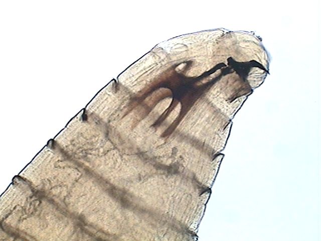
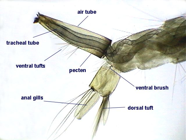
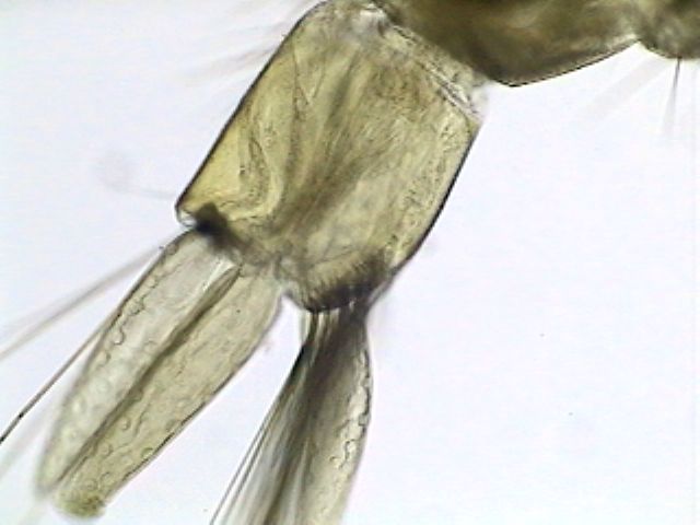
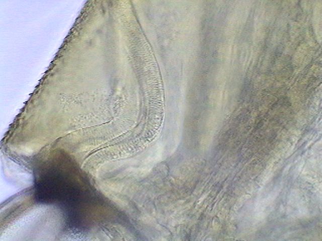
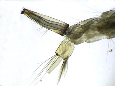
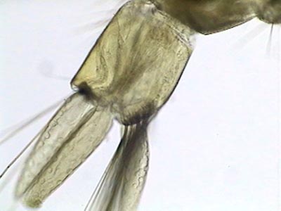
|
These four pictures show
the clearing power of lactoglycerol. The mosquito larva was fixed in 70%
alcohol, and cleared using the solvent density gradient of Pantin. The
solution layers used were alcohol 70% - glycerin - lactoglycerol. The sunken
larva was mounted in pure lactoglycerol. The third picture shows the nucleus
of the cells inside the caudal paddles. The fourth one shows two muscular
cells at the end of the body. The left one shows the nucleus. The right
one shows that these are striated muscle cells. Focus was a compromise
to show both details in one picture
Click the first photo to see a fully labeled
picture.
|
Another
method for using glycerol as a mountant.-
As you see above the biggest problems with glycerol and lactoglycerol arose
from their nature as being viscous and hygroscopic liquids. Microscopists
solve this by converting glycerol to a solid. The commonest solid form
is glycerin Jelly. I have two other useful formulae fructogel and glycogel.
All
of them are explained in the third part of this series. In the next (second
part), I will explore the use of gums and sugars alone or in several combinations.
REFERENCES
DIONI, WALTER.- About microscopy
and the chemistry of nail polish.
HOWEY, RICHARD.- Equipping a laboratory
(parts I to IV).
HOWEY, RICHARD.- Culturing and
collecting microorganisms safely.