This article
includes two avi format video clips. Click
here for an
animated gif
version if your computer doesn't support avi.
Radiolaria have always been a fascinating microscopic subject1,2,3. Some part of the most important historical literature and very good historical illustrations can be downloaded immediately and without costs at the website of Kurt Stueber4. There are also several fine papers in MICSCAPE, by Brian Darnton, Richard Howey, Mike Samworth ...
It took a long time for scientists to get a basic understanding of living radiolaria, e.g. how they are able to stretch and to retract their thin linear ray-form plasmopodia5.
Unhappily most of us will be
confined to look at the dead relics of these splendid
organisms which are nevertheless of an outstanding beauty and
interesting symmetry. This paper shows how to get some
additional visual impressions from the fascinating glass like
radiolarian skeletons. The techniques which are discussed
here might be used for many other objects as well.
Build your own virtual radiolaria exhibition slides
Radiolaria exhibition and circle pattern slides have a long tradition and of course will never be outdated or replaced by electronic gadgets. Nevertheless digital imaging offers the possibility to combine any of your favourite objects — which might reside on different slides at different places — to one gigantic picture.
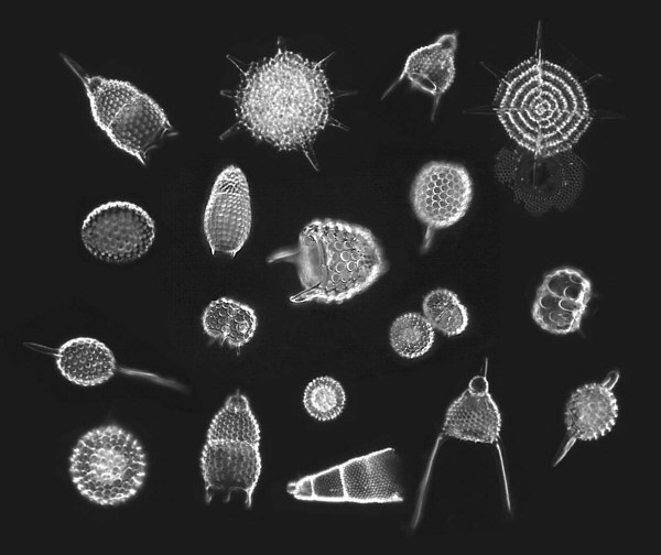
To illustrate this I have
chosen a so-called radiolarian strew slide6. There
are many beautiful shells on the strew slide which can be
photographed one by one at the best individual focusing
depth. I have merged 18 of these individual photographs taken
with a computer CCD camera7 (see figure below).
But this is just an example. You might combine hundreds of
objects to a much bigger picture just in the same way.
With standard digital imaging software8 in
particular, dark-field9 photographs can be easily
combined without seams, as the exact colour of the black
background can be measured by means of the software: e.g. a
measured R(ed)G(reen)B(lue) background color value 0,2,0 can
be set easily to 2,2,2 or 0,0,0 in order to fit into an
existing mosaic. On the basis of the measurements you can
even adjust photographs which have been taken under slightly
different conditions. Just create an empty black picture,
adjust the darkness of the background of your other
photographs and let them "drop" into the black
picture.
It is much more difficult to prepare normal bright-field photographs in order to build a combined bright-field image as the bright background is more susceptible to brightness and colour variations at the edges of the photographs.
Of course, when you start from
a strew slide you will not be able to change the orientation
of the objects like the diatom (radiolarian) laying artists
who are able to arrange their shells in arbitrary orientation
by means of guinea pig bristle hairs or extremely fine metal
needles. But when working with loose individual objects you
might even combine one and the same object with different
orientations in one picture !
Focusing into the inner radiolarian spheres
The computer software for this experiment, Dave´s Targa Animator (DTA), is an ingenious $35 shareware utility by David K. Mason. It is ready for testing and downloading from the Internet at
ftp://ftp.povray.org/pub/povray/utilities/dta/dta30.zip
The DTA software enables you to combine a series of image files (e.g. img0001.bmp, img0002.bmp, ....) to an animation. A simple command-line like "dta img*.bmp /FAVI" does the whole job and converts a series of image files to an avi-film. The size of the input pictures may vary within a broad range. The two small, low-quality avi-animations within this article, which were produced by help of DTA, will fit on an ordinary floppy disk. A medium quality 640 x 480 pixel animation in so called FLC10 format might take typically 10 MB of hard disk space depending on the number of frames. DTA offers even more: it is no problem to generate extremely high quality animations e.g. on the basis of classical photographs or photographs from consumer CCD cameras up to 1280x1024 pixels which can be displayed on the computer screen. Just have a try and feel free to give your TV set to the garbage afterwards ...
The following animation example
(291kb) documents the process of focusing to the inner sphere
of a radiolarian shell. For each of the series of photographs
the fine focusing screw of the microscope was moved a little
ahead.
Enjoy a microscopic radiolarian "walkaround"
If we were able to scale down
ourselves to a height of about 200 microns we might have a
closer eye-to-eye look at the radiolarian shells, we might
walk around them and perhaps knock carefully on the
glass-like network structure. Obviously most of us will not
be able to change size that much. But we can instead use the
microscope in order to look at a radiolarian shell rotating
around an axis roughly perpendicular to the optical axis of
the microscope. This will give a similar visual impression as
a walkaround (click to see 794kb avi-animation below).
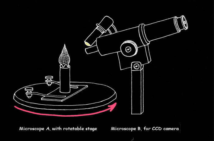
Fig: experimental setup for the "walkaround" photomicrographs
In order to generate the photographs for the animation the radiolarian shell11 was lifted with a thin metal needle and put on the blackened12 top center of a metal cylinder (see figure). Two shells preferred to jump on a nearby carpet instead of posing on the cylinder. I heard a very high "flick"-noise and they were gone. But finally I managed to drop a third one in a Mars-lander-tripod-manner on the metal cylinder. The cylinder had been glued to a slide on a rotatable microscope stage of microscope A. The shell had to be centered around the height axis cleanly by means of the microscope optics of microscope A. The radiolarian shell was then photographed by a simple microscope (microscope B) with an 8x/0.20 achromatic objective. A torch provided incident light13. For each photograph the rotatable stage was turned about 5 degrees further. In the end a series of photographs with continuously changing direction of viewpoint had been created which was fed to the DTA software as described above.
BTW: I am well aware that it is
not possible to outdate Ernst Haeckel´s beautiful 19th
century drawings of radiolarians. Perhaps one of the reasons
why they are so good is that they had just a pencil and no
computer at that time ...
Comments to the author Martin Mach welcomed.
References
1 Short and good introduction to
non-living radiolaria e.g. by William B. Carpenter in: The
Microscope and its Revelations. 7th ed. P. 770-778.
London 1891.
2 Ernst Haeckel: Report on
the Radiolaria, collected by H.M.S. Challenger during the
years 1873-1876. Zoology 18 (1887).
3 Roger O. Anderson: Radiolaria.
Springer-Verlag, New York 1983. (see Richard Howey´s comment
in his paper "Mounting Radiolaria Shells", here in MICSCAPE).
4 Kurt Stueber has put an
illustrated copy of Ernst Haeckel´s "Die
Radiolarien"-book on the web (http://www.mpiz-koeln.mpg.de/~stueber/stueber_library.html). This site contains also the
famous colour plates of Haeckel´s "Kunstformen der
Natur".
5 Jean Cachon, Monique Cachon and
Manfred P. Kage: Radiolarien - Orchideen des Meeres.
Bild der Wissenschaft 15 (1978) p. 10 - 47.
6 The strew slide used was bought
from Klaus D. Kemp in the United Kingdom. It has a good
quality and appropriate density so I was able to find many
shells to photograph.
7 PS 39 CCD computer camera by
the COMPRO company. It has only 640x480 pixels but a
permanent cable link to the computer, so the microscopic
image is being monitored and controlled directly on the
computer screen.
8 I have used Picture Publisher
4.0 by Micrographx (about $12 in Germany).
9 See MICSCAPE articles about
illumination techniques. A 1 cm diameter black paper disc can
be placed in the filter-holder to give a good dark-field
effect (only for small magnifications).
10 Special FLC animation viewers
are available in shareware collections or via CompuServe.
11 Many thanks to Herbert
Ostermeier-Reinhard for providing the radiolarian shells.
12 A black paper disc was glued to
the top of the cylinder.
13 Possibly polarizing filters
might help to improve the image quality.
Editor's note: to access Micscape articles on either Radiolaria or dark-field illumination, type either of these keywords in the search engine of the Article Library (see link below).
Microscopy UK Front Page
Micscape Magazine
Article Library
© Microscopy UK or their contributors.
Published in the January 2000 edition of Micscape Magazine.
Please
report any Web problems or offer general comments to the Micscape Editor,
via the contact on current Micscape Index.
Micscape
is the on-line monthly magazine of the Microscopy UK web
site at Microscopy-UK