Gallery of Desmids by Jan Parmentier & Wim van Egmond

Desmid locomotion


In many species you can see a layer of mucilage around the cell, it is easy to see when you add a drop of black ink to the sample or use phase contrast on your microscope.

Desmids can even locomote by selective extrusion of mucilage.

Because in this Xanthidium the mucilage was extruded from tiny pores, it appears as strands.
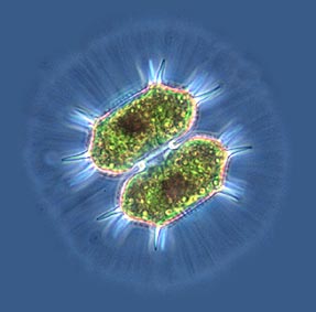


Xanthidium

NEXT DESMID PORTRAIT

Comments to the authors Jan Parmentier and Wim van Egmond are welcomed.

More pictures of microscopic subjects can be found on Wim's HOME PAGE and Jan's HOME PAGE

Microscopy UK Front Page
Micscape Magazine
Article Library


Image © Wim van Egmond

WIDTH=1


© Onview.net Ltd, Microscopy-UK, and all contributors 1995 onwards. All rights reserved. Main site is at www.microscopy-uk.org.uk with full mirror at www.microscopy-uk.net.