Editor's note: Aaron Messing sent us some splendid images of diatoms showing how a digital camera and image manipulation can help show the detail resolved by the microscope. He kindly agreed to let us share them on Micscape. Aaron writes:
I have acquired, recently, a Zeiss Standard WL microscope with Neofluor phase contrast objectives from the 70's and 80's with attachments for darkfield and DIC for both transmitted and incident light. I have a Nikon 990 with a Leitz eyepiece screwed onto the main lens of the camera which mounts onto a trinocular head. I use Corel Photopaint 10 to crop, orient, and otherwise enhance the digital images. The sharpening and contrast improvement routines are very impressive and the software itself is reasonably priced.
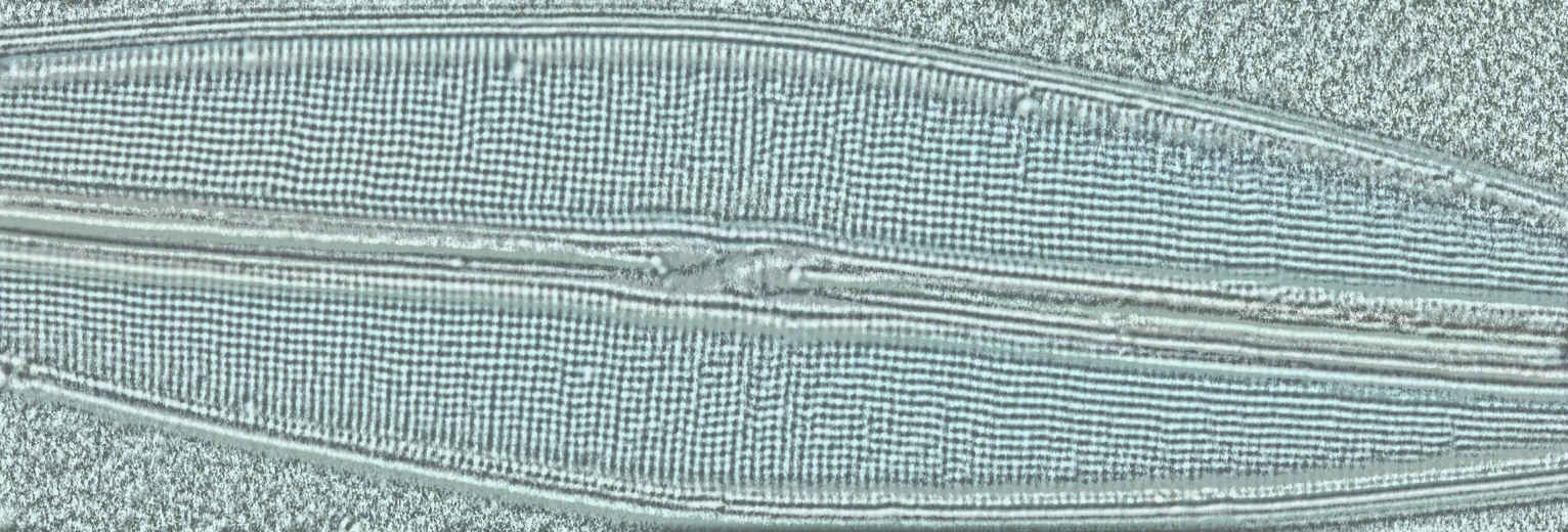
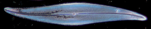
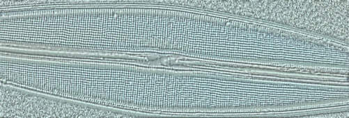
The Pleurosigma angulatum was imaged through a 40X Phase Neofluor objective with both DIC and darkfield techniques using routine digital processing to duplicate what I see in the eyepieces. The Amphipleura pellucida and Frustulia rhomboides were imaged with a 100X Phase Neofluor 1.30 oil objective with DIC. The contrast of the image at the eyepieces is very low, especially for the A. pellucida. Both these diatoms require a rather intense contrast enhancement routine to bring out the details. This routine unfortunately also creates some artifacts. The perforations focus at a different plane than the main structure of the diatom so the images seem a little out of focus.
Commments to the author Aaron Messing are welcomed.(The prepared diatom slides were obtained from Carolina Biological Supply).
Images (Note: If your web browser has set an 'auto-fit image to page' feature (Microsoft Explorer 6.0 offers this), the master image may not be displayed at full resolution. In this case, hold the cursor over the lower right hand corner of the master image to display the expansion icon; click the icon to view the full resolution image.)
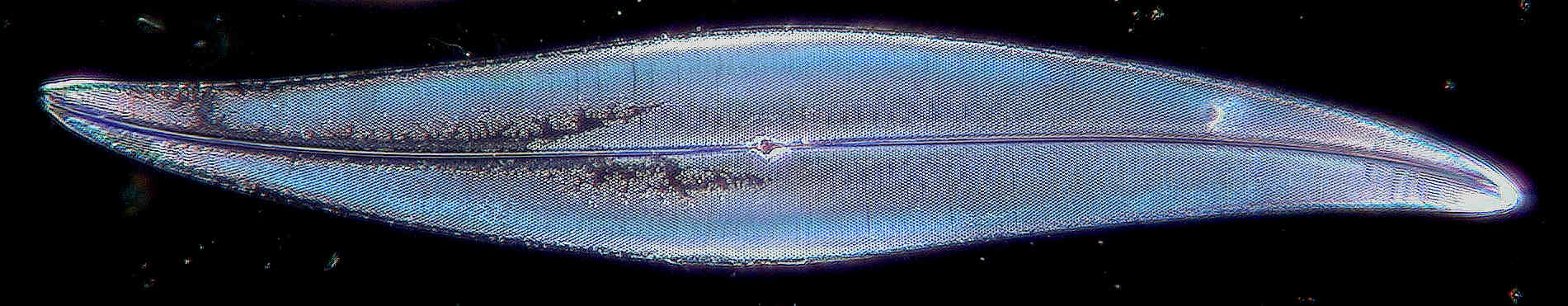
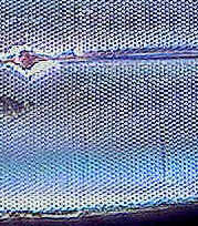
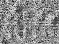
Pleurosigma angulatum. 40x objective, darkfield, image enhanced in software. Click image above to see master image. Section of detail cropped from master shown right.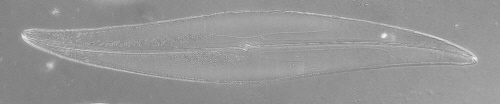
Pleurosigma angulatum. 40x objective, DIC, image enhanced in software. Click image above to see master image. Section of detail cropped from master shown right.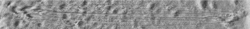
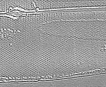
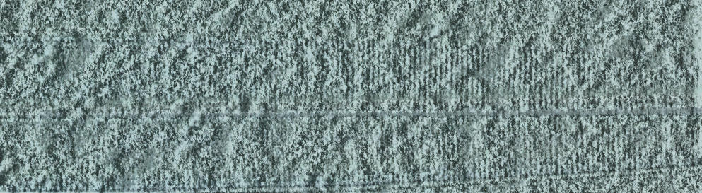
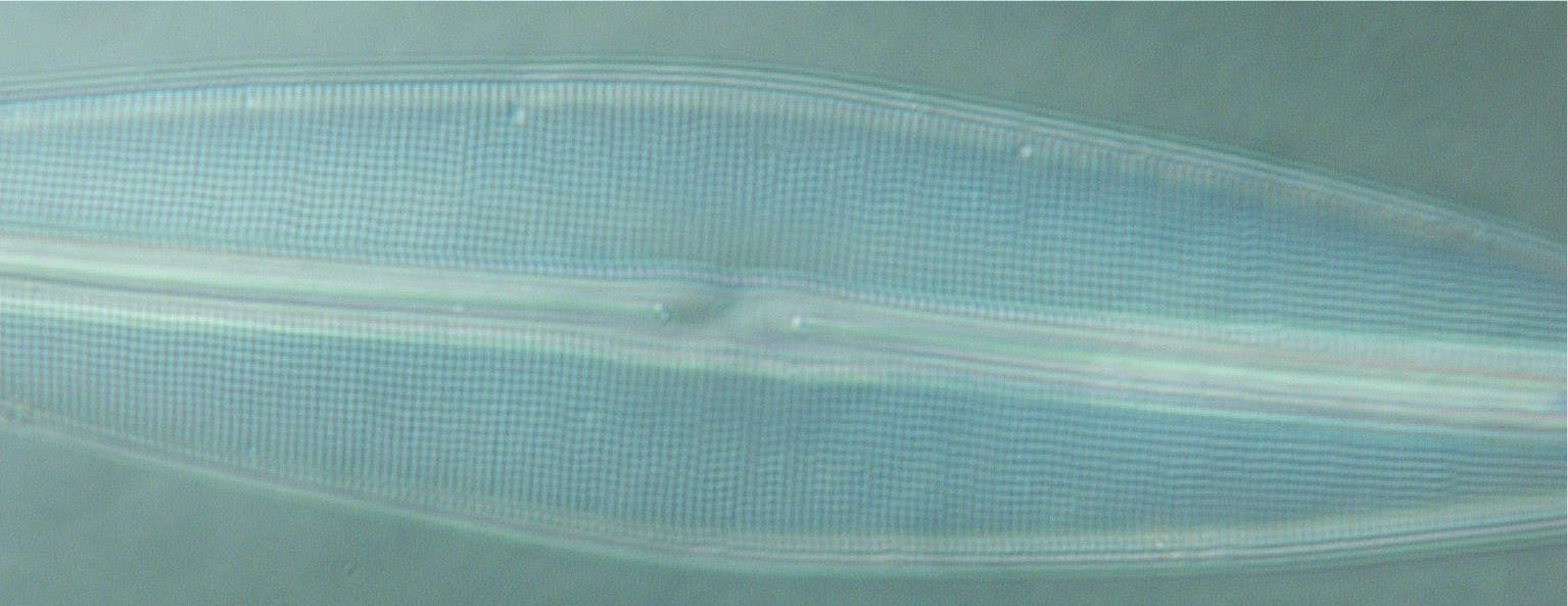
Amphipleura pellucida. 100x objective, DIC, image enhanced in software. Click image above to see master image. Section of detail cropped from master shown right.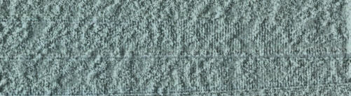
Amphipleura pellucida. 100x objective, DIC, image enhanced in software. Click image above to see master image. Section of detail cropped from master shown right.
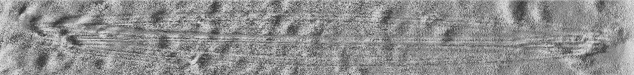
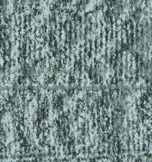
Frustulia rhomboides. 100x objective, DIC, image enhanced
in software. Click image above to see master image.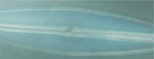
Frustulia rhomboides. It is a less processed image,
more typical of what I see at the eyepieces.
Click image above to see master image.Editor's note: Resolving the detail of diatoms is a challenging and rewarding aspect of microscopy. Related Micscape articles are as follows.
Counting the dots: giving microscopes a 'workout' with diatom test slides
Test diatoms - what you can expect to see even with modest optics