These are three old slides I've been enjoying recently and which may be of interest. They are from small sets of 3-9 slides bought on eBay. Extensive sets or slides made by the famous mounters understandably go for quite tidy sums, but the smaller sets especially of 'unnamed mounter' slides often sell for modest prices.
I chose these in particular as I
think they illustrate different aspects of the fascination of old slides
i.e. learning about an unfamiliar subject from microscopical observations
and background reading to learn more about the slide, which in turn can
often reveal interesting avenues of study. These are 'slide studies in
progress', so would welcome any information where queries are raised.
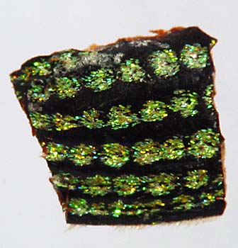 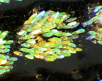
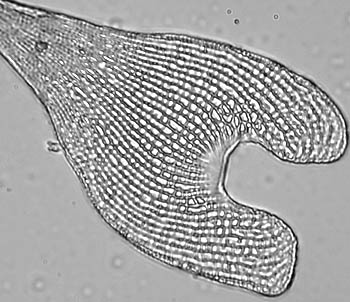
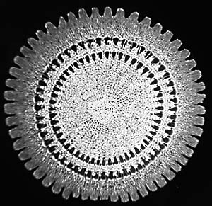
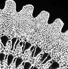
Left: The full mount ca. 5x4 mm of beetle elytra. The rows of scales are typically 1 mm apart. Right: One cluster of the scales under a 3.5x objective. The clusters of scales are ca. 0.9 mm in diameter.
|
| This slide had been badly broken
beyond repair in its past so I'm reviewing options for remounting the specimen.
However, this did give the opportunity to study at higher mags than a deep
cell mount would normally permit.
Although not obvious from the images, the clusters of scales sit along the 'furrows' of the ridged elytra. Comparing the view by transmitted and reflected light shows that the iridescence is a result of light interference and/or diffraction with microstructure on the scales rather than by pigments. I'm uncertain if the microstructure would be easily resolvable by an optical microscope and would be interested to hear of refs. to electron micrograph studies that give some insight into the scale structure. To learn more about the 'diamond beetle', I did a web search which brought up two species with this name, one Australian and one S. American. Some online dictionary entries note that the latter is a large weevil Entimus imperialis with iridescent scales, so the slide seems more likely to be the S. American beetle. The only image of it I found online was 'Diamond beetle, Samari, image no. 2692' in the Gallery Insects/Beetles on the Butterfly Alphabet Inc. website. This image shows its black elytra and scales. On the Olympus Mic-D digital microscope web site they have a superb 'Physics of light and color' section with an article on interference which mentions the diamond beetle. A downloadable illustrated article 'Colour effects in bright butterflies' by Vukusic and Sambles on the Society of Dyers and Colorists web site' is also a fascinating read. |
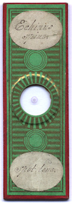 
Left: Slide scan. An unnamed mounter
but a well presented slide.
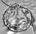
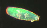
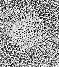
Left: Edge detail 9x objective,
darkfield.
|
| I've never owned a slide of sea
urchin spine to date, but this slide looks a good example. The mountant
is still very clear after all this time. It amazes me how such a
potentially brittle structure can be sectioned so well with little apparent
damage. I'd be interested to hear if anything can be gleaned on its identification
just from the cross section, as the label doesn't state which urchin it
comes from.
Richard Kerr's delightful book 'Hidden Beauties of Nature' (I have the 4th edition, early 20thC), has a chapter on sea urchins with a plate showing a wide range of different spine cross sections. Richard Howey discusses the wonders of sea urchins, including their spines and some preparation techniques in his splendid essay 'The virtues of broken and dirty specimens'. |
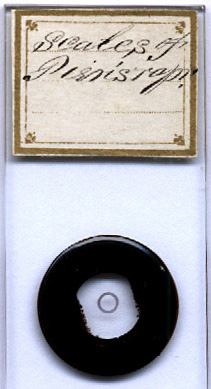 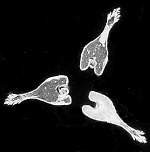
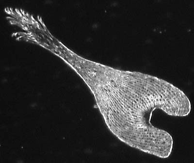
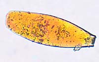
Left: Slide scan (crop). Right: The arranged three scales in darkfield. (Each scale is ca. 170 µm long, 60 µm wide).
|
| I believe the label should state
'Scales of Pieris napi'. If so, they are scales from a green veined white
butterfly, a fairly common species in the UK.
I have a modern mount of a Pieris species' wing scales and was expecting to see a similar looking mount. But I was intrigued to find three arranged scales of a shape I'd never come across. I gather they are the specialised 'scent scales', androconia, found on the males. I haven't found to date any extensive refs. either in books or online, so would be interested to hear of any refs. which discuss androconia. The only images found to date were in a delightful old book 'Butterflies Worth Knowing' by Clarence M Weed 1923 that has been carefully transcribed with illustrations by 'Kellscraft Studio's Web Text-ures: Public Domain Books On-Line' onto their web site. (The androconia are discussed and illustrated in 'Part I: The Scents of Butterflies'. One androconia illustrated does look similar to those on this slide.) In this mount there's a spore/pollen
in two of the 'receptacles'. Is this just a coincidence? In fact before
I deciphered the script on the slide I thought they were some sort of spore/pollen
bearing structure of a plant as at first sight to my inexperienced eye
they did look a bit botanical!
|