|
Bryozoans in colour! |
J.M. CAVANIHAC FRANCE |
|
|
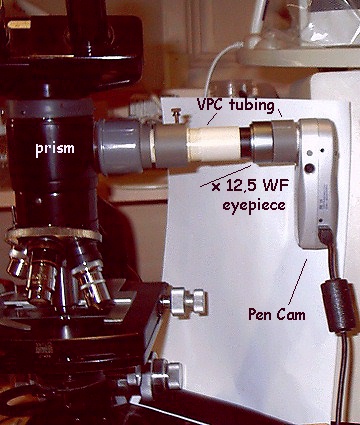
Some years ago, I presented a short article about bryozoans, but at that time I only took pictures in black and white or were manually coloured. Since then, I have improved my equipment and also my observation technique. With an analogue colour video camera (with CMOS sensor and capture board in PC), I can now take more attractive pictures and movie clips. But the main progress is the use of another small digital low cost USB camera (model: Pen Cam SD 2M by AIPTEK, 2 megapixels, price around $60 US!) Now, I can take pictures at a larger scale. These pictures are resized (to around 1/3rd of their true size of 1600 x 1200 pixels). The picture below shows the Pen Cam fitted on the phototube prism on my old Wild M20. I think it gives good results for cheap equipment! The USB cable is useful to monitor real time pictures and to adjust focus but must be disconnected when taking high resolution pictures. (All the animations below were taken with the analogue video camera, all other photomicrographs were taken with the Pen Cam.) |
|
|
|
|
|
|
|
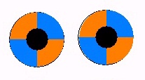
The second improvement I made was the use of Rheinberg lighting. Until recently I was not an enthusiastic user of this technique, but seeing the good results obtained by Walter Dioni, I decided to try it. In fact I use only one Rheinberg disk as shown in the picture below, which allows well separated vertical and horizontal structures to be shown when rotating or slightly decentring it. The disk is printed with an ink jet printer onto a transparent sheet and two copies are superimposed to obtain a better colour density. |
|
|
|
|
|
|
|
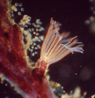
Bryozoans are intriguing but beautiful animals. They are classified as Ectoprocta because their anal tract is outside their lophophore. But what is a lophophore: it's a bush of ciliated arms, generally retractile which makes a sort of funnel with the mouth located at the bottom (smaller end) of the funnel. Cilia create water currents which attract particles towards the mouth. But if an unwanted particle (too large or not digestible) enters the stream, cilia are able to eject it! |
|
|
|
|
|
|
|
A short animation showing feeding (taken with analogue camera and x2,5 objective - picture not resized). |
|
|
|
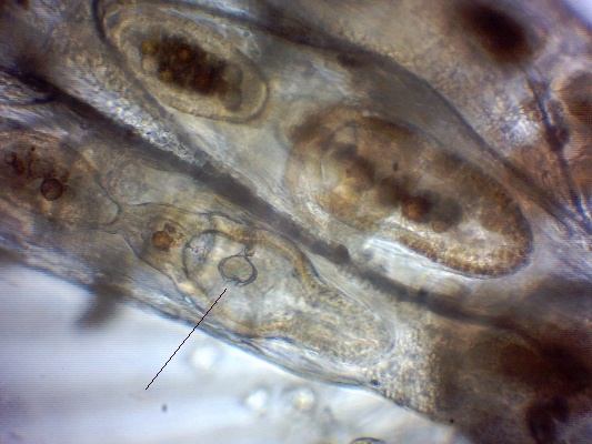
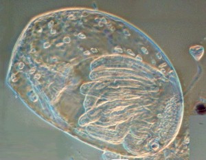
Two species are easy to find: they colonise rock, boat hulls or even the shells of mussels for example. The first species is in the genus Bugula with some sub-species: each individual can retract its lophophore into a rigid lorica which is either chitinous or calcareous. The colony develops by the budding of each zooid. One interesting feature is their polymorphism i.e. some individuals have specialized functions. Thus we can find: gastrozooids which are the feeders of the colony using their lophophore to eat. A closer look at a stomach shows a dinoflagellate (Protoperidinium) inside. The animation here (avi format, 940 kbytes) with Rheinberg lighting shows particles moved by cilia inside the digestive system. |
|
|
|
|
|
|
|
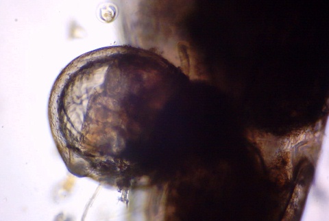
But a closer look shows other functions: first spherical structures called oecia which are reproductive individuals which give arise to a swimming planula: |
|
|
|
|
|
|
|
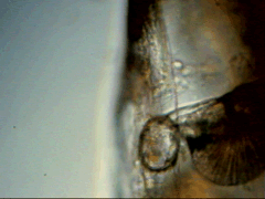
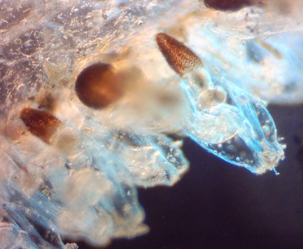
And more strange, a third type which defends the colony against predators: an avicularian. Well named because their shape looks like the beak from a bird of prey! Note the hairs inside which are probably a sort of trigger used to close the beak. When you move the colony gently, avicularians swing out as if they've detected a predator: see the animation left. (Taken with analogue video camera). The picture below shows a closer view of an avicularian. |
|
|
|
|
|
|
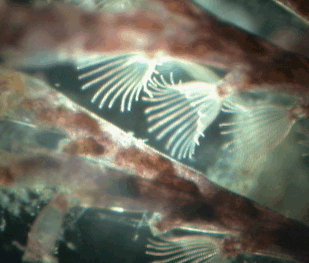
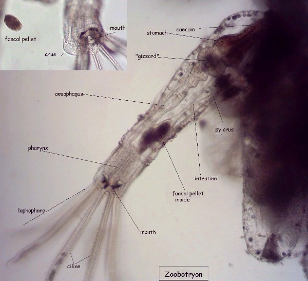
Another species is Zoobotryon, which doesn't have a rigid lorica but a sort of stolon with zooids attached and arranged in clusters. The stolon seems to be under pressure and if you pierce it, it becomes flaccid. (Pictures below taken with x6,3 objective.) It has a lophophore which possess only eight arms, but the cilia are very efficient! The video clip here (avi format, 1 Mbyte) shows the currents generated by the cilia in action. |
|
|
|
|
|
|
|
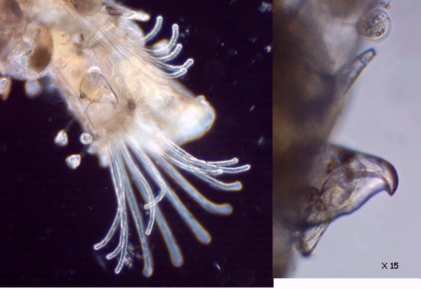
Below is an annotated picture of a zooid. In the top left hand corner, a detail (very rarely seen!) shows a dilated anus which has just ejected a faecal pellet. The brown mass you can see near the base, is a sort of gizzard with the ability to break siliceous frustules of diatoms! |
|
|
|
|
|
|
|
Below is a picture of a retracted zooid (Rheinberg lighting): the retractor muscle is well seen. |
|
|
|
|
|
|
|
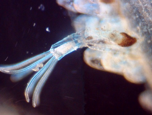
Sometimes young individuals such as the one shown left, fall from the stolon and are able to create a new colony (picture taken with Rheinberg disk and Circular Oblique Lighting (x15 objective). |
|
I think these pictures are able to show some of the beautiful aspects of these marine creatures which are easy to find and to observe if you use well slides or small Petri dishes to let them deploy their delicate lophophores. |
|
Comments to the author Jean-Marie Cavanihac are welcomed.
All drawings and photographs © Jean-Marie Cavanihac 2005
Published in the January 2005 edition of Micscape Magazine.
Please report any Web problems or offer
general comments to the
Micscape
Editor,
via the contact on current Micscape Index.
Micscape is the on-line monthly magazine of the Microscopy UK web site at http://www.microscopy-uk.org.uk