MEMORIES OF 2004
Walter Dioni Cancun, Mexico
(Part 2)
A mysterious worm
Key
words: polychaeta,
trochophore,
metatrochophore,
Sabellidae,
Sabellinae,
Fabriciinae
|
The picture below is the last image in my First Part of these Memories on the materials collected by accident after a storm on a beach in Cancun. Gliding between the smooth grains of sand, under the low power of my microscope, this little worm doesn't know that it would open a long journey through my books and the Web to identify its characteristics, and discover its identity.
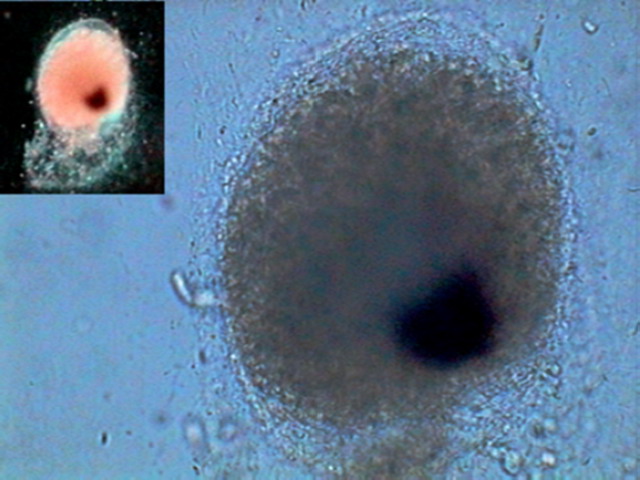
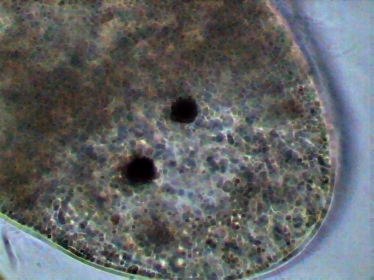
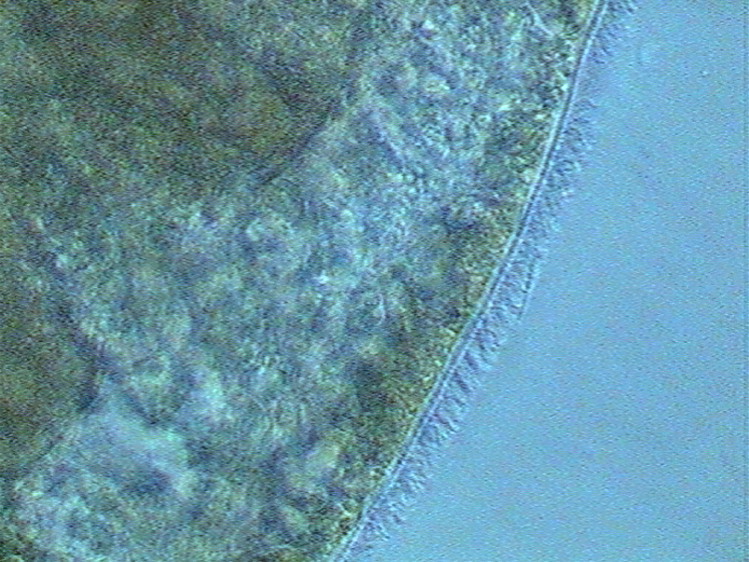
The little worm glided smoothly, without contractions of its small body, probably driven by a thin layer (invisible at the low power, but predictable, according to my experience with worms) of ventral cilia. At low power the appearance and movements remind me of a turbellarian. ( http://es.wikipedia.org/wiki/Turbellaria , http://en.wikipedia.org/wiki/Turbellaria , http://fr.wikipedia.org/wiki/Turbellaria ) www. earthlife.net /inverts/ turbellaria .html What makes it different was, above all, the opacity, the rigidity of its cylindrical body, the faint suggestion of segmentation, according to some side indentations, and the two dark areas it had on both ends.
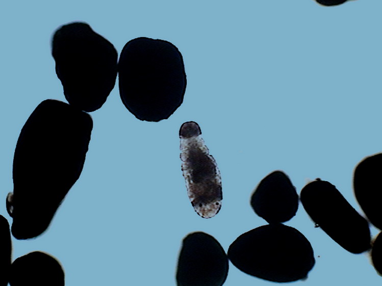
It was evident, not only by the direction of the movement, but by the presence of two black eyes, that it had a differentiated cephalic end, but I could not see any mouth, not any other specific organ. I soon found other specimens, a few gliding on the surface but most in the mass of the gelatinous cylinders that had been collected along with the algae. Were they inhabitants, or consumers of the jelly? Many different species of bacteria and protozoa shared the transparent substrate.
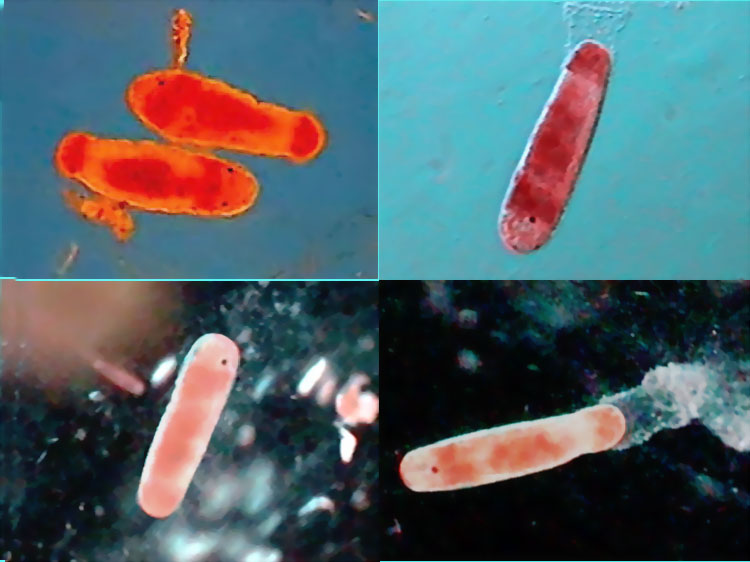
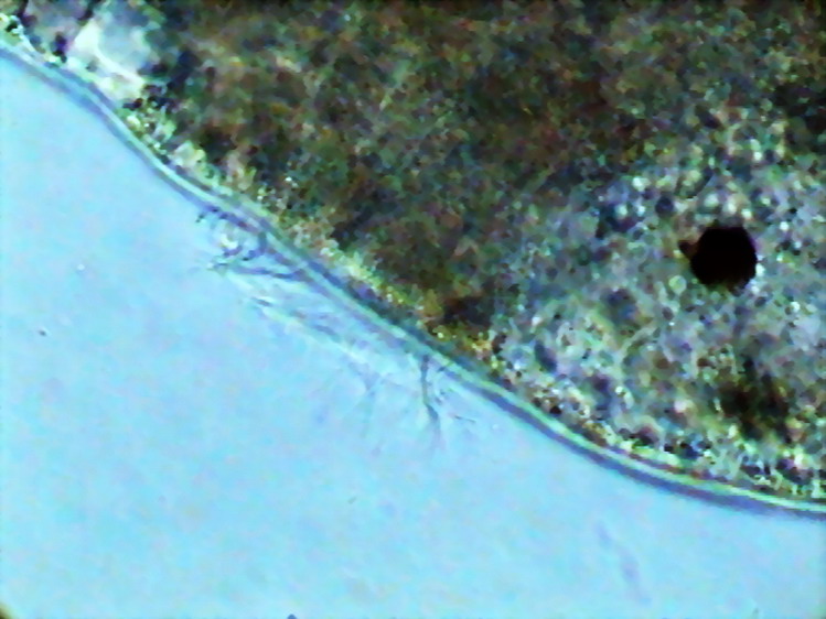
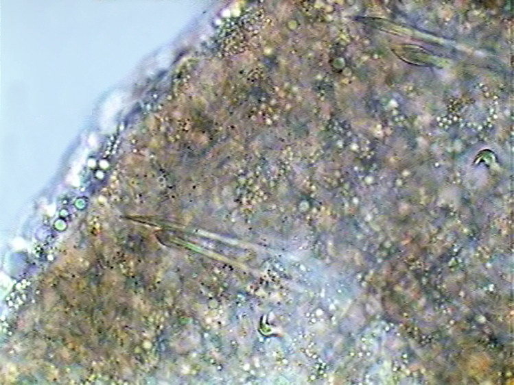
Worms in righthand image glide over the jelly. Those on the left are navigating within the same, as the steles that they make when "drilling" through the transparent substrate indicates. (Rheinberg illumination). But, apart from some areas now empty, suggesting that they had hosted some ovoidal bodies, I also found what obviously was a resting egg .
A logical inference was that the many, blunt ended, small, and mobile cylindrical worms, with ventral cilia, were born recently from eggs housed in the jelly like cylinders that were naturally floating on the surface, or were uprooted from the bottom by the storm surge of the previous night. Those were the "bad waters" (jellyfish) that my grandchildren suspected in the morning!!! The most important detail seemed to me the production of those jelly 'egg-carriers'. And, if I remember right from my old teaching on invertebrates, I taught that those marine worms that could produce jelly were the polychaetes . (It's not possible to describe here the polychaetes and relatives. So, for those who do not have enough knowledge of basic zoology, but would continue to read, I would advise them to open now SOME OF THE INTERNET ARTICLES I suggest at the end of this article.)
But, I also taught that these worms
develop from eggs that produce
trochophore
larvae
. Do you want to know what a
trochophore
larva
is? 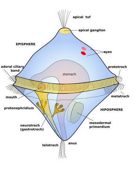
http://www.diatomloir.eu/Siteplancton/Vers.html
ON THIS SITE
you can see various kinds of
metatrochophores
,
planktonic
of course. What can be seen easily is
the amount of long bristles that
they
own.
These are just aids that increase the
surface of the worm without
significantly increasing its weight,
to give the little larva an easy
float.
http://www.pubmedcentral.nih.gov/articlerender.fcgi?artid=2440741
Obviously this is not about my little worms. These were born from eggs, without having even the aspect of a trochophore and they were never pelagic. But as you have seen trochophores have an interesting feature. Its body is divided into 3 parts, by the presence of two bands of cilia, and I thought seeing two bands of cilia, an anterior and a posterior one in my little worms. See them here:
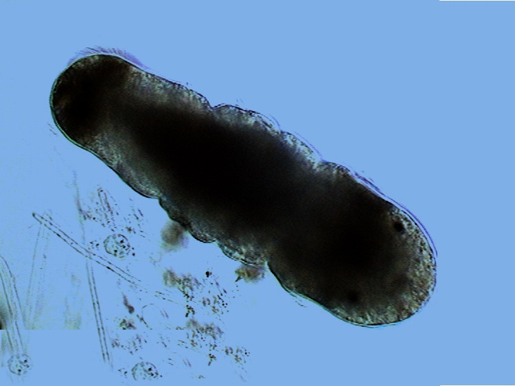
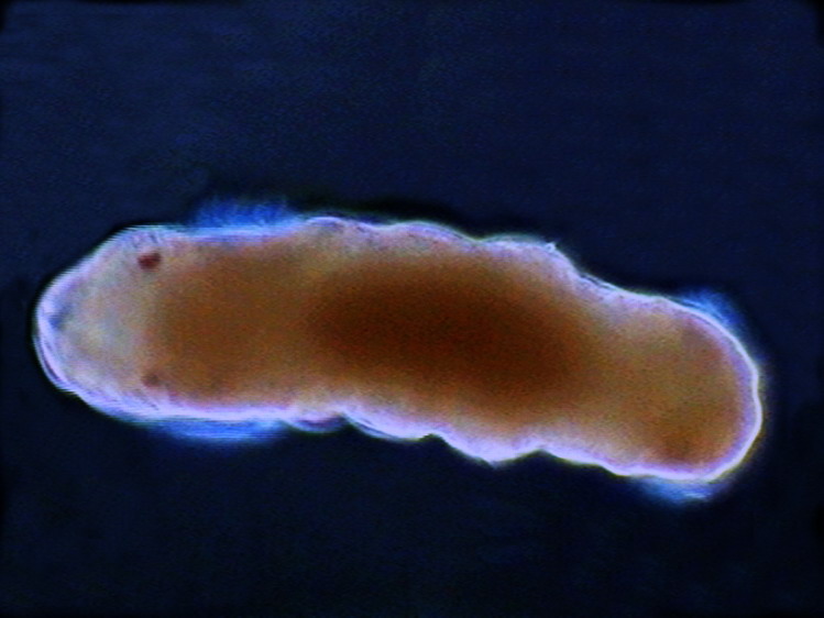
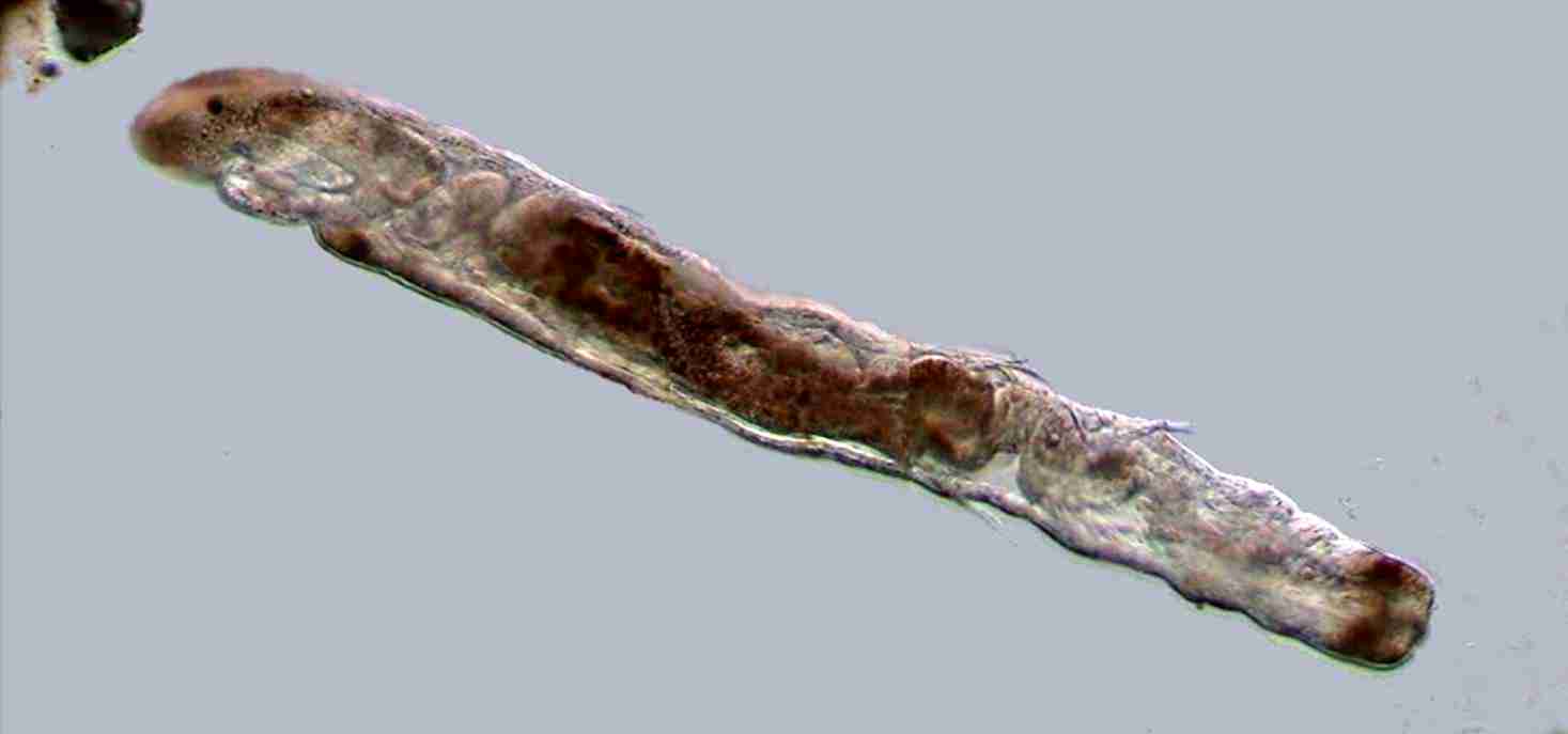
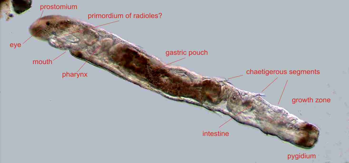
Little worm showing the prostomium, derived from the episphere, eyes, the prototroch ciliary band, the medium body with signs of segmentation (derived from the hiposfere). This segmentation begins in the dark zone immediately above the telotroch. Behind the telotroch, is the terminal segment called pigidium. (Rheinberg Illumination, dark field). The closest thing I found was this picture, labeled as "metatrochophores", whose address I did not file, for over-confidence, and which despite my later long searches I do not find again. (If the author, or a web surfer more fortunate than I, falls on this page, please, tell me the address to be included here)
Therefore I devoted myself for a while to find descriptions and pictures of trochophores of different shapes, and, above all, an indication that some of them might be fully benthic trochophores . It was absolutely obvious that some of my little animals had left the egg recently, and, yet, they have exactly the same structure and behavior of the elderly. But I never found a reference to a benthic " trochophore ", or one with that shape. So could these be metatrochophores , without a trocophore stage? They had the basic structures.
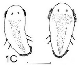
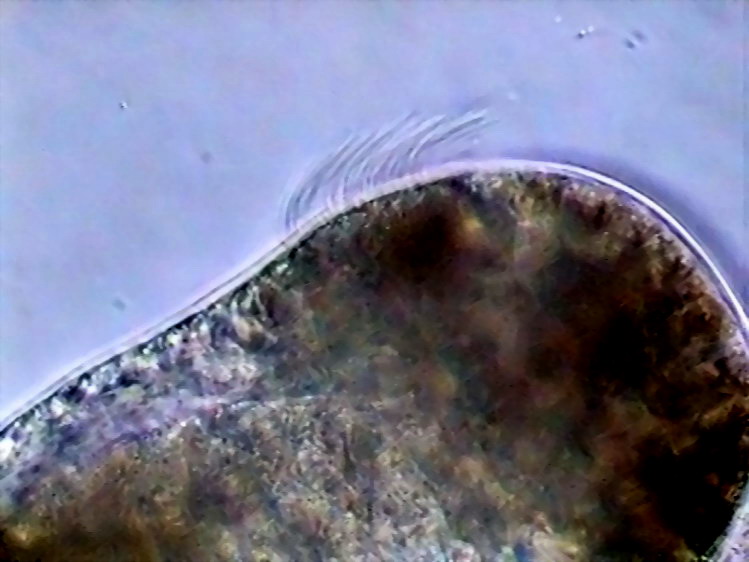
a) head , lateral view, b) head, top view, c) the prototroch d) the telotroch; e) and f) the neurotroch. The compact body and the low proportion of cilia clearly indicate that this is a benthic worm.
Therefore I left my books and surfed
the Web. Just as in my books, also
here I found references to the
production of a gelatinous mass with
included eggs. For example:
But they are not jelly cylinders, and apparently they are small.
One of the things I discovered is that
apparently the original works on
polychaetes
reproduction published on the
Web, are
very few. But there are a huge number
of replicas of each one, sometimes
published as if they were original
works, without the slightest
indication of the source that produced
the data. One gets tired, opening page
through page, which essentially
repeated the same data and sometimes
even the same phrases. Or which simply
cite the same work.
At last! Two genera that produce mucous cylinders loaded with eggs as those that arrived in my hands. There may be more, but at least I could now search data more efficiently.
The Natural History Museum in London has most interesting and informative web pages. Included is a visual key for the families of polychaetes . Diopatra belongs to the family Onuphidae , and Sabella to the family Sabellidae. In addition to pictures of the adults, each plate shows the type of bristles typical for the family.
Obviously the chaetae I show have nothing to do with the Onuphidae and they are very similar to those of Sabellidae. Please confirm that visiting THIS SITE
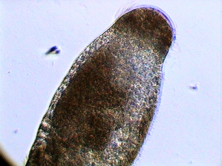
Placed in shallow aquariums, with some of the algae, the metatrochophores survived several days. In its fifth day of development I could take a series of pictures that allowed me to assemble a patchwork ( mosaique ) of one small polychaete that already may be regarded as a juvenile.
Dr. Maria Ana Tovar, a researcher at the ' Colegio de la Frontera Sur advanced research institute based in the south of the Mexican Quintana Roo state, to whom I made some consultations, had the courtesy to communicate the Pernet, B et al. Chapter 12 of the "Atlas of Marine Invertebrate Larvae" on the larval development of 21 polychaeta families, beautifully illustrated with photographs of larvae obtained with Scanning Electronic Microscope. I could see there that portrayed metatrocophores are all very different from those here illustrated. But 21 is only a quarter of the 81 families that are recognized for the polychaeta . And the pictures are for only a representative species of each treated family....
Nevertheless the most interesting thing is that, definitely, the chaetae on my young worms match well with the illustrated for Sabellidae in the plates of the Natural History Museum at London, and also with published papers on genera in the family Sabellidae such as these
http://scientiamarina.revistas.csic.es/index.php/scientiamarina/article/view/11/11 (PDF, 1.4 MB) http://www.mapress.com/zootaxa/2006f/zt01168p058.pdf
Of course I can not identify either the genus or the species, and is a gift of a single publication on the Internet, that it has brought me near the family, giving a little more peace of mind to my conscience. The systematic location, of the larvae, according to the categories often used in classic taxonomy would be as follows:
Domain: Eukaryota Whittaker & Margulis,1978 Kingdom: Animalia Linnaeus, 1758 Subkingdom: Bilateria (Hatschek, 1888) Cavalier-Smith, 1983 Branch: Protostomia Grobben, 1908 Superphylum : Eutrochozoa Phylum: Annelida Lamarck, 1809 Class: Polychaeta Grube, 1850 Subclass: Palpata Order: Canalipalpata Family: Sabellidae (Malmgren, 1867)
An interesting fact: Canalipalpata refers to the fact that the 'feathers' that make up the 'duster' that gives a so striking appearance to these species have its top surface grooved and ciliated. The movement of the cilia, capture and guides the food along these grooves, channeling it into the mouth, that wait in the center of the showy duster.
So, probably we can believe with some
confidence that what we present here
is the benthic
metatrochophore
larva, with direct development of a
Sabellid
that, as we shall see right away may
probably belong to the subfamily
Fabriciinae,
if were not that ... well we can
discuss this in a moment. Now take
note of
this
:
Well!! That also explains why my little worms had no mouth.
This also explains the size of the eggs, which must to host an abundant supply of yolk (larval food). These are called 'lecitothrophic' larvae for opposition to the 'planktotrophic' ones that feed on the plankton. And also explains the opacity of the body of the newborn, with the tissues still filled with yolk.
Dr Maria Ana Tovar informed me that the more primitive Sabelliinae (to which such genus as Chone pertains, and descriptions and pictures of adults of whom, and their chaetae, can be found in one of the works mentioned above) have a direct style of reproduction like Fabriciinae .
Therefore it is clear to me that I should stop here my attempts for a closer taxonomic approach. I have no more data to continue. I just want to stress that my 5 day young worms have no cephalic differentiations as palpes or radiolae, and that this is different from all that can be seen in the bibliography (printed or electronic) at my reach. I hope that at some time they can fall into the hands of a qualified specialist (which can dive and work in the underwater world that is forbidden to me) which can link the larvae with its adult parents and so be able to assign to them an identity .
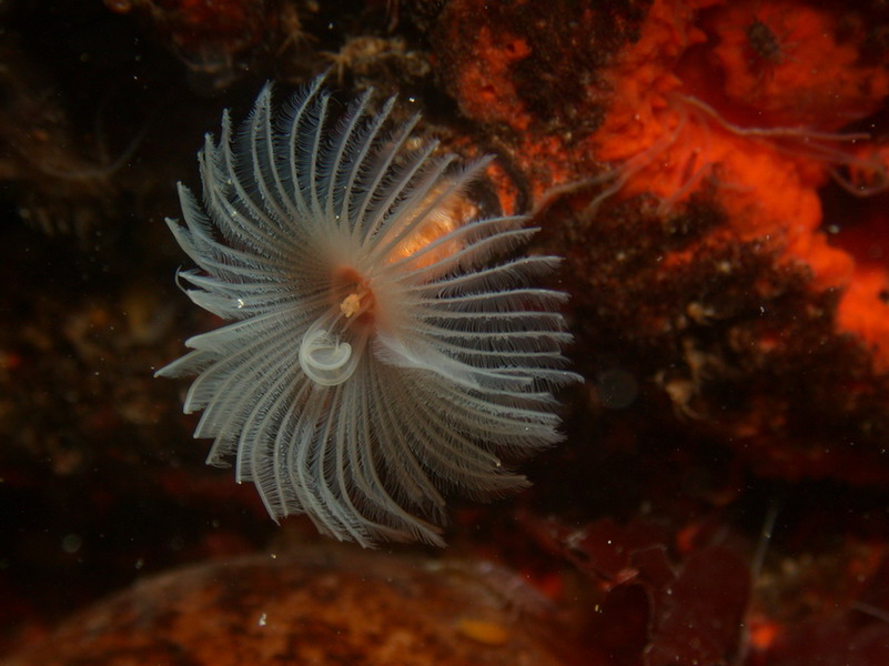
As the
Sabelids
are really beautiful animals, although
seafarers and port workers may find it
annoying because
of
"unduly" building their colonies of
intertwined parchment tubes on the
structures they wanted to see clean
(ships, port structures, buoys, etc.),
I show here, to reward those that have
followed this arid investigation, a
picture of an Antarctic
Sabelid,
(could it be that my larvae ends as
this?) on loan courteously by
Dr. Dirk
Schories
, who manages a beautiful
SERIES OF PAGES
on marine
life: http://guiamarina.com/gallery/main.php
SOME COMMENTS
Surely those who work with marine polychaeta, especially if they do so on reproduction and in the laboratory, must have seen many times some larvae as those presented in this article. They might therefore consider them so well known and common, that it's not worth uploading an image to the Web. But I took a couple of days to identify conceptually the specimens I had in my hands, a few weeks to find one or two more or less coincident images, and months to collect scientific information, that never came to be defining, not even dependable, about the family and their reproductive habits.
An interesting exercise for the reader
would be to put in his browser search:
"Polychaetes",
"Trochophore"
and
"Metatrochophore"
to see the harvest of images they get.
If you search for
Sabellidae
(now that we have an idea of whose
family can be our
metatrochophores)
you will be rewarded by the harvest of
many beautiful illustrations ... of
the adults.
This refers of course to the investigation of the Web Those who live in a city with good academic libraries might have better opportunities to document on this subject, especially if they had access to a specialist in polychaeta at some research institute, or a university.
In Spanish http://www.hydronauta.com/temas/biologia/vertebrados-infe/anelidos/poliquetos.htm#morfologia http://es.wikipedia.org/wiki/Polychaeta In english http://en.wikipedia.org/wiki/Polychaete http://www.earthlife.net/inverts/polychaeta.html In French http://fr.wikipedia.org/wiki/Polychaeta http://www.darse.org/v1/sciences/plan_annelides.html http://www.cg66.fr/environnement/espaces_naturels/reserve_marine/especes/annelides/index.html the latter has also a magnificent collection of pictures of worms.
|
Comments to the author,
Walter
Dioni
, are welcomed.
Microscopy UK
Front Page
Micscape
Magazine
Article
Library
© Microscopy UK or their contributors.
Published in the January 2009 edition of Micscape.Please report any Web problems or offer general comments to the Micscape Editor.
Micscape is the on-line monthly magazine of
the Microscopy UK web
site at
Microscopy-UK
© Onview.net Ltd, Microscopy-UK, and all contributors 1995 onwards. All rights reserved. Main site is at www.microscopy-uk.org.uk with full mirror at www.microscopy-uk.net .