|
Memories
from
2004
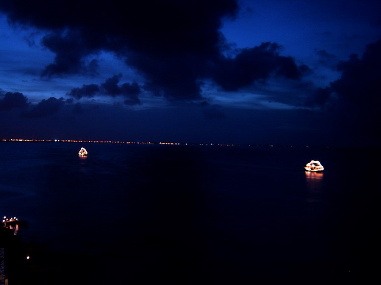
The first image is not mine. It's the artistic work of Lucy Nieto and I've used it with her authorization (and modified it somewhat) from her page in Flickr. But it fits very well in the mood of my old night at the beach. Photomicrographs were shot with the 0.4 MP camera on my National Optical microscope. I took the other pictures with my Canon Powershot A300. Most of the photomicrographs and the most important part of the text were posted, in 2004, at Microscopies, a French Forum for Microscopy.Key words: Cancún, coralline sand, formaminifers, polysiphonia, cladophora, cothurnia, Vaginicolidae
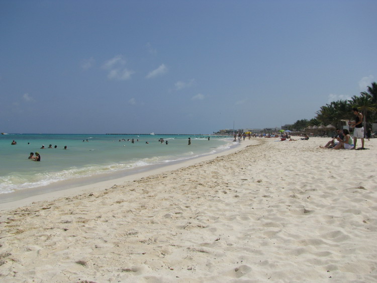
The night before, there was no moon. Seated on the tenuous sand of the beach, with the dim lights from the distant city over my shoulders, I had the sensation of being inside a black diamond. The absolute transparency of the atmosphere let me enjoy the incessant and caressing murmurs from the small and soft waves, and of the gilded twinkle of the stars on the black and perfect smoothness of the firmament. A pair of distant tourist ships were reflecting their lights on the wavelets, and, as if it was brought by the marine waves, a faint sound of music was arriving to me intermittently. I really enjoyed that moment on my first vacation in several years. And, feeling me happy, I sank my hands in the still lukewarm sand.
And, inevitably, my biologist conscience appraised the smoothness of the grains, and evaluated the slow, constant and timeless work of the waters on the dead coral skeletons, rolling them, time and time again, with infinite patience, to produce this strange sand that never burns you. Over which it is possible to walk with the naked feet for many hours, in the heat of the noon, with a torrid sun, without feeling nothing else than a comfortable tepidity. Of course! I took a sample to examine it later! And dreaming about the incredible blue of the oligotrophic Caribbean waters, which, I hoped would be waiting for me the following morning, I went away to sleep.
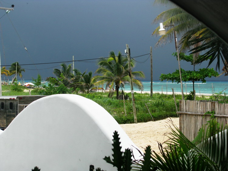
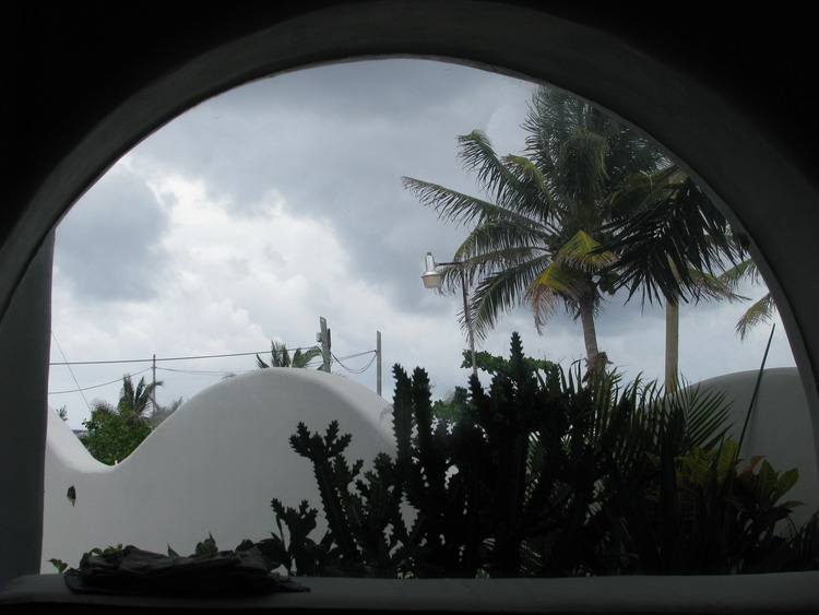
The wind began to blow at night. At dawn the waves struck and crowded on the beach, with a dull and threatening rumor of military drums. With the first light, gray and threatening shredded clouds could be seen, flowing swiftly in the sky over the lead colored sea. My day was going to ruin. But just towards mid-morning the sky was clear, the wind asleep, and the only witness of the nocturnal fury which remained was the still turbulent sea, with an opaque bluish-milky color due to the amount of suspended fine sand. I was seated again on the desolate beach that began to be filled with visitors. The crest of the waves, now calmed, had an untidy aspect, by the seaweed that floated whole or in pieces, uprooted by the violence of the waves at night. The sea did not invite to a visit.
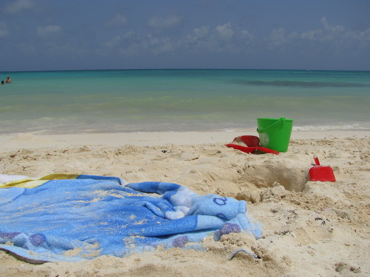
But for my grandsons this was really indifferent. And soon they were jumping around me, with their small buckets full of trophies robbed from the waters.
The sample tools I quickly saw the delicate hairs of the red and green seaweeds, and .... those cylindrical gelatinous bodies. 'Bad Waters', grandfather, 'bad waters'!, (Mexicans call 'bad waters' those containing jellyfish). Were the boys thinking that the small gelatinous pieces were the little stinger jellyfishes which often invade waters near the shore? What would they be? Because certainly, jellyfish they were not. Of course, I kept some samples to examine them later. Biology and microscopy have priority over vacations!!! I was aware of these contradictory desires before coming to the beach.
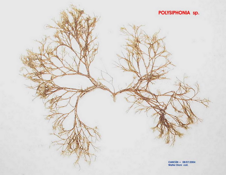
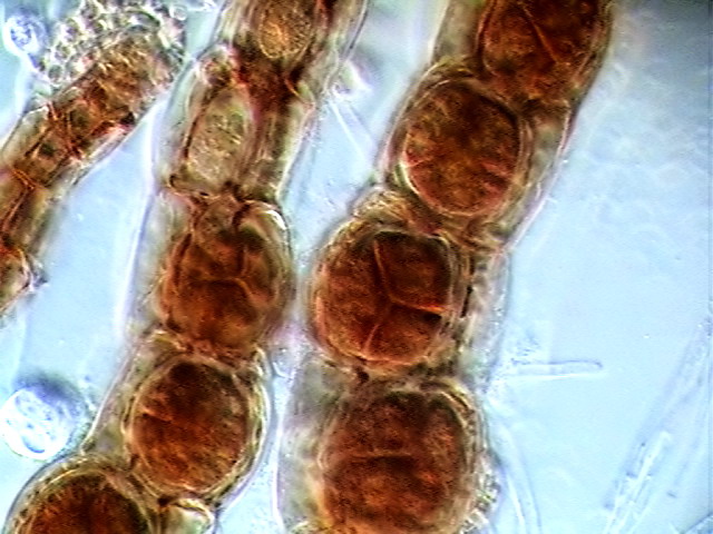
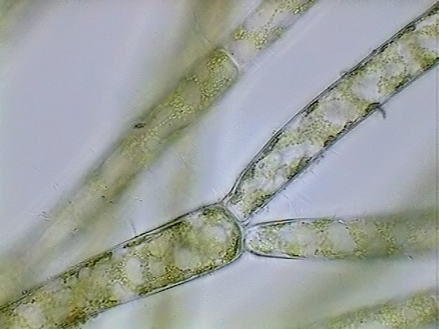
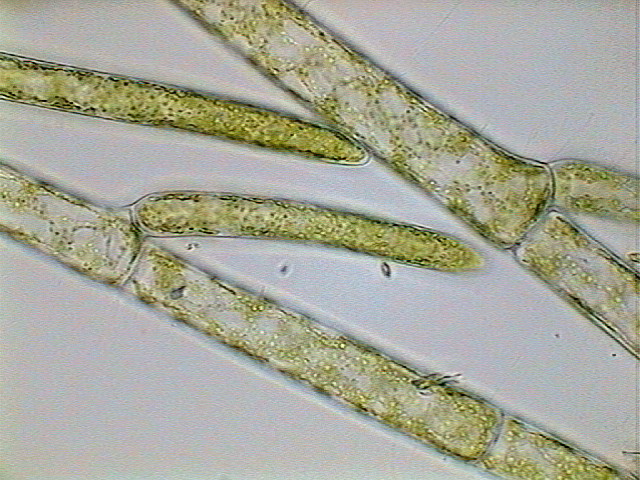
Seated, later, at the microscope I first examined the seaweeds. I could identify them quickly. The red one was a Polysiphonia of which I show an exicata of Polysiphonia and the most characteristic traits of its structure.
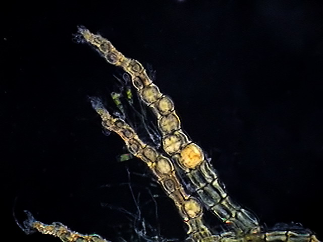
Images above in
order: 1 – A Polysiphonia branch with tetrasporangia.
2 – High power tetrasporangia view.
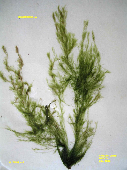
Jean Marie Cavanihac and Jean Parmentier, have presented two beautiful photographic studies of this genus, which can be seen HERE and HERE The green alga was a Cladophora. That I also show in the following pictures. See the characteristic chloroplasts and branching of the thin filaments.
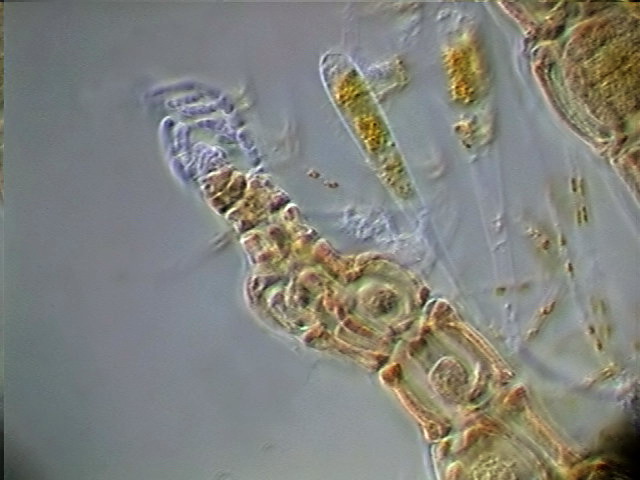
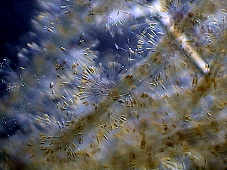
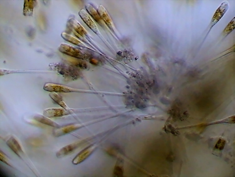
Most interesting, on the seaweeds, were the epiphytes. Abundant diatoms and protozoa use them as a substrate. I believe that the festive spirit of the previous night celebration had influenced its behavior, because, evidently, they imitate the fire-works (image below) that had arisen intermittently from the tourism ships. Mexicans love the fire-works, and they always include them in the tourist's 'Mexican nights'. Apparently, the exuberant architecture of those agglomerations is due to a special diatom of the genus Climascosphenia,) (thanks to Dominique Voisin for confirming the identification).
Clusters of diatoms fixed to the algae and a lateral view of one Climacosphaenia.
There were also some
Vorticella and
Epystilis,
peritrich
protozoans, scarcely distributed in
the spaces that the diatoms left
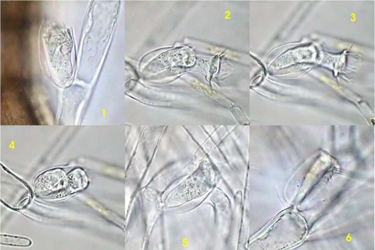
free. But the most interesting and abundant guest hid itself, imitating water by its transparency. In tiny transparent glasses, lodged transparent tiny bodies, extended like trumpets, with the widened end surrounded by a circular crown of cilia, generally clinging by pairs in their crystalline cockpits, firmly adhered to the algae by short stalks.
Six images of the transparent protozoa on its transparent substratum.
They prefer to adhere to the trichoblasts, also themselves transparent, of Polysiphonia. The corresponding genus is Cothurnia, and belongs to a group of peritrichs that normally amateurs have difficulties in identifying.
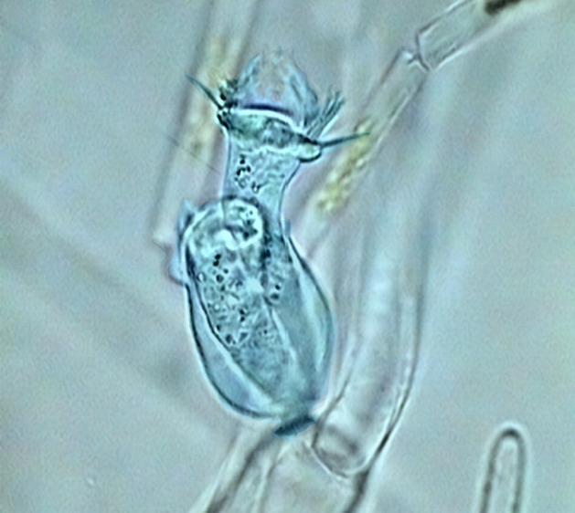
One individual colored electronically to make it more evident.
I present in an
Appendix a key, which
should allow the careful
microscopist
to clearly identify the genera of the
family
Vaginicolidae,
which are relatively frequent in the
samples taken from fresh water or from
marine water. The genera
Cothurnia and
Pyxicola
, are quite often mistaken, as
it can also happen with
Pseudocothurnia and
others. According to the illustration in the on-line facsimile of Kahl’s 'Wimpertiere oder Ciliata', perhaps the Cothurnia of Cancúns Polysiphonia can be identified as Cothurnia ovata.
As is the case for rotifers (see my article in the November 2008 issue of Micscape), microscopes with Differential Interference Contrast (DIC - Nomarski), and the electronic flash, could offer spectacular images of these cryptic inhabitants of my seaweeds. So far my cothurnias must be satisfied being known as Cothurnia cf. ovata. The abbreviation cf, interposed between the genus and the species means: 'Confer', that is 'Compare it with' or, efectively stating, it is a declaration of the biologist who implies: ' this species probably is ......, but I am not sure'.
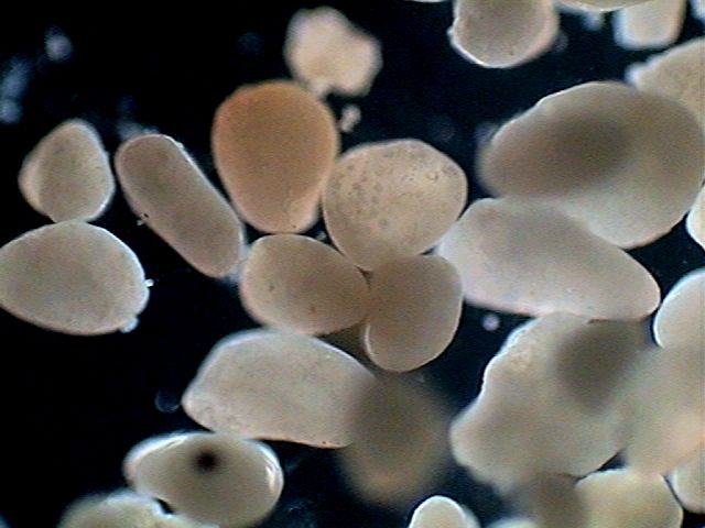
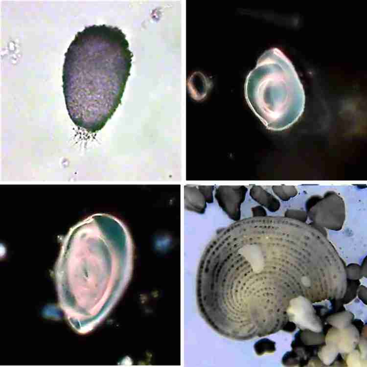
Finally, I dedicated my attention to the sand and I had my moments of excitation when I could identify between the blunt and light colored grains the characteristic shells of some dead Foraminifers, (with an exception, a single live specimen of Allogromia). But the small sample did not show a great species richness.
1 – Allogromia sp. alive. 2 and 3 – Probably Quinqueloculina. 4 – Probably Elphidium
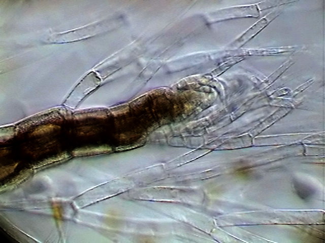
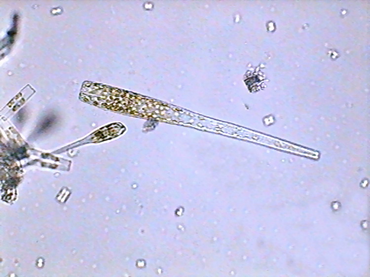
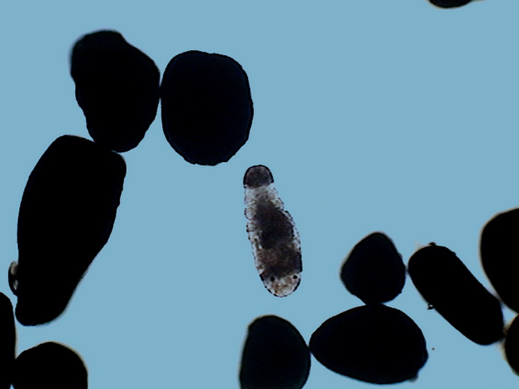
Nevertheless, gliding smoothly over the glass slide, between the small and irregular coralline grains, it appeared a vermiform organism of doubtful affiliation. So doubtful that it demanded days for me to reach a conclusion on its probable lineage. I enclose here the first snapshot that I took of it. And I will share next month my research on its micro-anatomy, even if that allowed me a taxonomic approach still less exact than that of my cothurniae. Sometimes this happens.
APPENDIX A KEY TO THE GENUS OF THE FAMILY 'VAGINICOLIDAE' Some of them are very rare, i.e. Cothurniopsis, Pseudothuricola and Pachytroca
|
Comments to the author, Walter Dioni , are welcomed.
Microscopy UK
Front Page
Micscape Magazine
Article Library
© Microscopy UK or their contributors.
Published in the December 2008 edition of Micscape.Please report any Web problems or offer general comments to the Micscape Editor.
Micscape is the on-line monthly magazine of
the Microscopy UK web
site at
Microscopy-UK
© Onview.net Ltd, Microscopy-UK, and all contributors 1995 onwards. All rights reserved. Main site is at www.microscopy-uk.org.uk with full mirror at www.microscopy-uk.net .