|
Notes on exploring two old slides labelled 'Nitzschia singalensis' and 'Nitzschia singalense' by David Walker, UK |
'This diatom is one of the severest tests for a 1/12th that we know of'. On 'Nitzschia singalensis', Spitta, 1920.
Additional text to accompany two new refs. added Jan. 14th. 2010.
Note added April 13th 2010: Frithjoff Sterrenburg has kindly explained aspects of the naming of the species discussed below and queries raised
on the interpretation of fine structure in his April 2010 article: Nitzschia singalensis v. N. firthii. What's in a diatom's name?
As a hobbyist I enjoy studying named, prepared slides of diatom frustules, especially if they are discussed as test objects in the older literaturenot because I'm obsessed with testing scope optics, but the literature serves as a useful focus (excuse pun) for studies with different optical techniques to see what can be reliably gleaned about their structure. As remarked by others, nowadays diatoms aren't regarded as valid tests for optics with the known variability in the frustule of a species and where the ability to resolve fine detail may be dependent on factors such as collection locality, specimen size, effect of mount, quality of mounting etc.An old slide that was of interest in my modest collection was labelled 'Nitzschia singalensis' mounted in 'Realgar' (arsenic trisulphide). Its very high refractive index (typically RI 2.549, ref. 1) gives high contrast with diatom frustules and certain diatom species in this mountant were popular with workers like Spitta when studying objective performance (1). Spitta discussed 'N. singalensis' as a test object and remarked 'This diatom is one of the severest tests for a 1/12th that we know of' (1). Spitta and a contemporary worker, Ainslie (3), both studied specimens in Styrax mounts and each made remarks suggesting Realgar mounted specimens weren't readily available, so this seemed an opportunity for me to study and photograph this Realgar mounted species and to compare with their studies.
But on inspection of the slide, despite the single species name on the label, I was faced with a busy strew of a variety of species beyond my admittedly casual level of ability to reliably identify to genus, let alone species. But I knew a UK seller, Brian Sneesby, of named Victorian diatom slides with an extensive online catalogue and bought 'Nitzschia singalense from Burma, Styrax' by 'W. Watson and Sons Ltd' (assuming that the Watson label was a slight spelling mistake). This slide had three selected specimens in a clean mount, so hopefully now had reliably identified specimens from a well known maker to compare with and seek out examples in the Realgar strew.
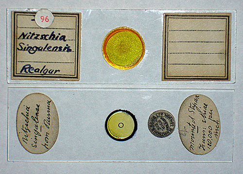
The two slides: Upper, the strew labelled 'Nitzschia singalensis' 'Realgar' by an unknown mounter and undated. The author's two other Realgar diatom strew slides (Amphipleura pellucida and Pleurosigma formosum) are in this label style and handwriting.
Lower, a slide labelled 'Watson and Sons Ltd 313 High Holborn, London.' ; 'Nitzschia singalense, from Burma.' ; 'Mounted Styrax. Trans: striae 120,000 inch.' The name and address label dates it between 1908-1957, (ref. 9).Supporting formal descriptions and drawings of the species from the literature would be invaluable, but after a search it seems that the diatom named 'Nitzschia singalensis' as discussed by workers as a test object (e.g. see refs. 1, 2 3) was never formally described by the noted professional diatom mounter W A Firth; Dingley Fuge in his 1937 paper remarked that the name 'must be considered illegitimate' and he formally described and named the diatom Nitzschia firthii and also discussed the history of its likely earlier naming (4). Bracegirdle in his invaluable book 'Microscopical mounts and mounters' notes that Fuge 'became an international authority' on diatoms (9a). Apparently some diatom slides by W A Firth of 'Nitzschia singalensis' were also inadvertently misspelled on the label 'Nitzschia senegalensis' (4). The latter is the valid name of a species described by Grunow in 1880 (5) but N. firthii and N. senegalensis bear no resemblance (4).
Hopefully, I've correctly summarised the diatom's background from the references cited and readers are not as confused as I originally was! Although apart from historical references and McLaughlin's 1990 paper (6), there seems almost no mention of Nitzschia firthii in online searches with Google, so uncertain if this indicates that it's either a less well known species to study or if still the currently accepted name post 1990. McLaughlin also discusses his reasons why he was of the opinion, that from a description of N. geitleri Hustedt published in 1988 (6a), that this diatom 'was identical with N. firthii Fuge'. There seem many more references to N. geitleri online than to N. firthii. Comments are welcomed on the current status of this diatom.
There seems to be at least two described collection localities of 'N. singalensis' (and misspelling variants) now Nitzschia firthii.
'From Amherst, Burma': W A Firth is reported to have made and distributed mounts from this source (9). Spitta is thought to have had examples from Firth (4).
From 'Chinese canned fish': the species formally described as Nitzschia firthii by Dingley Fuge were from this source (4). The sample discussed by Robert McLaughlin in 1990 (6) was also from a vial from this source.
This raised two queries regards my two slides.
1) Was the Watson slide labelled 'from Burma' the same locale as the 'Amherst, Burma' sample? If so was it possible that the specimens I was studying were close to that described by Spitta?
2) Was the Realgar strew also from Burma or could it be from elsewhere, perhaps the 'Chinese canned fish' source? Fuge and McLaughlin both mention the dominant typical other species in their strews so offered a long term project to try and compare.
Aims when studying the slides: I was interested to see;
1) if the striae could be resolved on my own microscope and under what conditions,
2) how the Realgar and Styrax mounted specimens compared for this species,
3) how did the striae spacing on these two slides compare with reported results,
4) a very speculative later addition after literature searches, was did the straie show any evidence of puncta on these examples, especially the Realgar mount.As the author's two slides refer to 'Nitzschia singalensis' or the Watson spelling variant, the present author continues to use this name in quotes below when referring to my own studies of these slides, rather than N. firthii.
Watson slide, 'Nitzschia singalense, from Burma', Styrax mount
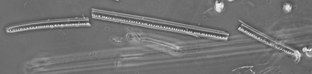
General observations: Styrax has a quite modest RI of 1.582 (1) but was struck by the low contrast of the diatom compared to other species I had in this mount. Even phase didn't really make them stand out. Despite good optics, the diatom detail also seemed to be somewhat elusive and rather soft. Photoshop Elements 7's quite strong 'Autosharpen' had to be used for the images below to crisp them up. The three specimens on the slide are shown below. Two of the specimen sizes lie within the range quoted by Fuge (4) for N. firthii , i.e. length 200-380 µm, width 9-11 µm.
Size 283 x 7.4 µm (as presented in mount)
Size 297 x 9.2 µm
Size 258 x 9.2 µm (estimate from pieces)Above: The three diatoms of 'Nitzschia singalensis' on the Watson slide, Styrax mount. Unless the specimens have moved with age, it is not one of Watson's better slides. One specimen is at a markedly lower focus level in the mount and two specimens overlap as shown in lowest figure. Zeiss 16x NA0.4 phase, Optovar 2x green filter.
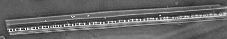
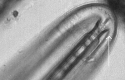
Detail of what is described by Fuge (4) as the 'eccentric raphe' in the broken specimen. Of the three specimens, this is the only one with the additional linear feature arrowed, the Realgar strew specimens also displayed this but not all. Diatom width 9.2 µm. Zeiss 40x NA0.75, phase, green filter, Optovar 2x.
Camera used for the images above, Nikon D300 DSLR.
Studying the striae: I've never found that reliably deciding the nature of low contrast diatom structure by eye to be easy, especially if deep blue was used. But one benefit the modern worker has over the past workers is the ready availability of sensitive cameras with live view, so the camera effectively becomes the eye to more objectively assess in real time the effectiveness of different lighting methods. I now routinely use a sensitive astronomy type C-mount USB monochrome camera (1.3 Mpixel, Opticstar PL-130M) with PC screen in real time to assess the effect of changes in wavelength, oblique, angle of oblique etc. Some enthusiasts successfully use multiple image shots and stacking to improve contrast and reduce noise, but prefer a single shot to minimise artefacts if studying the finest detail.
Straie on diatom frustules are widely reported as often best seen with oblique lighting directed along the axis perpendicular to the striae. Using this route with a strong 15% oblique (i.e only 15% sector of the full aperture as seen in objective back focal plane) using deep blue showed evidence of striae, but the low contrast made it the toughest striae to image clearly of the various classic test diatoms studied on the same scope in the past.
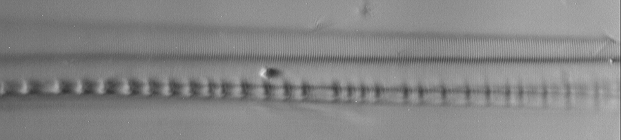
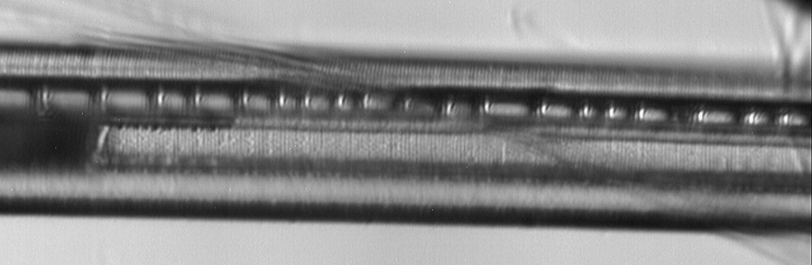
The striae visibility did improve by using a polarising filter with plane perpendicular to striae as described by workers like Spitta (1), see image pair below. It's only recently that I've started using this technique after being alerted to it by René van Wezel's Micscape article and the work of Osama Oku.
Zeiss 63x NA1.4 planapo, 1.6x Optovar, Schott BG3 deep blue glass filter. Oblique 15% bright sector along long axis.
Upper, with polar filter above objective at right angles to long axis. Lower, without polar filter.
To compare images the highlight level of the polar image grey background was matched to that of the non polar.
Realgar strew
General observations: I only have three Realgar diatom strews in my collection, but the increase in contrast of diatom frustules cf comparable examples in normal high RI mounts is quite striking. The first task was to establish if specimens of 'Nitzschia singalensis' (i.e. N. firthii) were present (it could be a misspelled slide of the validly named N. senegalensis or a misidentification by the mounter). After careful studies within my skill level, I believe there was a small population of 'N. singalensis' whose features matched those on the Watson slide and with those aspects described in the literature (4,6). Two examples of specimens are shown below from the strew.
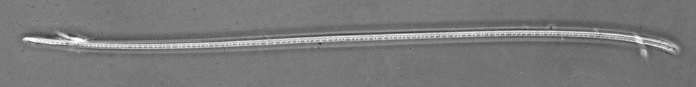
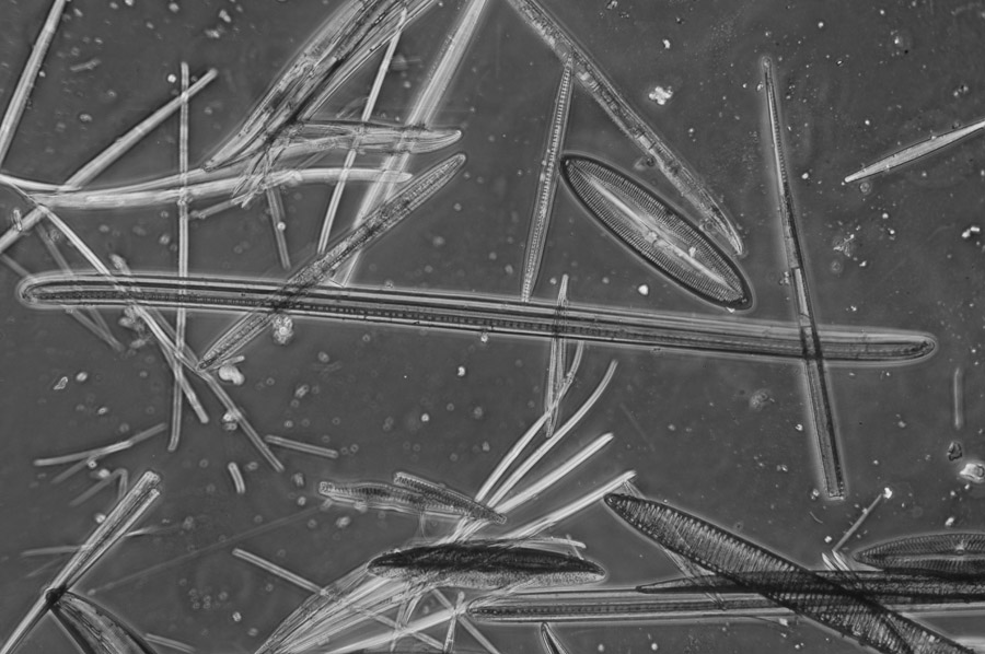
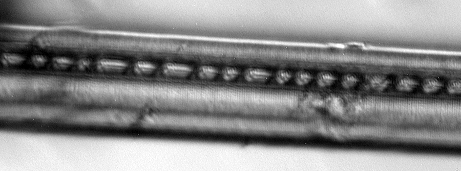
Realgar strew. Diatom believed to be 'N. singalensis', central horizontal diatom (size 304 x 11 µm). This species was present at low population but was noticeable by its size (this specimen length only slightly outside Fuge's stated size range for his specimens). Its eccentric raphe, additional linear feature remarked on and forked end detail were consistent with those of 'N. singalensis' on Watson slide. Most of the specimens larger than this diatom that were present were long, very thin and apparently featureless and may be e.g. spicules. Zeiss 25x NA0.65 Neofluar, phase, green lighting.
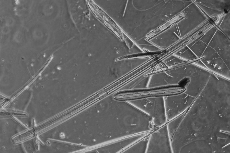
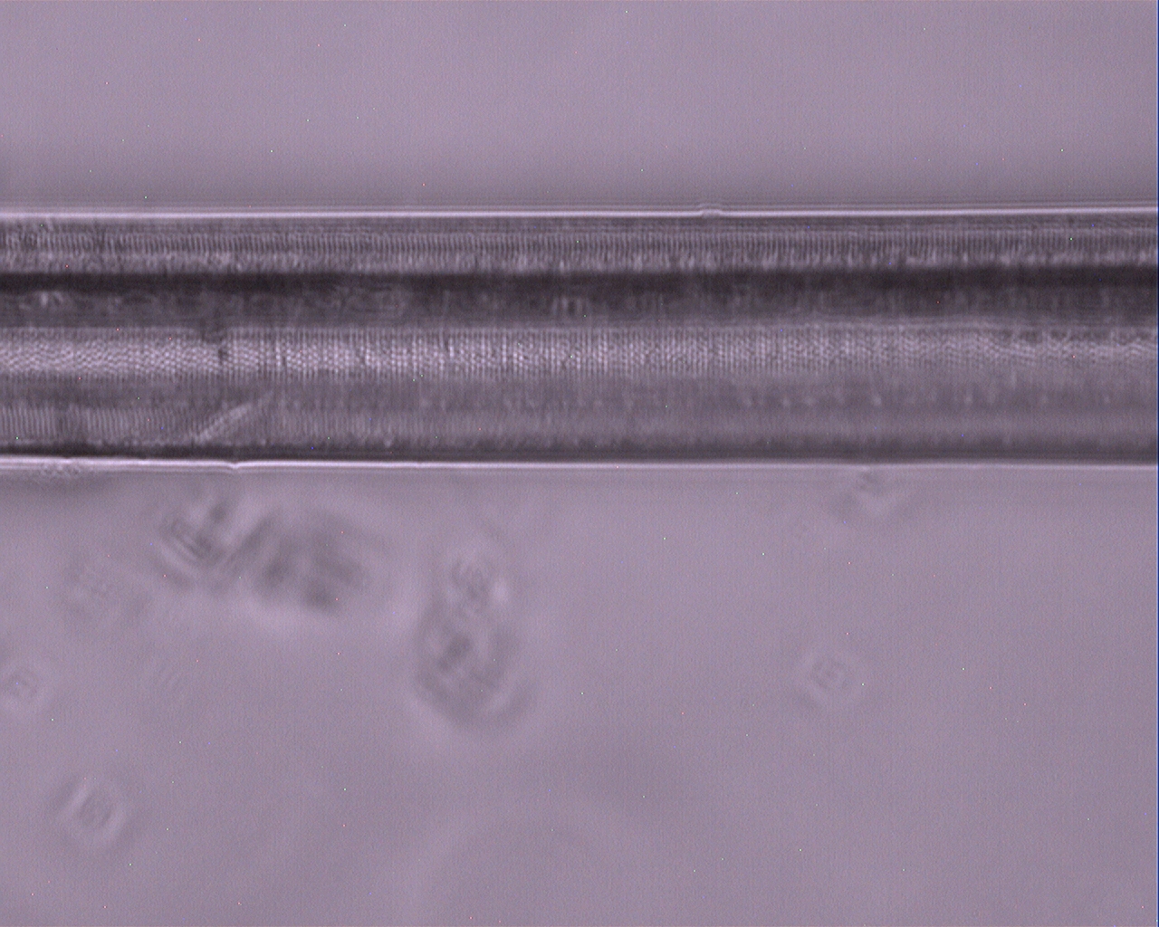
Realgar strew. A second example of a diatom believed to be 'N. singalensis', diagonal diatom (size 340 x 11 µm), size within Fuge's values.
Zeiss 25x NA0.65 Neofluar, phase, green lighting.
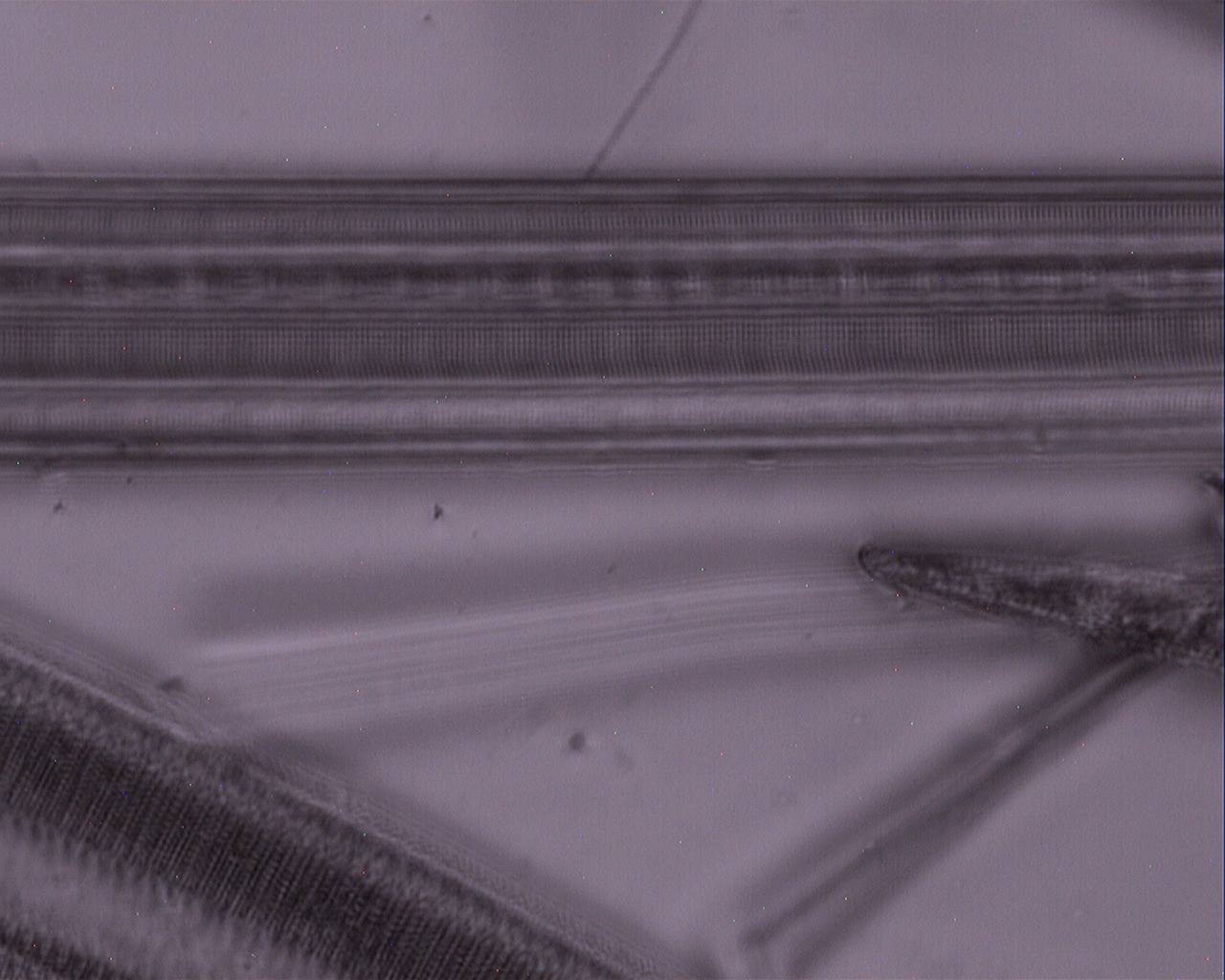
End detail of a typical Realgar specimen believed to be 'N. singalensis'. McLaughlin discusses a '"forked" termination' to the raphe in the 'Chinese canned fish' sample which he studied and illustrated; this feature seems to be present above (ref. 6). Just beyond fork, out of focus there is what appears to be an opening or feature (the 'carinal opening' described by McLaughlin, ref. 6?). The longitudinal feature remarked on earlier for the Watson specimen, arrowed here seems to curve round and form part of the termination of the raphe. Zeiss 100x NA1.3 Neofluar, Optovar 1.25x, brightfield.
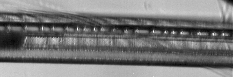
Studying the striae: The straie were much clearer in the Realgar mount, compare the tonal range of the images below with those for the Styrax mount above.
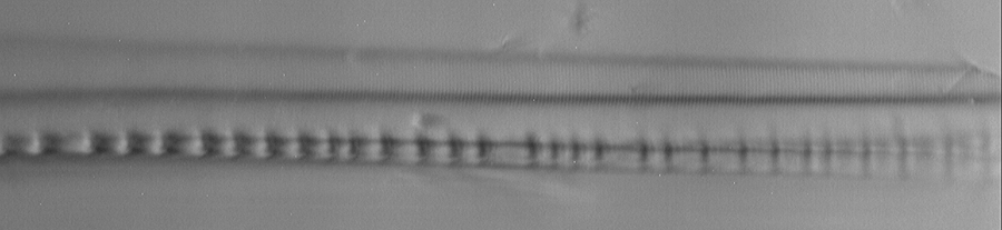
Striae with and without polar analyser, as with the Watson Styrax mounted specimen, the use of the polarising filter distinctly improves visibility of the striae.
Zeiss 100x NA1.3 Neofluar, 2x Optovar, Lee blue acetate filter. Oblique 25% bright sector along long axis.
Upper, with polar filter above objective at right angles to long axis. Lower, without polar filter.
The grey level of the polar image background was matched to that of the non polar. Diatom width ca. 11 µm
Counting the striae of 'N. singalensis' on Watson (Styrax) and Realgar mounts
Counting fine striae with an eyepiece reticle is tricky. I much prefer, as others do, printing out the image (in this case A4 sized images taken with a Nikon 12 Mpixel D300 DSLR) together with a micrometer slide under same conditions and counting striae on paper. (Merlin also used a photographic measurement method, 2). For more accuracy, 50 striae were counted and spacing averaged; the images had their tonal balance adjusted with Photoshop Elements 7 'Autocontrast' to improve visibility on print.
The repeatability of the present author's results was better than 1%, but accuracy was relying on the Motic off the shelf micrometer slide. Gradations of 0.01 mm are also a little coarse at the highest mags, 0.001 mm would have been better.
Worker, reference
Diatom source / Mounter / Mountant
Striae to inch (Striae in 10 µm)
Spitta (1)
Amherst, Burma? / Firth? / Styrax
'approximately' 110 000
Merlin (2)
Amherst, Burma / Nelson / Styrax
116 000 (eye) 115 200 (photo)
Ainslie (3)
Unknown / Unknown / Styrax?
115 000
Needham (8)*
'Chinese canned fish' / Unknown / Unknown
110 000
Present author
Unknown / Unknown / Realgar
115 400
Present author
'From Burma' / 'Watson' / Styrax
(120 000 to inch on label)113 800, 113 800 (45 in 10 µm)
(two places on same diatom specimen)
113 600
(different diatom specimen)*May be quoting a value from published work.
Considering the difficulties of reliably measuring such fine detail near the limit of the optical microscope and likely variability between workers, specimens, and collection locality, the striae count of the present author's are close to that quoted by other workers. The Watson slide states '120 000 to inch' on the label but uncertain if this had been measured for this specimen or was a literature source. I haven't found a reference to date that quotes the density that high, and would place the striae separation at 0.21 µma tough task for an optical microscope in visible light?
... but do the striae resolve to puncta?
I was pleased that the elusive striae had revealed themselves on both slides, and Spitta, who was one of the masters of high resolution diatom photography in visible light remarked 'They have, never, to our knowledge, been broken up into dots' (1). Merlin (2) and Ainslie (3) both wrote on this diatom as a test object but neither remarked on puncta being resolved. But after sourcing a copy of Fuge's paper (4) I was surprised to see that his own drawing of N. firthii (the now accepted name) showed the striae as crisp puncta! The figure caption notes the puncta but there's no mention in the text of the conditions used to achieve this feat.
I later obtained a copy of a paper on Nitzschia firthii by Robert McLaughlin (6). This is a fascinating paper published in 1990 and the most recent I've found to date on this species. He discusses both his own studies of a vial labelled 'From Chinese canned fish, Nitzschia singalensis', the history of the earlier naming and shares photomicrographs of his studies as well as Fuge's drawings and one of Spitta's photomicrographs. McLaughlin remarked that with his own prepared Naphrax and Cargille Meltmount slides he could 'only just approach seeing striae with the equipment at my disposal.' He also cast doubt on aspects of Fuge's drawings of N. firthii including the puncta and later stated that he 'must reject that particular portayal of the striae for two reasons.' One reason was that Fuge did not mention the puncta in his 'Latin diagnosis' of the species and the second was Spitta's remark quoted in the paragraph above.
A Google Book search has the powerful ability to recognise text in books scanned as images and can help seek out any further references; two additional references remarking on 'Nitzschia singalensis' resolution were found (7,8). N E Brown in his delightful book 'Arachnoidiscus' 1933 (7) discusses the setting up of an 'inverted gas mantle' for microscopy studies and remarks, 'I find it suffices to resolve with axial light both Amphipleura pellucida and Nitzschia singalensis not merely into transverse lines, but more or less evidently into their component cells'. Resolving both species in axial light, but particularly the latter seems quite an accomplishment. N E Brown was a 'plant taxonomist' as noted in a Wikipedia entry where his higher plant work was summarised. The 'Quekett Journal' has papers by him on higher plants but also on diatoms; two papers (11,12) reported his extensive studies of the fine structure of certain diatoms using various techniques* and a range of objectives are mentioned. The plate accompanying one paper (11) shows drawings of some Realgar mounted diatoms ('N. singalensis' was not discussed).
*One technique he described was the use of 'a Leitz dark-ground illuminator, from which, by decentering it, various modifications of oblique light and light reflected from the under surface of the cover-glass can be obtained.'
He also describes his use of a darkfield condenser with funnel-stop removed in the objective, i.e. likely to be annular illumination. (11)Were my own Realgar specimens (assuming I'd correctly identified them) able to throw any light on the presence of puncta as hadn't to date found any work published specifically describing Realgar mounted 'N. singalensis'? I spent some days restudying examples of the diatom on the strew, in particular under conditions known to reveal puncta at the limit of the light microscope in Amphipleura pellucida, ie oblique light in deep blue directed at approx. 45° to long axis to capture the higher orders of diffraction (Spitta discusses the theory behind using oblique in this way, 1).
Using an optical microscope to reliably remark on features at the limit of resolution is dubious, if not foolhardy; but present below the 'best' images I could manage to optimise resolution on my Zeiss Photomicroscope III in case of interest.
Off-axis oblique and deep blue light for both slides
'Nitzschia singalensis' on Watson Styrax slide. Zeiss 63x NA1.4 planapo, achromatic-aplanatic condenser with iris at full aperture of objective, Optovar 1.25x, 410 nm interference filter, oblique 25% bright sector at 45° to long axis. Oblique using opaque card in filter tray of condenser. Width of diatom ca. 11 um.
Master 1.3 Mpixel image resized to 900 pixel width and Photoshop Elements 7 'Autocontrast' applied.
Specimen believed to be 'Nitzschia singalensis' in Realgar strew. Zeiss 63x NA1.4 planapo, achromatic-aplanatic condenser with iris at full aperture of objective, Optovar 2x, Lee '119 Dark blue' acetate filter (transmission max ca. 440nm), oblique 25% of aperture diameter bright sector at 45° to diatom long axis. Oblique using opaque card in filter tray of condenser. Width of diatom ca. 11 um.
Master 1.3 Mpixel image resized to 900 pixel width and Photoshop Elements 7 'Autocontrast' applied.For off-axis oblique, despite the high RI mountant, deep blue and Zeiss' 1970's optics, there's not enough resolution and/or image not good enough with my limited skills at least to make any firm comments. To complicate matters, for focus planes just above the striae (and potential puncta if present) there appeared to be some sort of slightly larger globular structures or deposits in a random arrangement on the frustule. Some enthusiasts have been using LED lighting in the near UV and sensitive cameras to push the limit of optical microscopy, an approach the author has also used in past, but Realgar blocks light in the near UV (10).
However, when rereading René van Wezel's and Osama Oku's Micscape articles I was struck by how clearly they had both photographed the resolved puncta of Amphipleura pellucida with high NA annular illumination (i.e. circular oblique, COL) and crossed polars. Could this technique eke out any extra contrast / resolution from the microscope used?
Annular oblique, crossed polarisers and deep blue light for both slides
A Zeiss 'ultra darkfield' NA 1.2 - 1.4 condenser was used oil immersed to create this annular illumination. It gives quite a broad annular light ring at edge of full objective aperture with the 63x NA1.4 objective but a thin ring at edge of full objective aperture for the 100x NA1.3.
The Watson (Styrax mounted) specimens did not suit this technique at all, giving low contrast imagery; off-axis oblique as used above seemed much more suited.
But for the Realgar strew there was a noticeable improvement in the visibility (better resolution and/or contrast?) of frustule fine detail. In this instance it seemed more marked visually with 10x eyepieces and 2x Optovar than what my camera was able to capture. Normally I can't see fine detail by eye with a deep blue filter, but because the Realgar mount is a deep yellow brown the transmitted light is actually a green bordering on blue. No comment can really be made on the presence or absence of fine structure in the low contrast image above taken with off-axis oblique light, but for the annular lighting is there evidence for the possible presence of puncta? Given the potential pitfalls of trying to reliably identify real structure in visible light near the limit of resolutionI welcome comments! Again I must stress this is for the diatom believed to be 'N. singalensis' from comparisons with the Watson slide, and share these results for confirmation or otherwise of this identification.
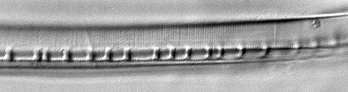
Realgar strew, detail of diatom believed to be 'N. singalensis'. Photoshop Elements 7 'Autocontrast'. Compare the contrast range of the fine detail with that shown in last section with off-axis oblique. Diatom width ca. 11 µm. Zeiss 100x NA1.3 Neofluar (phase) objective in brightfield, annular oblique with Zeiss darkfield condenser NA 1.2 - 1.4, crossed polars. Lee dark blue acetate filter. Click image to view out of camera master.
Realgar strew, detail of another specimen of diatom believed to be 'N. singalensis'. Photoshop Elements 'Autocontrast' 7. Zeiss 63x NA1.4 planapo objective, Optovar 2x, in brightfield, annular oblique with Zeiss darkfield condenser NA 1.2 - 1.4, crossed polars. Lee dark blue acetate filter. Click image to view out of camera master.
Comments to date
It would be interesting to know whether the two diatomists Fuge (4) and Brown (7) who both described resolving 'Nitzschia singalensis' (N. firthii) to puncta without further comment, used Realgar mounted specimens. In my own studies there was much improved contrast of the species in this mount cf. Styrax illustrated above, especially with annular oblique / crossed polars; perhaps these workers used annular oblique with/without polarising filters with Realgar mounted specimens*. As remarked earlier, Brown was known to have access to and reported on the fine structure of other Realgar mounted diatom species and he also described using a darkfield condenser without stops in the objectives that could potentially create annular illumination (11).
* I'm not certain of the earliest mention of the use of annular oblique with crossed polars, but both (a) annular illumination and (b) the benefit of a polarising analyser for studying diatoms with off-axis oblique was well known before Fuge's and Brown's 1930's reports; method (b) was discussed by e.g. Spitta in 1920 (1).
If puncta do exist in N. firthii, Spitta, although a master at this type of work, may not have been able to resolve to puncta because of the Styrax mounts he had access to. I could barely resolve the striae in the Watson Styrax mount; obtaining evidence for puncta if they exist seems unlikely with such low contrast on the author's slide.
I'd be interested if any readers have examples of 'Nitzschia singalensis' slides or spelling variants in their collection and how they are described on the label (e.g. mounter, date, mountant, locality), in particular the outcome of their own studies. To resolve (excuse pun) the present author's 'striae/puncta query' it would be very interesting to learn of any SEM studies of N. firthii.
But I won't lose any sleep not knowing if 'N. singalensis' (N. firthii) has puncta (!!); the main aim of these notes was to provide an example of how much fun even one or two slides can be to prompt our own enthusiast level studies and to enjoy reading the literature on the history of a specimen. As remarked, recent resources like Google Books with its ongoing scanning of literature, coupled with its text recognition, are powerful tools for the enthusiast without access to extensive literature to learn more about their slides. The author's two slides described and accompanying literature, certainly provided many hours of enjoyment on the recent long winter evenings.
Final note: The author is a hobbyist and casual studier of diatoms, so I would welcome comments and corrections of any errors e.g. of interpretation, misidentification, nomenclature or insights from literature I don't have access to. Thank you.
Comments to David Walker welcome.
Acknowledgements
Thank you to Brian Stevenson for kindly sourcing copies of two of the papers referenced. Thank you to Brian Sneesby for the sale of the Watson slide described. Also thanks to René van Wezel and Osama Oku for highlighting in their articles the potential of annular oblique with crossed polars. Members of the Yahoo 'Amateur microscope club' forum were also helpful with early leads on the potential misspelling of N. singalensis.
References with additional notes
1) E J Spitta, 'Microscopy. The construction theory and use of the microscope.', John Murray, London, 1920, 3rd edition.
Nitzschia singalensis is discussed as a test object on pages 393-394, and two photomicrographs showing the striae on Plate XXII are compared with the striae of Amphipleura pellucida.
Also discussed (in 3rd edition only) is the benefit of using a polarising Nicol prism to give an 'improvement in the definition' of striae on page 499.
Refractive indices of mounts of the time are listed on page 508-509.
The entire book with plates can be freely downloaded at www.archive.org.2) A A C Eliot Merlin, 'On Nitzschia singalensis as a test-object for the highest powers', The Journal of the Quekett Microscopical Club, 1916, vol. 19, p. 112-113.
Describes studies of a Styrax mount of specimens from Amherst, Burma supplied by E M Nelson. Plate 8 shows a photomicrograph clearly showing striae.3) M A Ainslie, 'Nitzschia singalensis, a note on Mr Merlin's paper', The Journal of the Quekett Microscopical Club, 1916, vol. 19, p.113-116.
4) D P Fuge, 'Nitzschia firthii spec. nov, a diatom from Chinese canned fish;, J. Royal Microscopical Society, 1937, vol. 62, p.183-185.
Formal description of the valid name for misnamed / misspelled diatom slides labelled Nitzschia singalensis or Nitzschia senegalensis.5) A Grunow (with notes by F Kitton), 'On some new species of Nitzschia', Journal of the Royal Microscopical Society, 1880, vol. 3, p.394-397.
Formal description of Nitzschia senegalensis, a dissimilar species but thought by Fuge and others to be the source of a later misspelling by W A Firth.6) R B McLaughlin, 'Nitzschia firthii Fuge, A test diatom', The Microscope, 1990, vol. 38, p. 304-311.
The most recent paper on this diatom known to the author.
The author also discusses his reasons why he was of the opinion, that from a description of N. geitleri Hustedt published in 1988*, that this diatom 'was identical with N. firthii Fuge'. There seem many more references to this latter diatom name online than to N. firthii.6a) *The cited reference by McLaughlin was: 'Krammer, K.,; Lange-Bertalot, H., Suswasserflora von Miteleuropa, 2 Teil: Bacillariaceae, Epithemiaceae, Surirellaceae. Gustav Fischer Verlag. Stuttgart, New York 1988.'
7) N E Brown, 'Arachnoidiscus. An account of the genus, comprising its history, distribution, development and growth of the frustule, structure and its examination and purpose in life, and a key to and descriptions of all known species, illustrated.', Watson and Sons Ltd., London, 1933, p. 23.
Apart from Fuge (4), the only other reference found to date where the author remarks that Nitzschia singalensis can be resolved to puncta ... and what a splendid book title!8) G H Needham, 'The practical use of the microscope including photomicrography', Charles C Thomas, Illinois, 1958, p. 225.
'Nitzschia Firthii (Singalensis)' is one of the diatoms listed as a test object. '110 000 lines to inch' is stated.
The index entry under 'Test objects ..' adds 'from stomachs of Chinese canned fish'.9) B Bracegirdle, 'Microscopical mounts and mounters', Quekett Microscopical Club, London, 1998, p. 44.
Under the 'Firth, W. A.' entry the author comments on the Nitzschia singalensis / N. sengalensis labelling by this mounter.9a) B Bracegirdle, 'Microscopical mounts and mounters', Quekett Microscopical Club, London, 1998, p. 37-38.
Under 'Fuge, D. P' entry.10) R M Allen, 'Photomicrography', van Nostrand, New York, 2nd edition 1958, p. 381, plate VI.
11) N E Brown, 'Some notes on the structure of diatoms', The Journal of the Quekett Microscopical Club, 1914, vol. 18, p.317-338 + Plate 23.
12) N E Brown, 'The structure of diatoms', The Journal of the Quekett Microscopical Club, 1914, vol. 23, p.52-65 + Plate 4.
Microscopy UK Front Page
Micscape Magazine
Article Library
© Microscopy UK or their contributors.
Published in the January 2010 edition of Micscape Magazine.
Please report any Web problems or offer general comments to the Micscape Editor,
via the contact on current Micscape Index.Micscape is the on-line monthly magazine of the Microscopy UK web
site at Microscopy-UK

© Onview.net Ltd, Microscopy-UK, and all contributors 1995 onwards. All rights reserved. Main site is at www.microscopy-uk.org.uk with full mirror at www.microscopy-uk.net.