 |
( using polarized light and dark-ground illumination ) by Brian Johnston (Canada) |
|
|
( using polarized light and dark-ground illumination ) by Brian Johnston (Canada) |
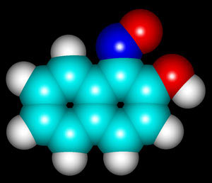
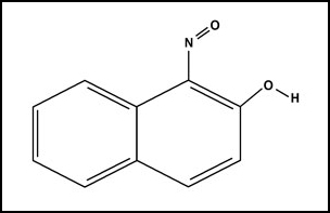
Most photographers have favorite subjects that they consider to be particularly photogenic. One of mine is the organic compound 1-nitroso-2-naphthol. This yellow-brown crystalline substance melts at about 107 degrees Celsius, and is a reagent used in analytical chemistry for the determination of cobalt. The usual caution should be exercised when handling this chemical, as it is considered moderately hazardous. The structure and molecular shape can be seen below. ( The molecular representation was produced using 'HyperChem'. )
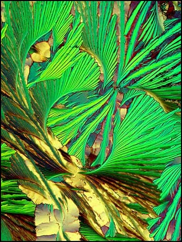
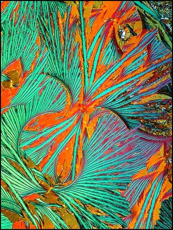
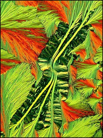
The relatively low melting temperature makes this an ideal candidate for producing a 'melt' specimen, by placing a very small quantity between microscope slide and cover glass, and heating using an alcohol lamp or hot plate. When the compound recrystallizes as it cools, interesting patterns sometimes result. Although experience has shown that it is fairly difficult to obtain a specimen with the ideal crystal thickness for polarized light examination, the effort is definitely worth making! 1-nitroso-2-naphthol produces an array of almost 'botanical' or 'leafy' crystal formations. Melt specimens of this compound are characterized by a textural quality that is highlighted by the use of plane or elliptically polarized light as an illuminant.
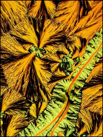
All of the polarized light photomicrographs in the article were taken with the polarizer and analyser adjusted to give a black background (extinction position). In many, compensators were used in the microscope to alter the colour range of the field of view. The two examples below illustrate how lambda/4 or lambda plates can change the appearance of a particular crystal field.
Choosing what area to photograph on a slide is, of course, the crucial decision. In my view, the slight change in field choice between the left and right photographs above resulted in one being 'better' than the other. The left image is superior, because the malformation seen in the top right corner of the right photograph is less obvious. Although the left image is less colourful, the 'leafy' texture seems to stand out more clearly, and this seems to improve the image.
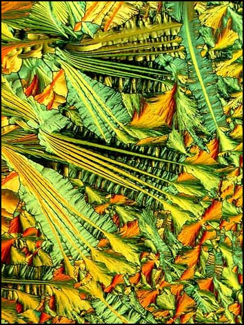
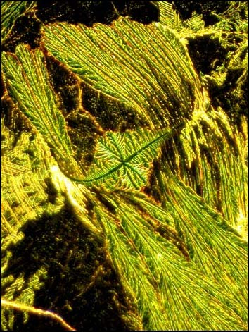
The example below shows a crystal formation viewed with ordinary plane polarized light, using no compensators.
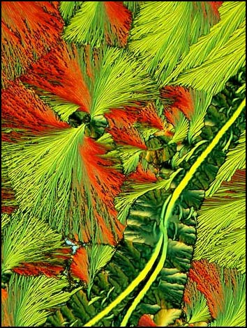
The two photomicrographs below are of exactly the same crystal field. The one on the left is illuminated by plane polarized light, and the other by elliptically polarized light produced by the use of compensators. Notice how the elliptically polarized light in the right photograph displays far more detail in the diagonal band. The black areas in the fan shaped structures are also removed by elliptically polarized light. In my very subjective opinion, the right photograph is the better of the two!
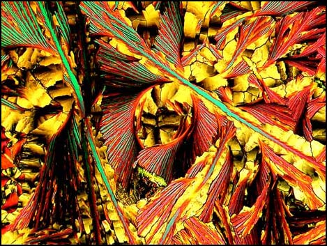
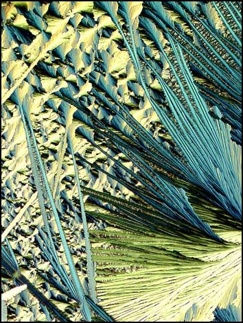
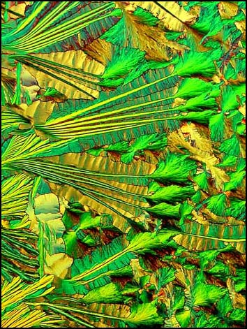
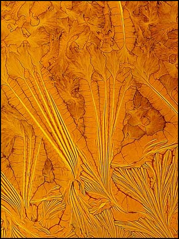
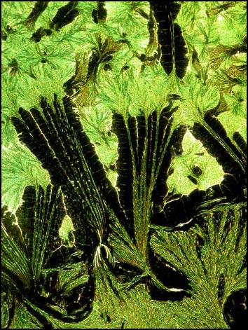
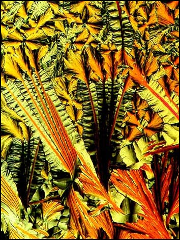
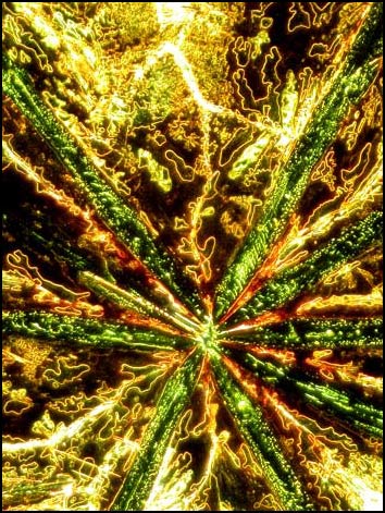
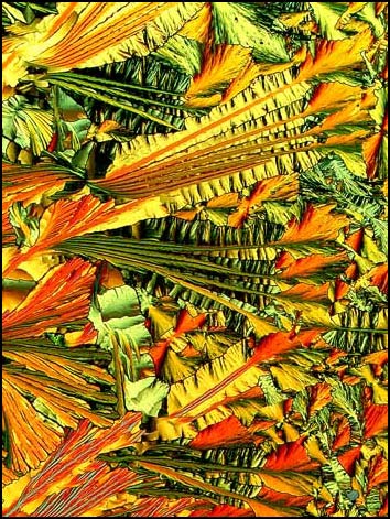
A polarizing microscope with its many adjustments allows the photomicrographer an amazing degree of control over the colours in the final image. All four of the images below were taken from the same edge area of one slide. Notice how subtle changes in illumination can completely alter the character of the resulting photomicrograph. Although I like all four, I must admit that all are a bit too 'busy', with perhaps too much detail to be good images.
So far, all of the photographs have used polarized light to illuminate the crystals. Here is what the crystals look like in ordinary white light (unpolarized). The deep yellow-brown colour is evidence that the crystal layer on the slide is quite thick. 1-nitroso-2-naphthol requires thicker layers to produce interesting crystal structures than many other compounds. I mentioned earlier that getting this 'ideal' thickness is one of the problems when working with this substance.
Here is exactly the same field as above, viewed with dark-ground illumination (left), and plane polarized light (right). The thickness of the crystal layer reduces the quality of the dark-ground photograph. This type of illumination is best used on relatively thin crystal layers.
The two photomicrographs below use dark-ground illumination. This method is often utilised to show inclusions in crystals and the right hand example shows numerous trapped air pockets. ( Or are they bubbles of oxides of nitrogen formed during the heating process? )
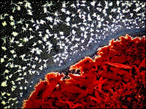
As an example of what can, and often does, go wrong, I offer the following photograph. This slide was reheated several times in an attempt to obtain satisfactory results. It never produced any fields of the quality of the earlier examples, but the tiny stars adjacent to the red lump were interesting.
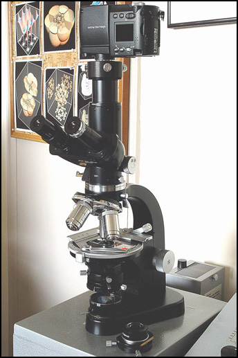
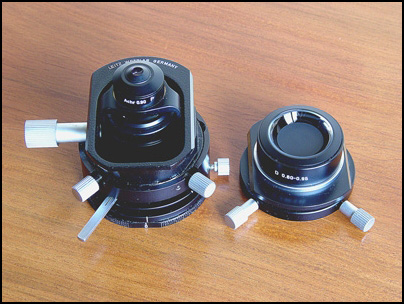
A Leitz SM-Pol microscope and Nikon Coolpix 4500 camera were used to take all of the photomicrographs in the article. Both the polarizing condenser (left) and dark-ground condenser (right) are also shown below.
The colours, textures and forms of crystals viewed under the microscope are almost addictive! Every bottle on the chemical shelf offers promise of an even more unusual photomicrograph. I hope that this article encourages you to try compounds that are not on most photomicrographers' lists. As long as you have a copy of the Merck Index, or equivalent to check melting points and DANGEROUS CHARACTERISTICS for the compound, you should be fine. If you can't find the information about a compound, then don't use it! I emphasize that compounds should not be handled and especially heated, unless you know that they are not going to explode or release poisonous gases! ( Sorry for ranting, but my chemistry teaching background just takes over at moments like this! )
For a more detailed explanation of the photomicrographic setup, and the use of compensators with polarized light, please see my earlier article concerning ascorbic acid.All comments to the author Brian Johnston are welcomed.
Please report any Web problems or offer general comments to the Micscape Editor.
Micscape is the on-line monthly magazine of the Microscopy UK web site at Microscopy-UK