001-coccoloba
COCCOLOBA UVIFERA
The leaves and a parasite

WALTER  DIONI                       Durango (Dgo) Mexico

click the picture to see a larger version of the grapes
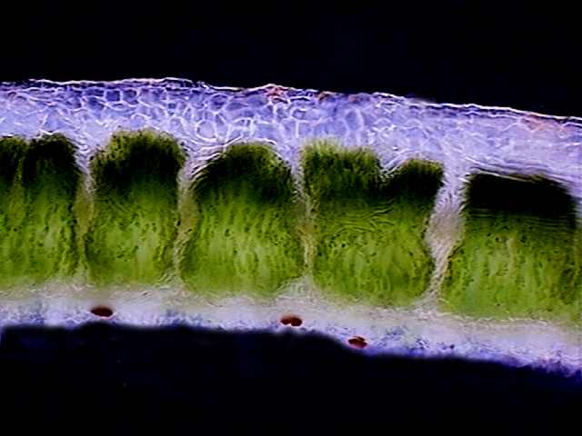
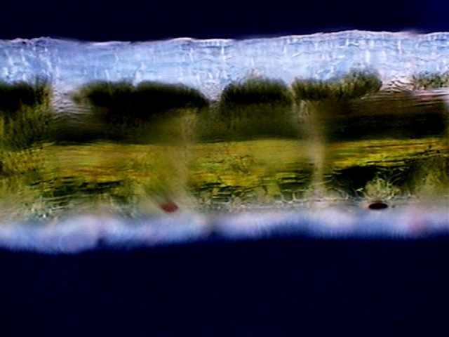
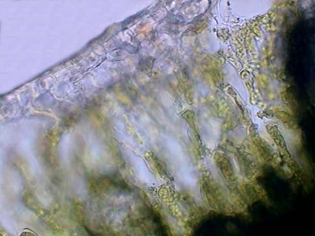
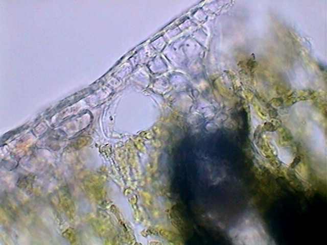
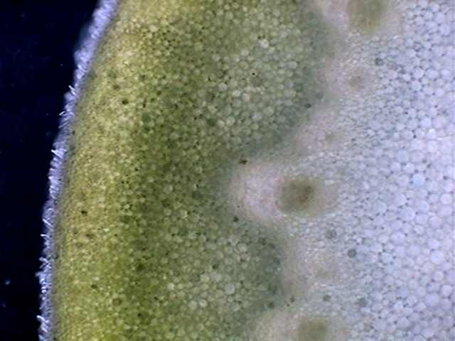
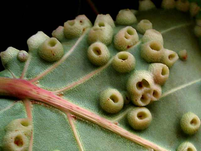
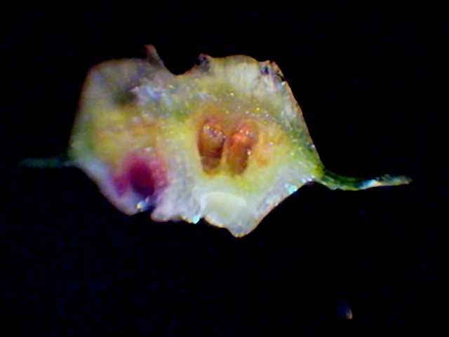
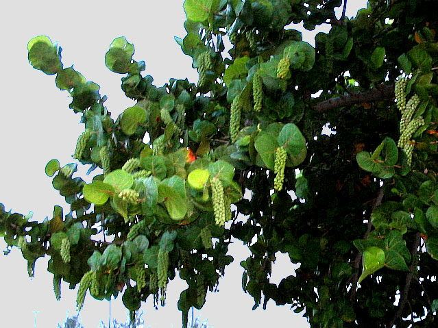
There grows in Cancún a so-called Sea Grape or Beach Grape tree. Reasons for the common name will be easy to imagine seeing the attached photo. The grapes are edible.

Coccoloba uvifera
, as it is called scientifically, is a salt resistant species now used as an ornamental tree, that has very large and beautiful round leaves, with a leathery texture. These seemed to me an excellent material to continue trying the possibilities of the mesotome (the name I propose for a homemade slicer made from double edged razor blades, see footnote 2). The results of this test are illustrated in the following images; the leaves show a strange architecture, with narrow spongy parenchyma and what would have to be the palisade parenchyma distributed in bundles compressed at the base(?) where the cells appear dark and colored. Among such bundles run identifiable vascular packages easily seen with polarized light, but feebly visible with normal illumination.

002-coccoloba
003-coccoloba
Transversal section-obj x10 - darkfield  - chlorophyll "palisade" parenchyma in tight bundles. The small dark disks in the inferior epidermis are stomata.
with polarized light a long bundle of vessels could be seen

The hardness of the leaf made it difficult to obtain the sections, mainly because at first I tried using razor blades of the "thin" type. Changing to blades of normal thickness, the sections were possible without problems. 
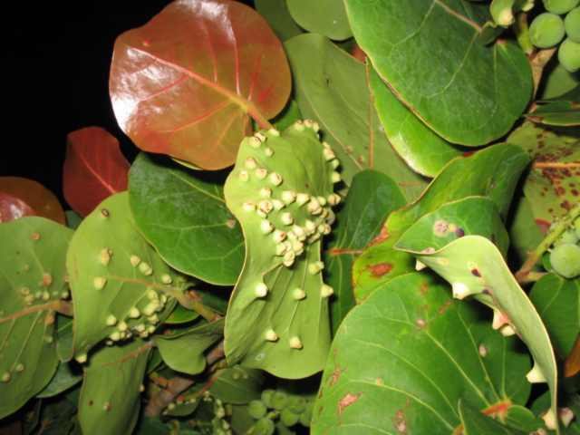
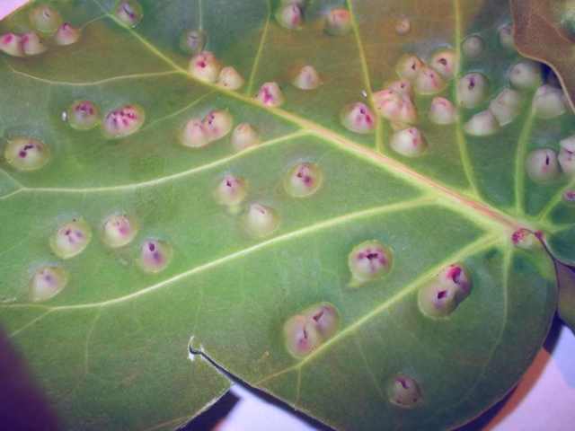
004-coccoloba
005-coccoloba
one minor vascular bundle, obj x10, darkfield
the minor inferior bundle with the x40 obj

006-coccoloba
007-coccoloba
 spongy (aerophyll) parenchyma, obj x40, brightfield
a stomatal chamber, in the spongy parenchyma


008-coccoloba
A section of the petiole from a young leaf. It was recorded as 6 individual pictures of 640 x 480 px. The individual pictures were stitched in PhotoPaint and the mosaic was reduced to 500 px to be inserted here.

But most interesting was that many of the leaves showed a surface marked by innumerable galls that protruded from both faces of the leaf and which I thought could either be caused by fungi (but not very probable due to their structure, which were small volcanoes in both sides of the foliar lamina) or, almost certainly, by parasitic insects.
 

009-coccoloba
010-coccoloba
There is a large pith in the petiole, surrounded by a continuous "stela" with a lot of small vascular bundles. Cortical parenchyma and cholenchyma are reduced to a narrow strip.
A sector of the petiole seen under the 40x objective. Notice the regular cells in both pith and cortical parenchyma. The blue 'halo' which are short hairs covering the surface.

It was a magnificent opportunity to try the mesotome to aid an investigation of vegetal pathology. But although I do not doubt that it will have utility on other occasions, I had to give up after breaking two instruments. Dishonorable failure!

I could only cut the incipient galls in young leaves, which are where the insect apparently deposits its eggs.

011-coccoloba
012-coccoloba
A better view with the 40x of a similar sector to the one shown above with the 10x. This time with a blue-white Rheinberg filter.  You see the small xylem and huge phloem system in the vascular bundles, the cambium as a dark strip, the clear cortical parenchyma, and the mostly yellow chollenchyma.
A view with the 40x objective of the chollenchyma to show the sclerified walls of the cells, and the row of small crystals (spherical aggregates of calcium oxalate) in the epiderm. Short "hairs" cover all the surface of the petiole.

 The completely developed galls have a fibrous structure and a hard ligneous consistency that ended up defying even my better scalpel. Finally, with it sharpened very well for each cut, I managed to discover not only the anatomy of the galls in longitudinal section, but also its inhabitants. 

014-coccoloba
015-coccoloba
this drew my attention to the parasite
a closer view from the underside of the leaf

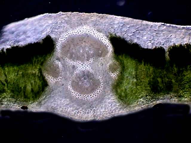
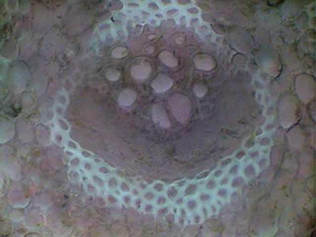
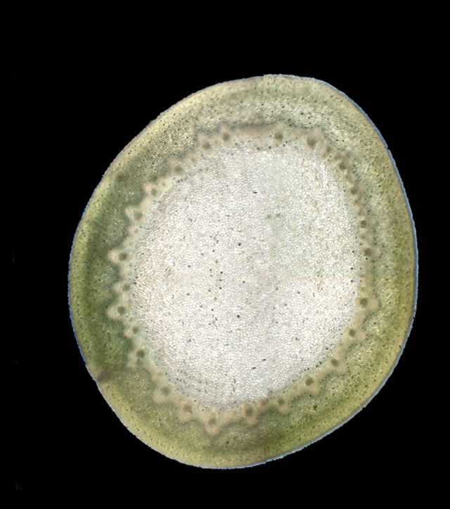
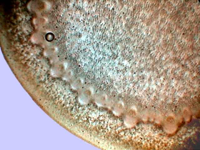
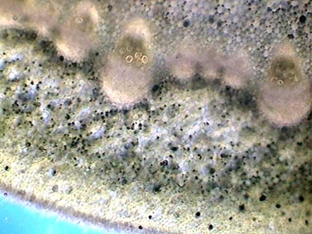
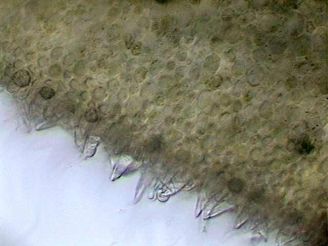
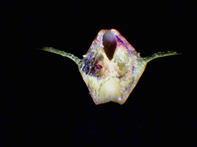
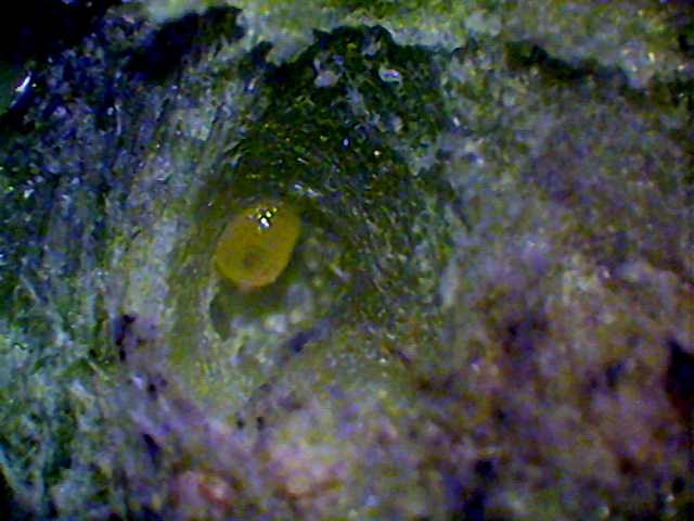
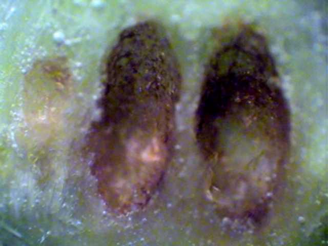
With incident light we can see in the two images below right and with a magnification of x25 the structure of the galls in a vertical section.

016-coccoloba
018-coccoloba
the upper side of the leaf
showing that the morphology of the galls varies
A cut through the gall, showing its relation with the foliar blade. In the dark sector at left is a larva. See below.

019-coccoloba
021-coccoloba
 one larva discovered by the scalpel x4 objective
a pair of contiguous galls - the right-hand one has two pupae
022-coccoloba
the two pupae seen with the x4 objective - incident light

In one empty gall I found the shed skin of a pupa, and at once verified that the guest 
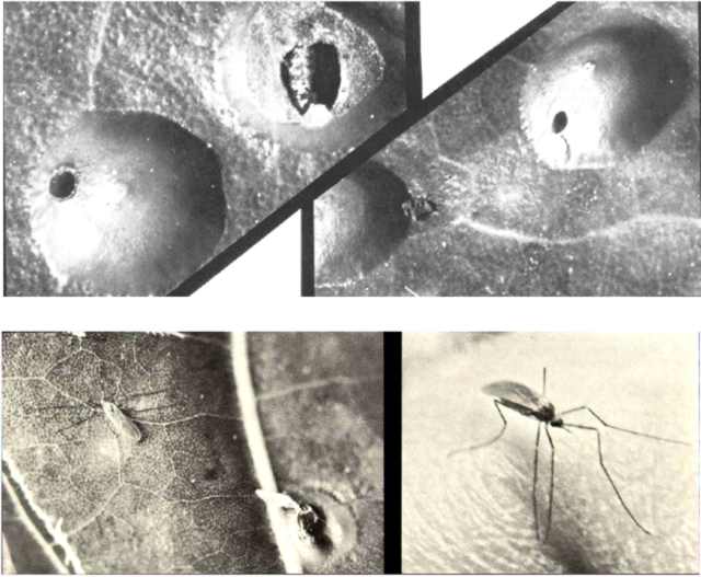
was a dipteran. I looked for the possible identity on the Internet. The only work that I found is dated 1970 and it identifies a dipteran of the Cecidomyidae family, from a group called "gall midges", with a North American species (from Florida, specifically) known as Ctenodactylomyia watsoni, whose nutgalls and maker can be seen in the following pictures taken from Circular 97 (1970) of the Florida Department of Agriculture & Consumer Services. 

023-coccoloba
The galls of Ctenodactylomyia watsoni, parasite of Coccoloba from Florida, EEUU. At upper left, one of the galls was broken up to show the larvae. The shape, structure and exit openings are very different from the Cancún material. In the lower images two views of the adult midge.

 Although I do not doubt that the genus of the Cancún midge is the same one, it seems to me that the shape of the galls and the double incubation chamber shows that it must be a different species, in spite of the relatively short distance between the Florida peninsula and the Island of Cancún.

To break two mesotomes can also have its compensations.

NOTES:

1)      I am neither a phytopathologist nor an entomologist. Even if I could obtain the adults which would allow identification of the parasite to species, I could not determine it. If there is amongst the readers somebody with knowledge and suitable bibliography to do it, I could provide the graphical materials and all the biological materials that can be needed. My e-mail address appears at the end of this article.
2)      I described the device in Micscape as a slicer (see this article). In French it is now named “tranchette”, in Spanish it would be “rebanador” but that is really cacophonic. It seems to me that it is better to adopt a name more easily identifiable with its function. I propose “mesotome”. Of course it is not a microtome, but the sections it provides, to be studied with a microscope, could not be called macrosections. From mesotome it is easy to derive other languages derivatives.


 Comments to the author, Walter Dioni , are welcomed.


Microscopy UK Front Page
Micscape Magazine
Article Library


© Microscopy UK or their contributors.

Published in the July 2004 edition of Micscape.

Please report any Web problems or offer general comments to the Micscape Editor.

Micscape is the on-line monthly magazine of the Microscopy UK web
site at Microscopy-UK


© Onview.net Ltd, Microscopy-UK, and all contributors 1995 onwards. All rights reserved. Main site is at www.microscopy-uk.org.uk with full mirror at www.microscopy-uk.net.