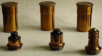
Supplied as standard with microscope
shown in Fig 4. are from left to right, [magnifications for
160mm tube length], a 1" [6x NA 0.21], 1/2" [13x NA 0.34] and 1/6th"
[42x NA 0.74] parachromatics. The parachromatics also seen in
catalogues with the name simply shortened to 'para' were one step up
from the basic Argus range from Watson, above these were the Holos
objectives
requiring matching Holos eyepieces and apochromats to be used with
either compensating or Holos eyepieces but never the less these optics
are good with bright contrasty images aided by the simple monocular
design of the microscope and one interesting point is the lack of
thickening and soft edge definition when looking at diatom type slides
with the 1/2" objective that often accompanies modern 10x NA0.25
achromats, instead, crisp edges not unlike those from Wild's high NA
phase fluorites are seen which is most pleasing.
The 1"objective provides a reasonably flat field but as you go further
up in magnification the field flatness and highest resolution becomes
more localized to the
centre where the subject should always be placed for best image quality
in these older design of lenses,
Pleurosigma
angulatum is quite easily resolved with
the 1/6th" but is very dependent on the quality and brightness of the
illuminating source for the microscope and provided better results when
fitted to a Russian LOMO stand with aplanatic condenser and external
LOMO filament lamp with field iris. The parachromatic
objectives provide best contrast with their aperture reduced to 65-75%
using the Abbe condenser unlike many modern computer designed
objectives
with special lens coatings which provide optimum contrast and
resolution with the condenser set at
90-95% of their full aperture. The objectives and cases are beautifully
made, heavily lacquered and miniature works of art in their own right.
The
Watson Catalogue of 1906 is rather confusing since they have reverted
to stating magnifications for the older 10" tube length [image distance
of 10"] so the 1" objective is stated as a 12x, the 1/2" 20x and the
1/6th" 65x but the Watson Edinburgh is primarily being used at 160mm
tube length and would require the draw tube fully extended to obtain
these magnifications which I think very unlikely so why they continued
to quote them like this I don't know. The magnifications change again
somewhat for the same objectives in a later condensed book 'The Book of
the Watson microscope' so whether the design of each objective
was changed I don't know but from my experiments the figures given in
the main paragraph above are reasonably accurate and agree with the
later book. Using low to medium power objectives the draw tube was a
convenient way of continually changing the magnification without
changing the eyepiece and quite a lot of leeway was possible
without due problems but this is not the case as you approach 1/8th"
and oil immersion objectives where the correct tube length should be
strictly adhered to.
Additional High Power Optics used
with the
Microscope.
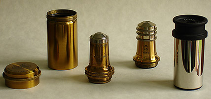
Fig 5.
Additional objectives I use with the Watson are a 1/12th"Apochromatic
immersion NA 1.30 by P. Koristka, Milan Italy in superb condition with
one of
the heaviest and finest tooled cases I've seen, again with a deep gold
lacquer and a
1/8th" Watson 'para' [51x at 160mm NA 0.87] which is shown in the
background of Fig 5. both are of excellent optical quality, the 1/8th"
is a later
design to that supplied with the microscope. A high quality Beck 8x
compensating eyepiece is used with these higher power objectives rather
than the simpler 6x and 10x eyepieces supplied. Francesco Koristka as
he later became known was originally from Poland and his business had
links with
Zeiss, if this objective is typical of his output then it is a
fine range of optics from the Italian catalogue, this maybe an early
sample since he moved from Milan in 1895.
A Simple Illuminator.
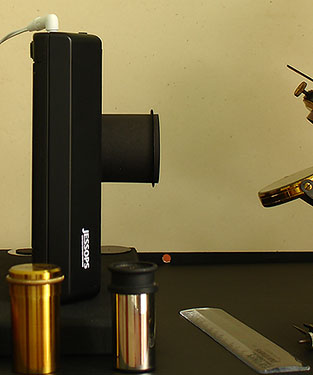 Fig 6.
In Fig 6. a simple cold light source
is shown using a Jessops fluorescent light box with home made baffles
removes glare especially to the unused eye whilst observation is taking
place. The light provides a good white balance being designed for slide
transparencies and needs no adjustment for luminosity and is supplied
with a low voltage DC source from a wall type adapter. Brightness is
sufficient from the lowest to highest powers provided the light box is
kept close to the microscope and condenser focus is aided by simply
holding a pencil point touching the light box illuminating surface and
focused with the condenser. True Kohler illumination from a quality
external lamp would no doubt improve contrast and edge definition of
diatoms at high powers but I find this light source cheap and
convenient to use with reduced eye strain which often accompanies
critical illumination using high wattage filament bulbs, even so I
prefer observation periods of about 15 minutes followed by a
short rest and then resume to reduce fatigue.
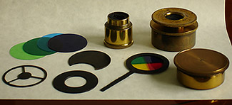 Fig 7.
Fig 7. shows some of the original
accessories supplied with microscope including a metal dark-field stop
for low power objectives and oblique illumination plate together with
their brass case and original polarizer-analyzer which I don't use
preferring modern Polaroids fitted to the eyepiece and condenser. Added
to
these are a set of acetate colour filters I made and my special colour
wheel with long actuating arm which can be used with or without
dark-field or crossed-polars for special effects and can be rotated
easily whilst fitted in the filter tray.
Name plate and Serial Number.
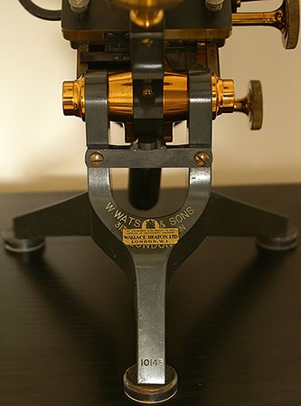
Fig 8.
Fig 8. shows the Watson & Sons
inscription and overlay by Wallace Heaton Ltd, London. W1 who supplied
the microscope originally to the first purchaser also showing the coat
of arms and rather official
'Appointment to his Majesty the King', Suppliers of Photographic
Equipment. The fading white lettering
of the serial number can be seen just above the nearest foot, 10143
placing it around 1907.
The Mahogany Case.
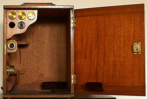 Fig 9.
A simple mahogany case shown in Fig
9. with lock and key was supplied with the microscope holding the stand
and three objectives, polarizers and accessories, some water
damage near the bottom panel is evident which thankfully doesn't seem
to have affected the microscope maybe because it was outside the box at
the time of damage, the original maroon felt is in good condition.
Mounting pins for a card holder, the holder itself and the card giving
details of objectives and magnifications with the supplied eyepieces is
missing from the door.
Condition
and Maintenance.
As received the microscope was in
very good condition with the natural lacquer in excellent order and
although the scope had been used extensively in the past some very
small adjustments to the focus, stage and sub-stage adjusters was all
that was needed to allow consistent and smooth operation. Some of the
lubricants had dried back so this was cleaned off and a trace of new
lubricant applied and with this type of design with close machined
tolerances much lower viscosity lubricant is needed. The optics were
quite dirty and needed a clean and my usual method of using a blower
brush to remove abrasive particles and then using photographic cleaning
tissue dampened with a little of the supplied cleaning fluid brought
them back to excellent working order. Typical for this age of
microscope the optics can suffer from fungal attack on the glass
elements and indeed the 1/6th" showed quite bad signs of this but
fortunately it
was on the back focal plane and removed effectively.
Sample Pictures.
The pictures below were taken with a Sony
P200 digital camera handheld onto the Watson eyepiece, unfortunately I
have no easy way of connecting my camera to the Edinburgh and my
patience ran out trying to set up a tripod and get the spacing right
between camera and eyepiece. Obviously with slow shutter speeds,
optical zoom on the P200 set around 1.5-2x and almost certainly camera
shake these are only a fair representation of what is seen visually,
however what can be deduced is acceptable field flatness from the 1"
objective, good detail and contrast from the 1/2" objective whilst the
image of Gyrosigma balticum can only be described as poor compared to
visual observation was included to show that the Abbe condenser running
full aperture can just about cope with high numerical aperture
objectives. As I summarise in the conclusion this is a microscope
ideally suited for visual observation and unless an easy method of
connecting a camera is found I prefer doing my pencil sketches and
drawings. All the images were taken with the Edinburgh illuminated by
my modified Jessops light box and the camera set to auto white balance,
I like black and white photography so three of them are in this mode.
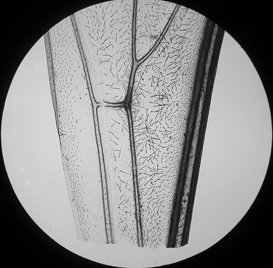 Fig 10a.
Bee wing mounted by Mike Samworth.
Watson & Sons 1" objective, acceptable field flatness considering
100% coverage by the camera.
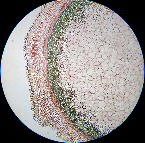 Fig 10b.
Old and faint transverse section of Elder mounted by Richard Suter.
Watson & Sons 1" objective, 100% coverage by the camera.
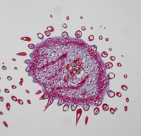
Fig 11a.
Yellow Sage, a section through flower buds mounted by Biosil.
Watson & Sons 1/2" objective, good detail capture aided by above
average NA.
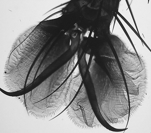
Fig 11b.
Foot of a Blow fly mounted by C.M. Topping.
Watson & Sons 1/2" objective, a difficult subject due to the depth
and density of the subject.
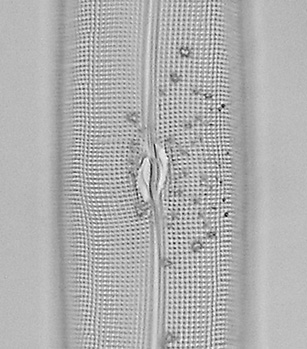
Fig 12.
P. Koristka 1/12th" oil immersion objective, much better seen visually.
Conclusion.
The Watson Edinburgh 'H' student
microscope is as usable today as it was 100 years ago, the fine
mechanics and optics plus ergonomic design are a delight to use and put
many modern microscopes to shame. The parachromatic objectives which
normally come with the stand are of good numerical aperture
providing bright crisp images with all subject matter including
botanical sections and specially diatom strews and type slides. I
recently purchased a large number of mixed diatom slides and this
microscope has been used throughout to make my usual notes on subject
matter and provide the images for making my small pencil sketches to go
with the slides. I don't think its forte is for photographic use where
modern plan objectives are more useful but if the main use is for
visual pleasure of subjects down the microscope together with
admiration of a finely
crafted instrument it is a sound investment and highly recommended.
Recommended reading: Brian Johnston's article in Micscape
The
Watson "Mint" metallurgical microscope with excellent images and
facts of
this superb stand.
Further reading and for comparisons:
A
Personal Review of a Smith & Beck microscope ca 1856.


