|
A Mystery About Micro-Technique: Part 2 by Richard L. Howey, Wyoming, USA |
When I concluded Part 1, I commented that I didn’t want to be hasty in judging the potential of the technique of using mica strips as mounts in relation to polarized illumination. So, let’s look at yet another slide, again brightfield, then polarized. This is clearly sections from plant material.
Unfortunately, I come to the same conclusion–no significant improvement.
However, this did set me to wondering whether the specimens were really mounted on mica strips or if these strips are something else. I know that in addition to mica, selenite was also used as a compensator and when I started poking around, I discovered that selenite is a form of gypsum and I remembered that on some of the specialized compensators by German manufacturers, there appear the descriptive identification “Rot 1" (Red 1) and other gypsum compensators are labeled “Gyps” or “Gips”. In fact, it has a variety of crystal forms : it is frequently flattened as twinned crystals; there are flower-like forms that have sand combined in them and are known as selenite roses.
In addition, there is a satin spar which has a fibrous character and is barely translucent. A very fine-grained compressed form is known as alabaster and it can have very delicate colors. Because Gypsum/Selenite is very soft–only 2 on the Mohs scale–it is easily carved for a wide variety of decorative purposes.
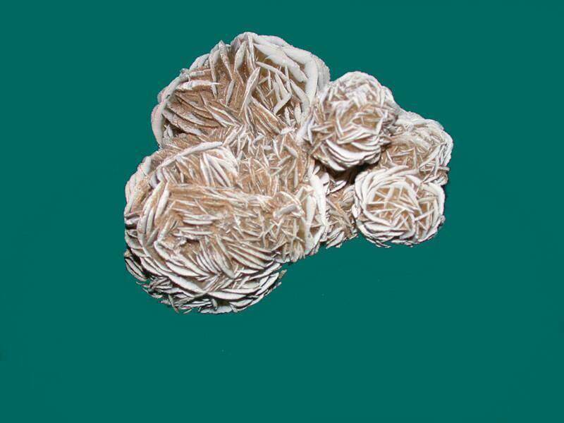
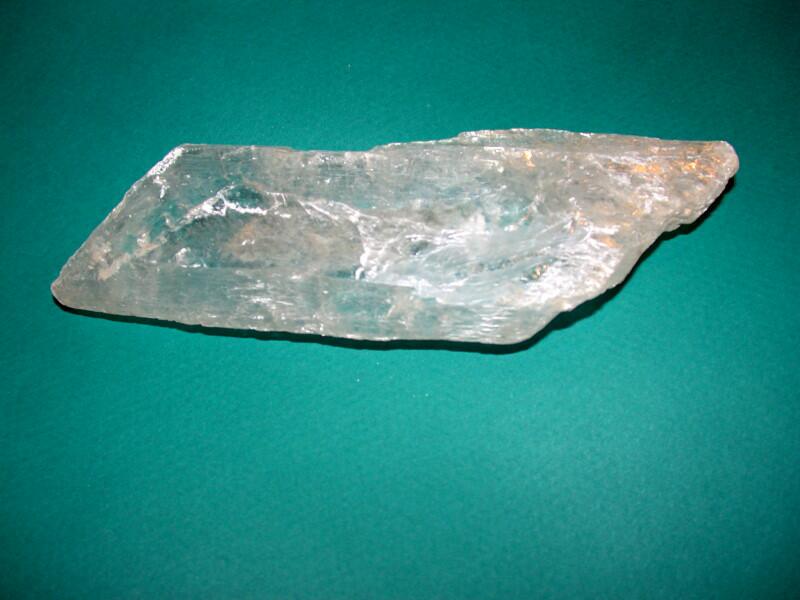
I have a sizeable piece which is called Ram’s Horn Selenite, and is, I believe a variant of the satin spar form.
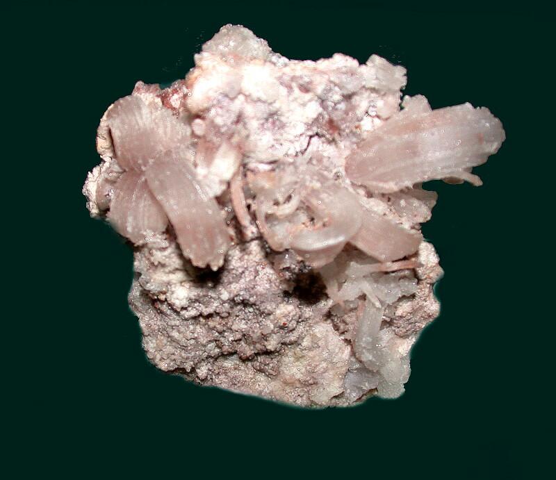
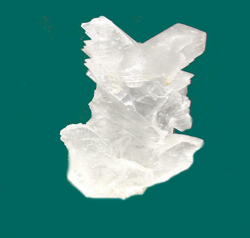
Here is an image of another type of Selenite which is one of the twinned forms.
Most all of us have encountered gypsum in another quite utilitarian form–namely “burnt “ gypsum. At one time, large quantities were quarried in Paris’s Montmarte district, treated with heat and became world famous as Plaster of Paris.
Selenite/Gypsum is not now regarded as having much worth yet, individual pieces can have a form or size that make them desirable to collectors. They form some of the largest crystals known and there is the famous Cave of Swords in Mexico (fortunately restricted–otherwise instead of Crystal Meth gangs, there would be Crystal Selenite gangs)–which have enormous bladed crystals extending from the surfaces that are over 50 feet long! I can well imagine a Victorian aristocrat adding on a special room to his estate to accommodate a few of these.
I knew that somewhere I had a 5 inch piece of gypsum squirreled away and I also knew that it was layered in a fashion that made it easily cleavable with the careful use of a sturdy scalpel or small knife. Remarkably, I was able to find it after a search of only about 10 minutes. (Usually locating something specific which I want to use requires days.)
So, now after all of the meandering, we are at the point where I can actually test whether or not a gypsum/selenite strip had been used in these old slides rather than a mica one. Well, the strip isn’t selenite/gypsum I assert dogmatically. (Can one assert catmatically?) Let me rephrase that. It very likely, probably maybe, isn’t selenite/gypsum but possibly could be, although, I don’t think so. How’s that for a definitive, assertive statement? The main basis for my assertion is that this piece is too photoactively birefringent.
The difficulty is that selenite/gypsum, mica, and calcite (all of which have been used in optical systems) are anisotropic, that is, they behave differently when their position angle, or orientation is changed. Rather than venturing into the complex discipline of the physics of polarizing optics, let’s limit ourselves to a few simple cases where we can use these slides to investigate along with playing with a few mica and gypsum fragments which I took from my mineral samples.
The images which I am going to present are mostly rather rough and unenhanced, which as you will see is, in the end, an advantage in making some basic points. As we proceed, we need to remember that position, angle, thickness, and orientation are all issues to be kept in mind.
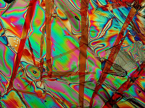
With standard polarization, we place a specimen, say a slide of birefringent crystals above the polarizer which is set to zero degrees and then rotate the analyzer above the specimen until we get extinction thus creating a black background against which we get a dazzling display of crystals. In this case, the image is of a mixture of Ascorbic acid, Magnesium sulfate, and the biological stain Orange G.
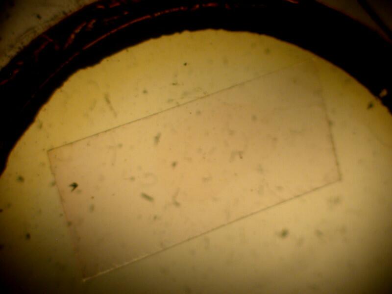
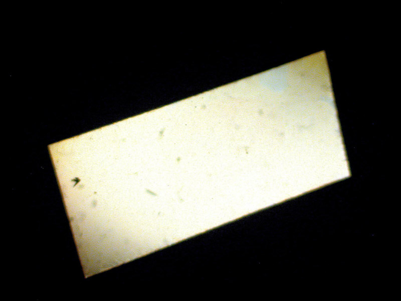
I mentioned earlier, before you dozed off and had a nice nap, that of these 21 slides, one had no specimen but did still have the small rectangular strip which makes the polarizers go bonkers. First, let me show you a simple view of the strip which is in a mount that had a circular cover glass ringed with black varnish, some of which has flaked off as you can see, again suggesting that this slide is very likely, probably, maybe nearly 100 years old. The cover glass is still intact and there is very little yellowing of the balsam. When the polars are not crossed, you can see the mica rectangle as a clear strip and you can also see part of the varnish ring around the edge of the cover glass. This is a brightfield image.
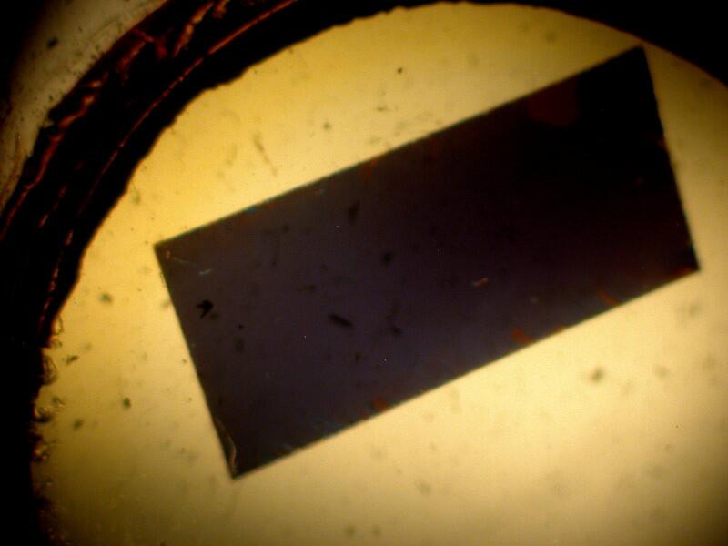
However, when we cross the polars, the ring disappears and the mica rectangle is now white.
In spite of the bits of flaking, the cover glass is well sealed and yet there is no sign of a specimen. This could mean that this was an early experiment just to test out using the strip.
If the polars are only partially crossed, the rectangle takes on an opaque brownish appearance.
Now, let’s look at some examples of slides that do have specimens on them (even though, I have only the vaguest ideas as to what they might be).
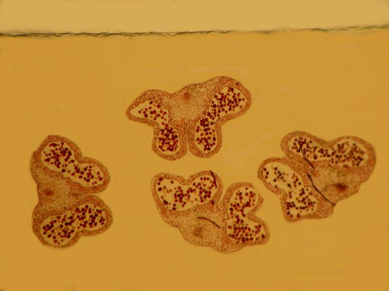
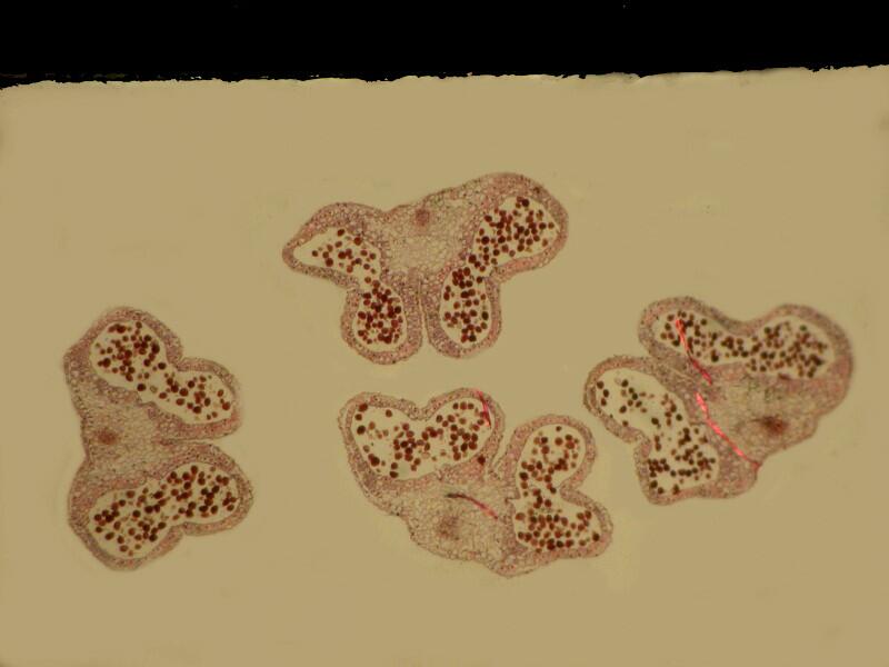
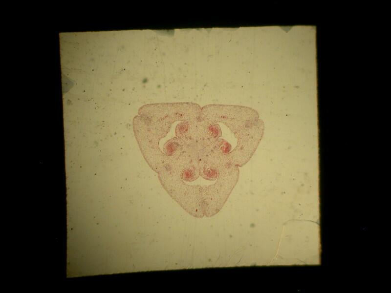
Let’s start with a nice plant section. First we see the section on a mica square with the polars not crossed.
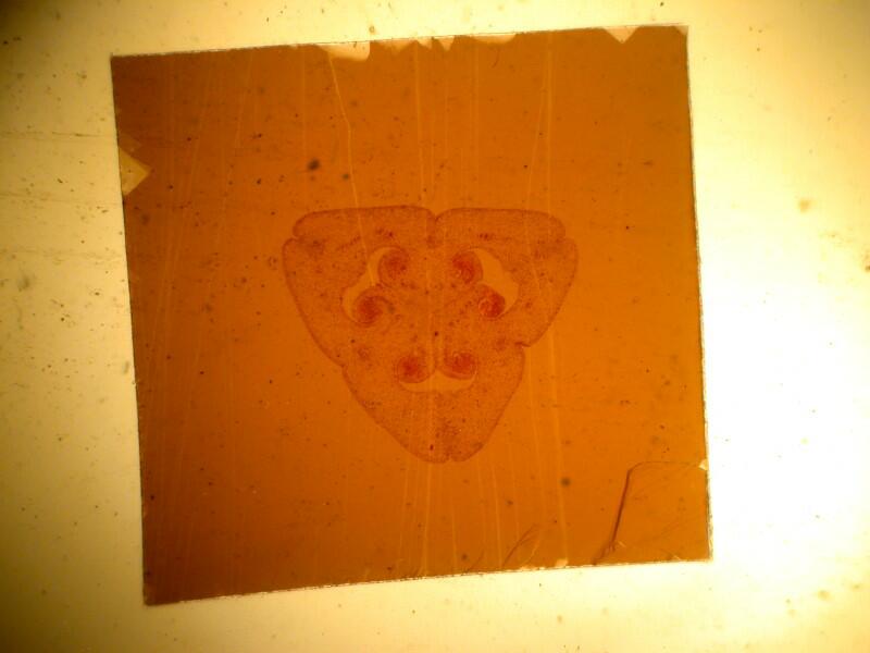
Here we have the mica producing a pinkish background which certainly doesn’t enhance contrast or resolution and there is also a white background outside the square. When we cross the polars, the white background becomes black and while the stained plant sections shows more contrast, the mica sheet is of a color such that there is no significant gain in terms of detail.
Another slide will show two problems quite dramatically. This section looks like it might be a section of a parasitic worm but, for all I know, it could be the cross section of intestinal villi from a Martian warthog. The first image shows you what the untouched slide looks like (that is, no computer enhancement). On the left, you can clearly see the vertical line of the edge of the mica strip. In that area, it is evident that there is crackling in the balsam mountant.
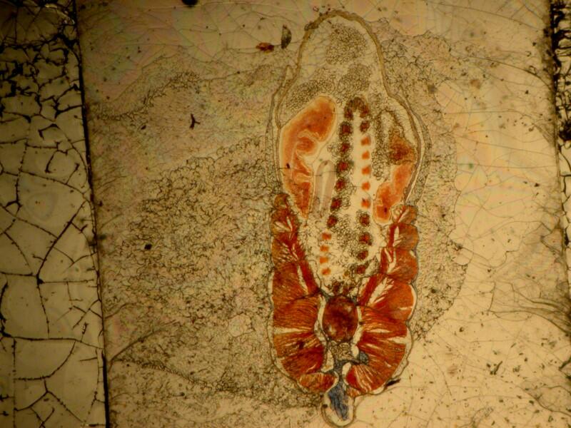
On the strip itself, there is a good bit of detritus around the specimen which detracts significantly. “Praise the Lord and pass the graphics software!” Here, the wonders of digital technology manifest themselves. With a bit of computer magic, we can become digital char-maids and go in, clean up the mess, and get an image like this.
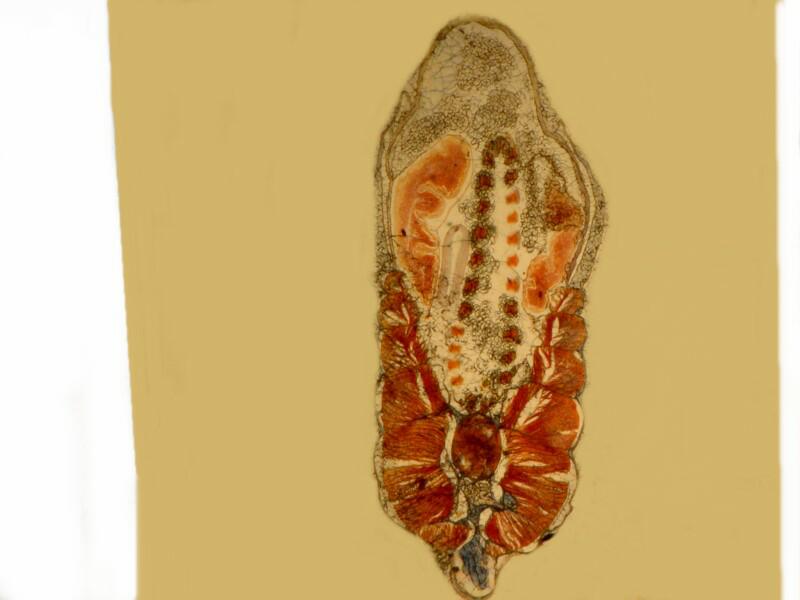
Now, it may still be only a cross section of the hemorrhoid of a Venusian Venture Capitalist but, in any case, it’s certainly much more distinct. We sometimes forget how much visual noise can distract us from seeing the detail we are seeking.
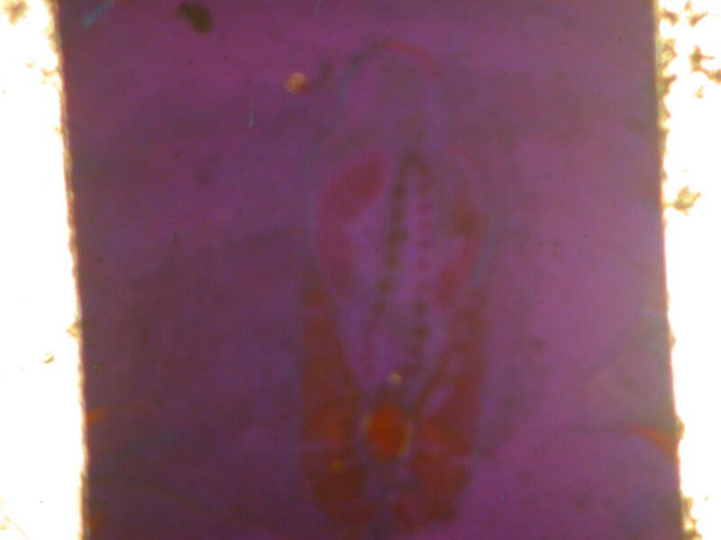
When we cross the polars, we have the edges of the mica strip, as expected, as white strips but, the mica strip itself has become a color that obscures virtually all detail in the specimen; in fact, the outline is barely discernible.
To be fair, let’s consider another example where the shift is not so dramatic and then do a side by side comparison.
Here again, the specimen is unidentified but, it looks to me much more likely to be animal rather than plant tissue. The first image is brightfield and, as you can see, the section has been nicely stained and shows a fair amount of detail. (Remember, this is at a fairly low magnification on a stereo-dissecting microscope.)
The second image is the same section at the same magnification, only this time, it is polarized and the polars are fully crossed.
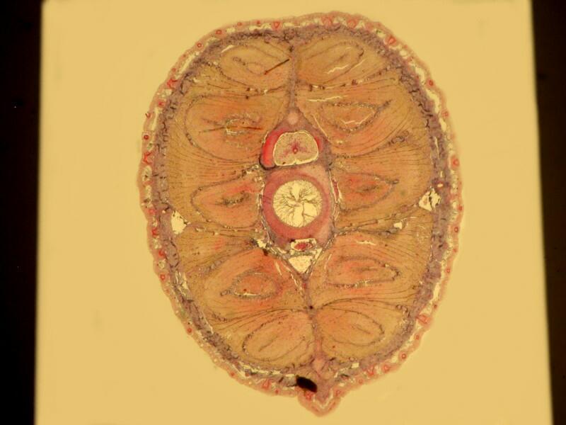
Here the image is a bit less vibrant and there certainly isn’t any enhancement of contrast or resolution that would provide us with more detailed information.
Now, let’s put them side by side. My graphics program has a function called “stitch” which allows one to place 2 (or more) images next to one another for comparison. The individual images are somewhat reduced in size but, they are still of sufficient size to provide what we need.
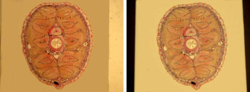
On the left is the brightfield image and on the right, the image with the crossed polars. There is certainly not an enormous difference but, the brightfield image is sharper and this brings us once again to that basic question: why go to the extra effort to use a technique that doesn’t seem to provide any real benefit in terms of microscopical information and, in some cases, seriously impedes any investigation?
Some might say that the motive is not important; I disagree. We’ll never really know what prompted this individual to take on such a project and expend considerable time, energy, and resources trying out this technique. However, if we stop and think about it, so many interesting discoveries and insights have arisen out of that wonderful, and largely hidden dimension of the human mind–curiosity. For every success, there are many blind alleys and failures. I know from my own experience that one can spend many frustrating hours, days, even weeks, pursuing a line of investigation that doesn’t succeed in producing the results one had hoped for. Nonetheless, in that process, one can still learn things both about oneself and about the methods of investigating a series of problems.
As far as this particular project of mounting specimens on mica strips is concerned, I suspect that the reason I have never encountered it before is that it was a dead end. However, that perverse element of curiosity embedded in my psyche has already begun to speculate about possibilities of modifying both the technique and the specimens to attempt to use this technique to produce some interesting new approaches. I guess when it comes to microscopy, I am an incurable optometrist, as Mrs. Malaprop would say.
All comments to the author Richard Howey are welcomed.
Note added August 10th 2012: Thank you to André Fuhrmann, Frankfurt who comments:
I read with much interest your paper on mounting sections
on mica. I believe that many mounters who did this did not think at all of
polarization. It was a common practice in microscopy classes that the preparator
would prepare large sections on mica. He would then walk from table to table
and cut a small piece for each student. The students would then mount the
pieces on slides. Typically in the student labs there were no polarizing
facilities at all. See e.g. this link describing the famous Tübingen
preparator Heidenhain at work:
http://www.mikroskopie-forum.de/index.php?topic=13210.0
I
dont know when mica ceased to be used for the purpose. But in the early 60s this
was still practiced at least in some places, as my father told me from his own
student days.
Editor's note: Visit Richard Howey's new website at http://rhowey.googlepages.com/home where he plans to share aspects of his wide interests.
Microscopy UK Front
Page
Micscape
Magazine
Article
Library
Published in the July 2012 edition of Micscape Magazine.
Please report any Web problems or offer general comments to the Micscape Editor .
Micscape is the on-line monthly magazine of the Microscopy UK website at Microscopy-UK .
©
Onview.net Ltd, Microscopy-UK, and all contributors 1995
onwards. All rights reserved. Main site is at
www.microscopy-uk.org.uk .