The following notes might help to redress this imbalance, though I have to stress that I am no expert in this matter, but offer some logical notions and experience which I believe to be pertinent to this issue, and hopefully might bring some insight into how our daily microscope observational routines might be improved from our eye's perspective.
The Eye
The eye, a wonderful
piece of organic engineering, together with the aid of the brain have perfected
the imaging of everyday views encountered over the various extremes of
landscape and climate for eons. What is not fully appreciated, is that
the eye is capable of detecting amazingly wide tonal ranges of light intensity,
such that photo cameras of all kinds fail miserably to emulate. How often
have we, in ignorance taken a photo of a very sunny and contrasty scene,
only to be disappointed with the resulting photo? Yet we saw more at the
time.
 |
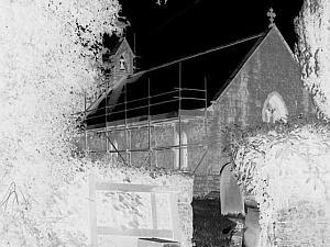
| Here is a typically contrasty
scene. The eye detected both the
extreme highlight detail in the roof slates, and also darker details than this digicam shot revealed in the foreground shadow areas. |
However limited
in an optical sense the eye may appear to be, it is an incontrovertible
fact that the images we see with it could not be as clear and information
saturated without the intervention of the image enhancing powers of our
minds, whose computational capacity is truly astonishing. The eye is also
capable of detecting extremely short duration light bursts in the order
of about 1/1000000th second or even less, though it must be said that its
ability to differentiate between tonal ranges of the darker shades is rather
poor. The positive and negative images demonstrate this effectively
below, in which the shadow areas in the foreground display more tonal information
when in negative form simply because our eyes are more sensitive to subtle
variations in lighter tones.
 |
 |
To understand in what circumstances the eye performs at its best, we should simply assess the conditions in which it has evolved. I shouldn't imagine that the scenes that existed many millions of years ago would be materially different to the ones we see today.
Basically, the eye focusses reflected light of varying colours and intensities off a multitude of objects, illuminated in mostly diffuse lighting conditions, and so it stands to reason that any instrument designed to display images to the eye should provide similar conditions to perform to its best.
Telescopes and binoculars simply gather and amplify the naturally lit scenes which we observe, and our eyes accept these images because they are effectively the same. The harsh fact is that the compound microscope does not provide idyllic visual imagery. In brightfield especially, which is very commonly used, the images we see are effectively silhouettes, from specimens made visible by either diffraction and or the blocking of light by varying degrees. Whilst this might seem rather simplistic, the fact is that we also observe specimens surrounded by, in many cases, large areas of bright field conditions, which will always cause the eye's iris to close down and so reduce the specimen's image intensity as a result. In short the total microscopic image in brightfield, can be far too contrasty and intense, lacking in tonal range in which circumstances the eye never evolved to deal with.
These are not natural circumstances for the eye, and we should therefore make some efforts to improve the situation if we can. Fortunately we can make an image more eye friendly, and there are several ways of improving matters.
Making the compound microscope image more eye friendly
To try to improve viewing by simply reducing illumination intensity will certainly feel more comfortable in many circumstances, but the inherent contrast is often still there, and all we will have achieved by reducing the source intensity is to lower the overall brightness of the image. This can however be beneficial, but not really rewarding.
It is of paramount importance that the illumination intensity suits the eye's optimum tonal range 'window'. This can only be arrived at through experimentation, but my experience has lead me to believe that illumination levels are far too intense on the majority of microscopes I have used over the years.
What we have to do is to broaden the tonal range by artificially introducing light by scattering or diffusion. Unfortunately the very word 'diffusion' which sounds so vague, causes alarm bells to ring amongst a number of microscopists, for this is unfortunately a contentious issue indeed. (See the author's Micscape article 'Illumination variants: Diffuse lighting'.)
The implementation of diffused light through the substage condenser using for instance ground or 'frosted' glass is very easy and convenient, but the way this affects the image is not so easy to explain, and I do not pretend to fully understand the way it improves the image in an eye friendly manner. I can guess that it not only dims the image by light scatter, causing much of the coherent light from the field condenser to be 'wasted', but also causes the light to become multidirectional so reducing diffraction effects and therefore providing images of specimens with finer boundary details much more in accord with their actual structure. Also since the light is scattered inside the condenser, any chromatic light separation caused in Abbe designs is effectively reduced and this in itself is, in my humble opinion an added bonus resulting from the use of ground glass diffusion techniques.
Just because the use of diffusers in optical systems might appear to nullify the designers skills and intentions, by scattering light in an apparently random fashion, does not mean that the resulting imagery is desultory and worthless, far from it. Ansel Adams, the large format photographer, made a case for images produced from uncoated camera lenses by reducing contrast, which can be a real problem with modern coated lenses. Similarly, brightfield imagery can be improved in a broadly similar way by diffusion, though I have to point out that not all brightfield conditions warrant this, and the observer has to experiment and find out which suits them best. As a rough guide diffuse illumination works well at lower powers, but is less effective in general terms above about x 200. But contrary to general opinion it does not reduce the resolving power of a fine objective, though photographically speaking it may appear to because contrast is reduced.
It must make at least theoretical if not practical sense to try to emulate imagery which our eyes have become accustomed to seeing, when using the microscope. How many observers take the trouble to try and improve imaging by just thinking for a while about what is going on ?
Surely our eyes deserve sympathy and some efforts to make imaging a pleasant and more natural experience, so here follows some simple tips on how to improve images.
Methods of making brightfield imagery more comfortable and enjoyable.
Image intensity levels are very important and need to be adjusted especially if too bright. I suspect that many beginning to take up microscopy might, understandably, use the substage iris control to reduce light intensity. Of course this can be done, but it causes much diffraction by thickening the specimen's detailed outlines and reduces resolution as well, though it increases depth of field and hides any shortcomings in the quality of the objective and eyepiece too. The better way is to use one of four simple methods :-
1) Reduce lighting intensity using the 'scopes control ( rheostat/ dimmer). This is very basic and unfortunately adds considerable amounts of colour, but nevertheless has its uses.
2) Using polarising filters in a pairing, ie both filters used above the field lens. By rotating just one we can attenuate the light considerably, but whilst there is little or no denigration of the image, the inherent contrast in the image will not change. Normal polarising 'scopes will elicit colouring from certain materials, simply because of the position of the polarising filters.
3) Graduated photographic intensity filters as used on cameras. These work very well but tend to be a little too severe or not as the case may be, since they are only made in 2 or 3 grades of light attenuation. Again the contrast is not reduced.
4) Groundglass ( OR translucent plastics ) as a diffusion aid which automatically scatters light so reducing the total input of light from the lamphouse to the condenser. I thoroughly like this method because it is cheap, but more importantly it also imparts qualities of viewing which I personally appreciate, and especially its eye friendly imaging. The iris adjustment with diffused lighting is as critical as in any other situation.
The iris diaphragm's aperture must always be adjusted to bring about a balance between the effects of diffraction and the resolving power of the objective, and in some circumstances the depth of field. It should never be used to reduce illumination intensity, which should be dealt with before the light reaches the condenser.
If you have never bothered to experiment with your lighting intensity other than using the dimmer or iris diaphragm, I recommend it heartily, and especially using groundglass .
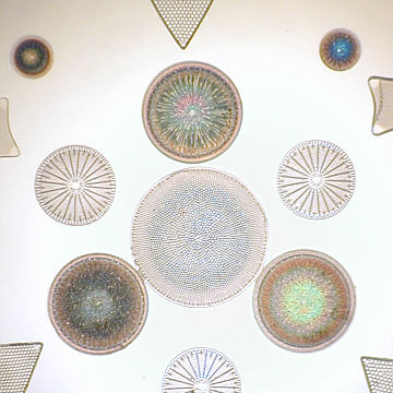
The following
images of diatoms demonstrate hopefully within the limits of my digicam
that diffuse illumination does not materially affect an image, but the
eyes are much happier!
 |
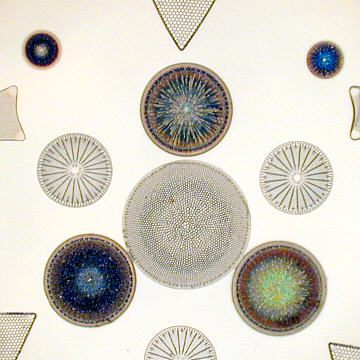 |
| Standard Brightfield | Diffused Brightfield
The diffraction colours are dependent
|
Darkfield observations are very enjoyable, but how many have reduced the light intensity whilst observing their favourite slides in a dark room as well? Sounds odd I know, but is probably the best way to observe with this technique. Ideally, the specimen should be only sufficiently illuminated to be seen comfortably, which many standard darkfield setups fail to do, as they tend to provide overly bright images which are far too contrasty for the eye and the camera.
All photo's taken with a Nikon Coolpix 800
digicam.
| All comments welcomed to Paul James. |
Microscopy
UK Front Page
Micscape
Magazine
Article
Library
© Microscopy UK or their contributors.
Please report any Web problems
or offer general comments to the Micscape
Editor,
via the contact on current
Micscape Index.
Micscape is the on-line monthly
magazine of the Microscopy UK web
site at Microscopy-UK