|
Micro-Hitchhikers: A Look At A Tropical Sea Urchin Spine by Richard L. Howey, Wyoming, USA |
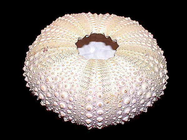
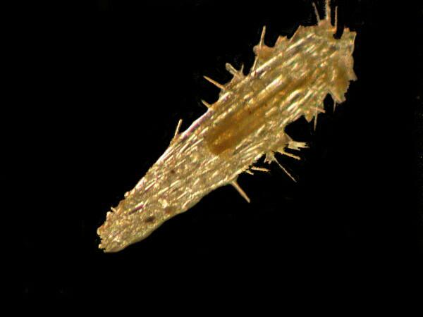
Recently I found a source in the Philippines for various sorts of dried sea life and most especially some wonderful sea urchins. One sizable specimen arrived with one spine dislodged. I like to order two specimens of each type, if possible, one for display and the other to slowly dismantle to study its structure bit by bit. I could have glued this spine back on and it would have made a perfectly fine display specimen, except in this case I was only able to obtain one. However, I set the spine aside for later examination, because it had some odd bumps and textures that didn’t look like they belonged to the sea urchin.
When you look at the surface of a sea urchin, it doesn’t seem to be a particularly inviting environment for creatures to try to colonize. Not only are there the larger spines in most species, but there are many smaller ones, and odd little projections from the hard calcareous plates that comprise the test (shell), not to mention the extraordinary pedicellariae. These are essentially defensive weapons designed to discourage and destroy any larvae that have an idea of settling in and occupying territory. These remarkable bits of natural technology possess elongated, fenestrated, calcareous plates that oppose each other to form “jaws”.
Some have only 2 plates, others have 3, 4 or even 5; some are hooked at the tips, other are relatively flat and some even possess poison glands. Also some are located almost directly on the surface, whereas other are stalked and have a thin calcareous rod running through the stalk as a support. Now, suppose some little larva of a sponge or barnacle or tunicate drops down looking for a nice protected spot to call home. The pedicellariae go into action and nip or crush the intruder, thus helping to keep the nuisances away. The pedicellariae are, however, of no help with regard to the spines because of the way they are positioned. How do they “know” that an there’s an invader? To me, one of the great joys of studying such organisms is that I am constantly encountering complex puzzles which force me to learn new things and to try to make rational deductions to explain such phenomena when there isn’t sufficient data to answer definitively.
Unless you’re fortunate enough to live near a sea coast, you’re most likely to encounter sea urchins that are dried and may have had their spines removed to show the intricacies of their tests. As a consequence, we often tend not to think of the fact that the surface in the living organisms is covered with a thin membrane which is very likely to have a complex network of cells which signal each other biochemically and biophysically. I suspect that there may also be some sensory receptors, probably tactile ones, associated with the membranes, but I don’t actually know that. In any case, something very intriguing happens. A tiny larva, let’s say one of a barnacle, come into the area where there are pedicellariae; its presence triggers a response from the pedicellariae and like some primitive micro-dinosaur, the jaws crush the larva.
But back to the spine. Sea urchin spines are attached to the test in a fascinating fashion. On the surface of the test, there are numerous smooth, hemispherical projections and, if you examine a spine, you will observe a depression at its base, so what we have is a classic ball and socket arrangement.
It is well-documented that when a sea urchin senses a threat, it will move the battery of spines in the direction of the perceived danger. Here’s another one of those intriguing puzzles; how does it perceive a potential threat? Part of the answer must involve biochemical sensors which detect specific compounds in the surrounding current that signal a potential predator. There may also be some sensors which are light/shadow sensitive, but these would be of use only at relatively shallow depths.
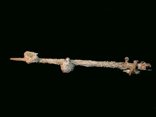
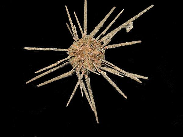
In almost all of the sea urchins I have ever had the opportunity to examine, the spines were mostly free of hitchhikers, so this one piqued my curiosity with its little bumps and protrusions. I went back and looked at the whole urchin and noticed that there are other spines which also have foreign items on them. So, on this original , single spine, here’s what I’ve found.
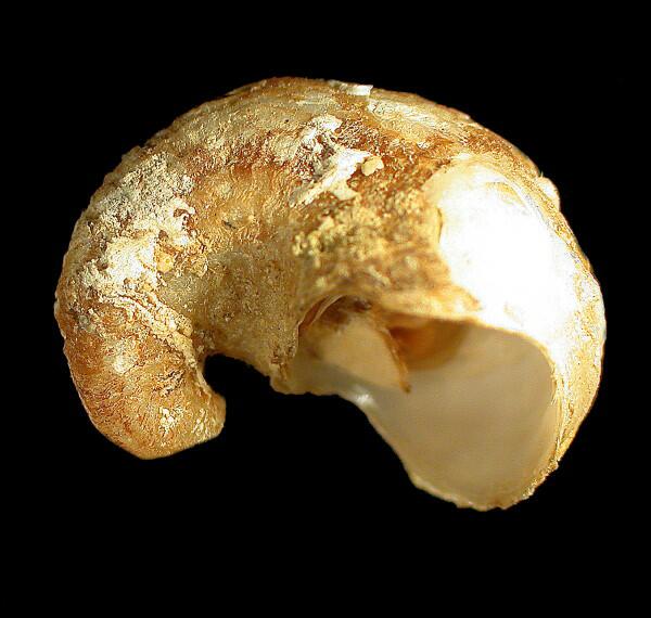
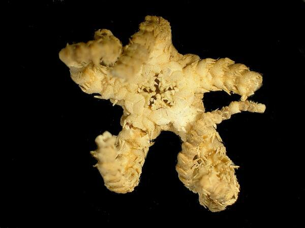
And here is a view of the urchin from which this spine came.
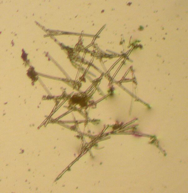
On the surface of the spine, in many areas, there is what appears to be a mesh of extremely thin rods which I suspect are from a sponge. When I scraped the surface and put some of the material on a slide in water and examined it at 400x, I found that they do indeed look like minute, clear, siliceous rods typical of many types of sponges. However, I also found what appears to be a tiny fenestrated spine typical of sea urchins. Furthermore, on this one there were some even smaller clear spines projecting from it and one longer one which seemed to have tiny bumps on it, as you can see from the image below.
Ah, these are the times when one might wish for a scanning electron microscope.
I took the slide back to my stereo-dissecting microscope and added a drop of dilute nitric acid. [CAUTION: Even in dilute solutions, nitric acid is a corrosive, dangerous, and highly reactive chemical which must be handled with great care.] As expected there was a good deal of bubbling on the slide as the acid began to attack and dissolve the calcareous material. However, to my surprise, the small spine with its clear needle-like, knobby spine had not dissolved after several minutes, so I decided to leave it overnight.
I’m also planning to take another sample and treat it with a 10% solution of hydrochloric acid. [Again, the same cautions apply!] I had originally intended to use this in the first place, since the results seem overall to be more reliable than those with nitric acid, but I couldn’t find my little dropper bottle of hydrochloric, so lazy twit that I am, I used a solution of nitric acid from a small bottle which I hadn’t even bothered to label with the concentration.
Now that’s inexcusably sloppy science, but hey, my excuse is that when I made up this solution, I was still teaching, going to deadly dull committee meetings, and reading dismal student essays, so I was very busy. Sorry, that’s still no excuse! Fifty lashes for the procrastinator. So to mend the error of my ways, I went to my chemical cabinet and made up a new dropper bottle of 10% hydrochloric and made some additional scrapings to treat with this solution. The results were conclusive and showed that the spicules of this sponge are silica.
Further down on the spine is a worm tube, possibly a serpulid, but not spirally coiled like Spirorbis. Rather this one rather meandered as though attempting to fit itself into the grooves along the length of the spine. It is smooth and white and looks like it is made of porcelain.
A bit further along is an interesting gastropod (snail) shell of a sort I’m not familiar with.
Yet further along, a barnacle of the Balanus type and on it are 2 small gastropod shells, 2 worm tubes, and an operculum from a third gastropod that got dislodged. Talk about maximal space utilization!
For those of you who are not snail fans, an operculum is a sort of built-in door which the snail carries around with it. (I was going to tell you that an operculum is a very short opera, but I decided against it.) The structure is composed of a hornlike substance and may possess calcareous layers as well. It is attached to the anterior end of the snail. If the snail is threatened, disturbed, or removed from water, it withdraws into its shell in such a way that the operculum seals the opening, thus helping to protect it from predators and to slow the process of dessication. Generally gastropods don’t attach to objects by means of opercula, but this kind apparently does, for when I pried the 2 shells from the spine, the opercula remained cemented to the spine’s surface, but the shells came off intact.
The barnacle was, however, an altogether different matter. I may be wrong, but I don’t think we humans have yet invented a glue that equals that of the barnacle. My attempts to remove the barnacle from the sea urchins spine resulted in a number of pieces, most of which fractured along the lines of the plates that comprise its “shell”. The barnacle is an extremely odd creature; it has thick calcareous plates which constitute its protective shell inside which it lives upside down. The “tentacles” which appear when it is feeding are, in fact, its feet, thus the name of the group to which it belongs–cirripedes or “curled feet”–so, it is a marine yoga master living its life standing on its head. The strange characteristics of barnacles are probably one of the reasons that Darwin spent 8 years studying them and produced a classic monograph on these organisms.
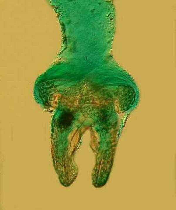
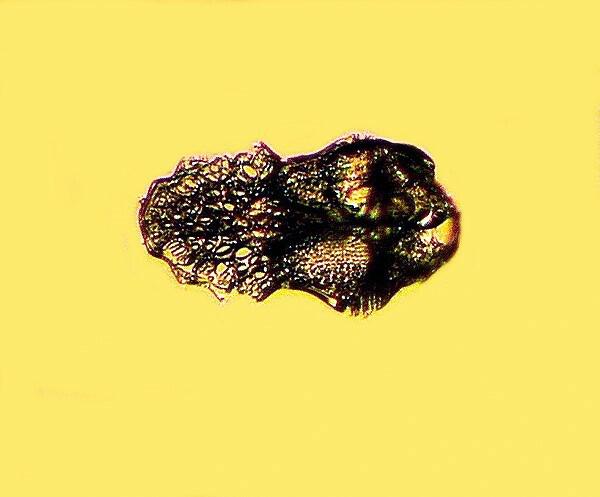
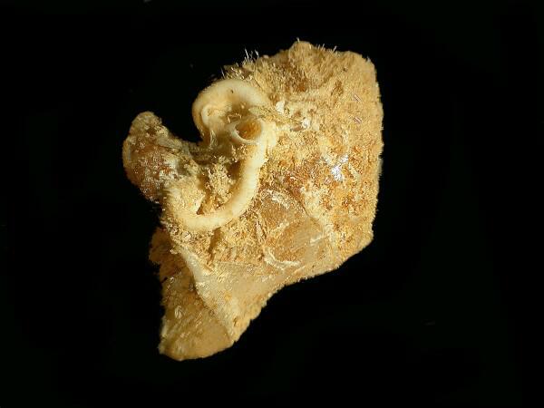
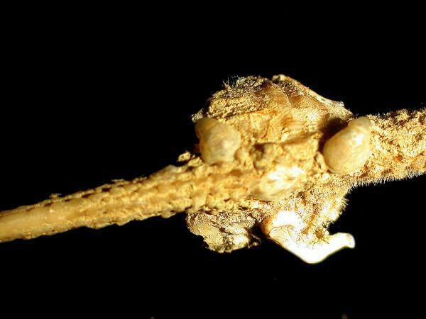
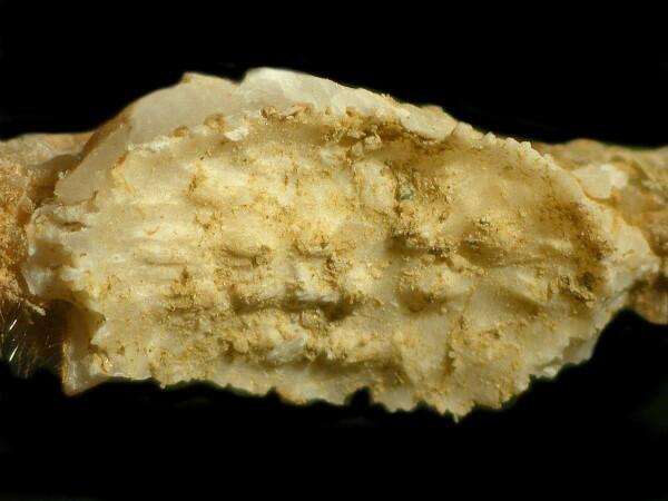
As I moved on down toward the base of the spine, I saw something very weird indeed. It looked like some miniature pieces of gray rope tied around the shaft of the spine. When I turned the spine to see the other side, I immediately recognized a wee brittle star embracing the spine.
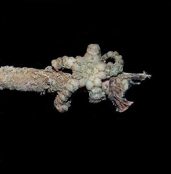
I had to break a couple of sections of the arms to remove it for closer examination. Here are 2 closeups.
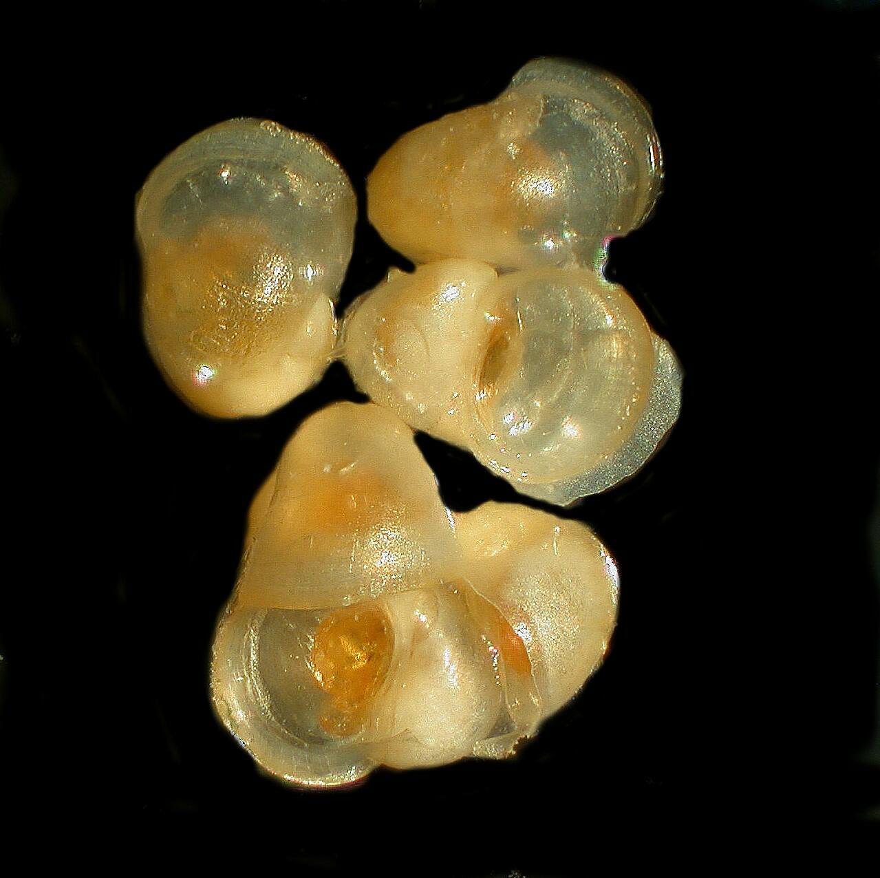
This is a view from the aboral or topside.
Here is the oral view and you can see in the center where the mouth is located.
I mentioned previously that my attempts to remove the barnacle from the spine cause the plates to break apart along the suture lines. Apparently this sponge just grows over everything in its path. The spicules are single acicular (needle-like) rods, some of which are slightly curved.
In the image above you see the result after my attempt to remove the barnacle; the basal plate by means of which it attached itself to the spine remained.
The adhesive which it secretes is a sort of super-marine epoxy. The only way to remove this plate from the spine would be either to chisel away the calcareous material of the spine itself or dissolve it out by the judicious use of a weak solution of acetic acid.
I mentioned 3 gastropods, 1 large one and 2 rather small ones and that these latter were attached to the barnacle.
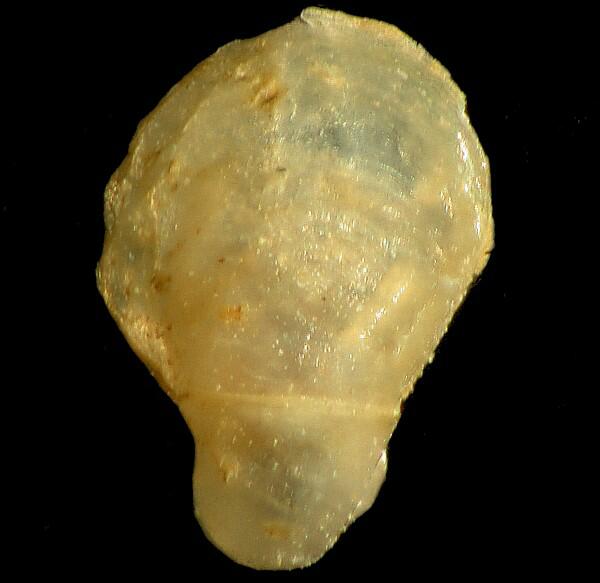
When I pried this one off of the spine, the operculum stayed attached and so I got to peer inside the snail shell and there were 2 clusters of baby snails, 8 in each cluster, being protected until they developed a bit more.
As I consider nature’s countless experiments, I am always amazed by the endless adaptations by means of which life exploits virtually every available niche. On the round, thin, and rather inhospitable surface of a single sea urchin spine–who would imagine that such a range of life forms could find refuge there?
ADDENDUM
Since I wrote this article, I have acquired some additional specimens of tropical sea urchins from the Philippines and I have found that the more elaborate the spine, the more likely it is to have hitchhikers. Some of these are both unusual and unexpected and I will be writing another article or two about these intriguing opportunists.
All comments to the author Richard Howey are welcomed.
Editor's notes: Visit Richard Howey's new website at http://rhowey.googlepages.com/home where he plans to share aspects of his wide interests.
Jean Marie Cavanihac's Micscape articles in July 2000 and May 2002 on sea urchins show some images and animations of pedicellariae.
Microscopy UK Front
Page
Micscape
Magazine
Article
Library
Please report any Web problems or offer general comments to the Micscape Editor .
Micscape is the on-line monthly magazine of the Microscopy UK website at Microscopy-UK .