Haupt's adhesive
Dissolve 1 g gelatine in 100 ml water at 30oC. Add 2 g phenol crystals and 15 ml glycerin. Stir, cool and filter.
I then place the sections in a desiccator, which contains formaldehyde vapour, and further helps adhere the sections, if left for 24 hours.
Sections are stained with haematoxylin and eosin.
When examining the sections, it must be remembered that the section may not always be exactly transverse to the body, so the parts will not be completely symmetrical as seen in a textbook.
On account of the thinness of the sections only parts of some structures will be included.
The series of sections shown are a TS of the body at segment 10, which is in the reproductive region and shows the lateral vesiculae seminales and the seminal funnels.
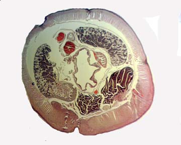
Section 1
The second shows the body wall consisting of the epithelium, circular muscles and the feathery longitudinal muscles.
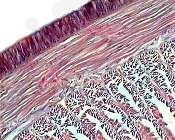
Section 2
The third section is of the nerve cord. This shows the giant fibre, the lateral fibres, the lateral neural blood vessels and the large sub-neural blood vessel.
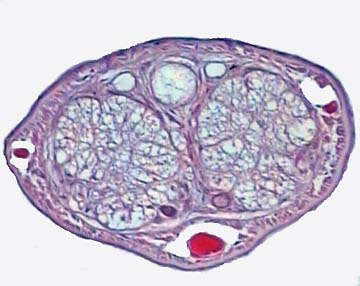
Section 3
Sections 4 and 5 show the seminal vesicles which are filled with the sperm morulae and spermatozoa.
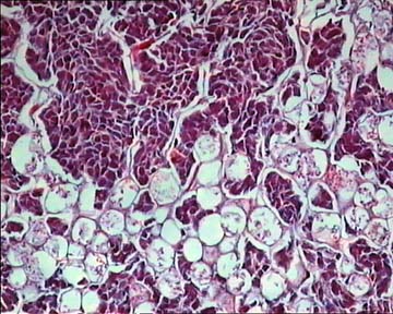
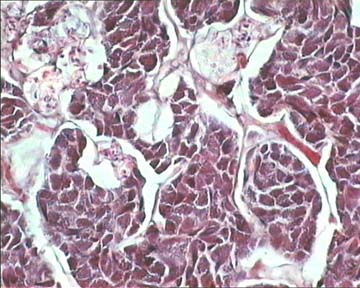
Sections 4 and 5
Lately, I have been having difficulty in finding decent sized worms in the wild for processing. I have found for mature worms that Blades Biological is a good supplier, with an on-line catalogue: www.blades-bio.co.uk. Either live or preserved specimens are available.