by |
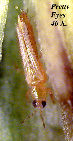
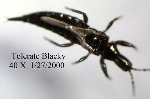
I recently built my own fiber-optics light source, (see Micscape article January 2000), and it is fun exploring different ways of applying this form of lighting. Now I can use it as an immediate source of incident light for my compound microscope, just like the QX3 Intel Play microscope which I also own. Incident lighting is good for low power observations and photomicrography of opaque subjects, like small insects.
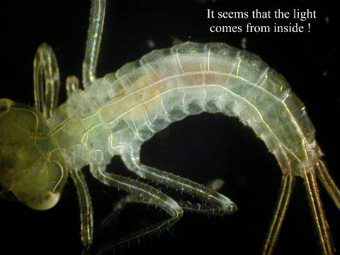
Looking at pond life under the compound microscope, I have found a way to create dark-field illumination with the fiber-optics i.e. illuminate the animals on a black background and make them stand out very brightly, so they look as if they where lit up from the inside. (See image gallery below).
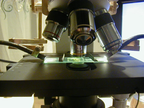
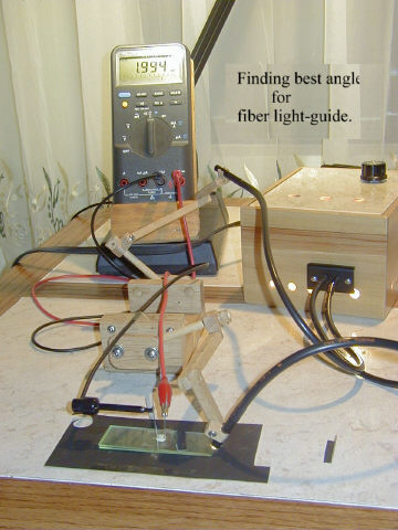
To obtain this dark-field effect I switch off the transmitted light of my compound microscope, and point the two fiber light-guides in a ca. 43-44° angle at the ends of a cavity microscope slide (see above). This is the most efficient angle, but a little off will still work. With a photosensitive resistor connected to my ohm-meter (see right) I tried various angles, and this is the best set-up I found.
In order to place the light-guides down onto the stage and touch the slide, a small spacer is needed on my mechanical stage clip. A 10 x 3 x 3 mm piece of aluminum or just a piece of match-stick, taped to the stationary part of the slide clip gives enough room.
An observation on using the Olympus D-450 Zoom digital camera.
Taking photomicrographs with this camera is very practical, once I found out how to do it. I do not want to profess expertise, but after many trials to get the focus right, I noticed that the camera gets tired of being switched on too long. When it is freshly switched on, I obtain a very nice sharp picture, but later on I was unable to repeat that. I blame it perhaps on the CCD in the camera getting too warm, and then even when it is focused right, the picture is not sharp at all.
The remedy I have tried is to plug the camera's AC-adapter into an outlet with an on/off switch, and use no batteries in the camera. That way I keep it switched off most of the time, and not using the cameras on/off switch, the lens stays extended, and when the power comes back on, it is instantly ready for use, but the LCD has to be turned on again.
Incident illumination of two insects.
The video connection to a TV set lets me see what the camera sees, and I don't have to look at the small LCD screen on the back; I wish this could be done with the computer monitor, because the TV shows all these lines, and it is sometimes hard to focus exactly (see Editor's footnote).
It is wonderful to use a digital camera system, because I can take many pictures and then use only the best ones, deleting the rest and all that at no extra cost and instantly I can see the results of my experiments.
Comments to the author Rudolf Baumueller welcomed.
(Editor's footnote: Installing a video capture card/TV card in a PC enables the computer monitor to display the video signal from the digital camera for critical focusing. Dedicated video monitors can be expensive and the PC card with video input offers a cheaper way of using the PC's usually high quality monitor.)
Image gallery
Aquatic insect larva (mayfly?) under dark-field illumination
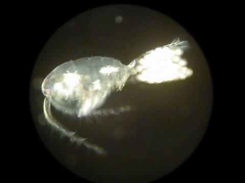
Dark-field illumination of a female copepod with egg sacs.This is an example of the camera changing the focus, when it is on for a long time. On the screen, this was in perfect focus, but snapping the picture, it changed for the worse.
Microscopy UK Front Page
Micscape Magazine
Article Library
© Microscopy UK or their contributors.
Published in the March 2000 edition of Micscape Magazine.
Please report any Web problems or offer general comments to the Micscape Editor,
via the contact on current Micscape Index.Micscape is the on-line monthly magazine of the Microscopy UK web
site at Microscopy-UK