|
Part II by Roland Mortimer,
|
 |
Part I of this series was presented in Micscape, February 2000.
When looking at diatom frustules in dark-field illumination one rapidly discovers that dark-field does not show as much detail in some diatoms as other types of illumination do. This is mainly true when looking at rather thick, rounded specimens such as Biddulphia.
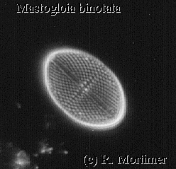
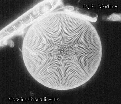
| The image of Mastogloia (right) only shows the illuminated surface whereas in transmitted light or phase contrast one can see the two elongated chambers which run along the sides of the frustule, one each side. In the image of Coscinodiscus (below) there is also an image of Climacosphenia moniligera in valve view (the elongated diatom with an internal 'ladder'). |
 |
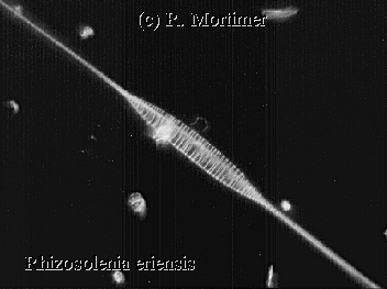
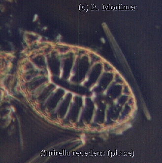
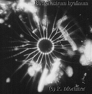
The following images were taken of specimens found near Gua¡ba island, Rio de Janeiro at a depth of about 12 metres, water temperature being around 20ºC. The island is used as an iron ore deposit and terminal for loading the same onto ships for export. Obviously the loading of ships with ore produces clouds of mineral dust which finally deposits into the surrounding sea area. This area is particularly rich in diatom species as compared to other parts of the region.
 |
 |
I have taken many photographs of the types
found here but obviously too many to include in such a small space as an
article of this type. The most difficult part in writing such an article,
as can be appreciated by anyone who has attempted a similar article, is
the identification of the diatoms especially when one is an amateur and
has limited literature to which he can refer. Therefore, any errors found
in diatom identification would be much appreciated.
 |
 |
I would like once again to thank Klaus Kemp of Microlife services for help in identification and Osman Medeiros Neto for braving the depths of the South Atlantic and collecting the samples for me.