Image gallery:
Images captured with an Olympus C-2040z 
digicam on a microscope

by David Young, Iowa City, USA


Hello. Enclosed is an assortment of images I took over the winter. The homemade adapter for taking these images has been described in my earlier article 'Adapting an Olympus C-2040z digicam to a microscope'. A variety of techniques were used including Rheinberg, polar and oblique illumination as well as some image montages. These details are mentioned in the image captions.

All images where photographed using an AO Spencer monocular microscope with Abbe condenser and a desk lamp as a light source.  I used the 10x objective with a relay lens in my home made adapter which gives an additional 10x magnification or slightly less.

The slides were homemade; some notes on their preparation and obtaining supplies are as follows:
Fructose has been recommended as a mounting medium. An alternative to fructose if it's hard to source locally is corn syrup which works very well as a mounting medium.  It can be used straight from the bottle or diluted with water and is easier if not a bit cheaper to obtain than fructose, which tends to be sold only in specialized stores.  (Corn syrup is mainly fructose sugar.)
 
A source of blank slides, cover slips and lenses is American Science and Surplus who sells bulk slides and cover slips.  A box of 100 cover slips costs about $1-50 and they charge about $5 for a box of 75 blank slides. (The slides come individually wrapped in plastic sleeves and are clean and ready to use. They are about the best I've found anywhere!)    They also sell lenses, magnifiers and complete microscopes. They do have a minimum order of $10 but shipping is reasonable starting at about $6 or so. They do only ship within the United States. Their web address is http://www.sciplus.com.

Comments to the author David Young are welcomed.
 

Images by the author.

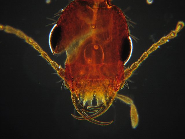
Image 1: A beetle with rather impressive jaws. Darkfield illumination.
The species is uncertain; it may possibly be one of the rove beetles. The whole beetle was about 1/3 of an inch long with a very slender body. (It was almost snake-like in form.) 

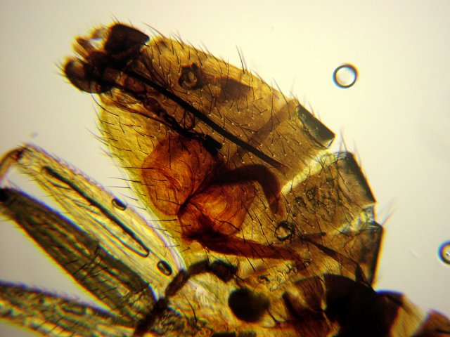
Image 2: Fly abdomen. Brightfield illumination.
The fly species is uncertain. It was about 1/8 to 1/6 inch long and had the overall body form of a fruit fly. Though definitely not the coloration of a typical fruit fly.

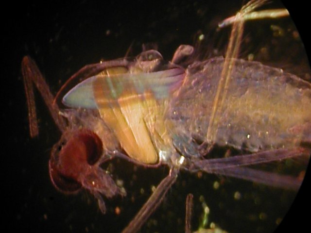
Image 3: Fly head. Crossed polars and darkfield illumination.

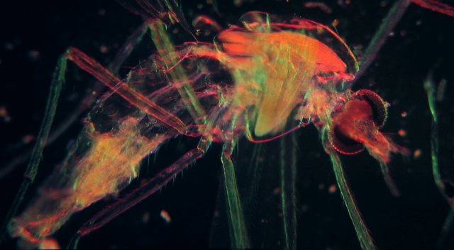
Image 4: Fly. Rheinberg illumination.
Images 4 and 5-7 were made using two or more separate images, which I then stitched together using the software program Camedia (which shipped with my Olympus camera and has a function for creating panoramas).  There are other similar programs that do the same thing. I basically took a picture, then moved the slide a little and took another picture and so on.  Until the entire specimen had been photographed.   I then just ran the images through the Camedia program to stitch the separate parts together.

Image 5: Stinger of the western harvester worker ant. Oblique illumination.

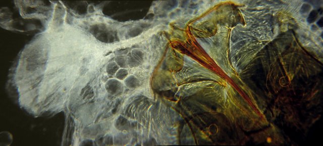
Image 6: Stinger of the western harvester worker ant. Darkfield illumination.

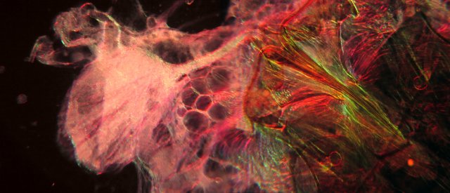
Image 7: Stinger of the western harvester worker ant. Rheinberg illumination.

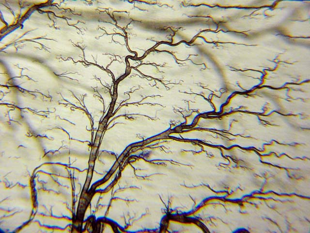
Image 8: Tracheal system of a mealworm. (Larva of a beetle; often sold at pet stores for feeding birds, small lizards etc). Oblique illumination. Optical mag. 100x.

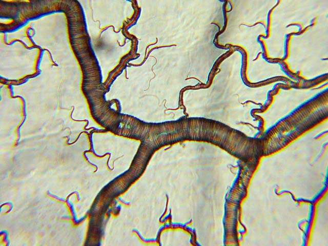
Image 9: Tracheal system of a mealworm. Dark field illumination. Optical mag. 430x.


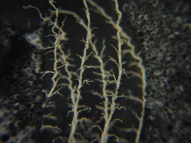
Image 10: Tracheal system of a mealworm. Dark field illumination. Optical mag. 100x.

Microscopy UK Front Page
Micscape Magazine
Article Library


© Microscopy UK or their contributors.

Published in the February 2002 edition of Micscape Magazine.

Please report any Web problems or offer general comments to the Micscape Editor,
via the contact on current Micscape Index.

Micscape is the on-line monthly magazine of the Microscopy UK web
site at Microscopy-UK

WIDTH=1


© Onview.net Ltd, Microscopy-UK, and all contributors 1995 onwards. All rights reserved. Main site is at www.microscopy-uk.org.uk with full mirror at www.microscopy-uk.net.