...The Strange And The Beautiful...
By Ian Walker. UK.
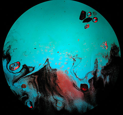
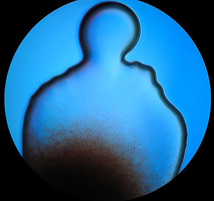
Areas around the cover glass edges, bubbles, drops of oil and poorly preserved specimens can all look interesting under the microscope, especially with dark-field illumination or crossed-polars. Sometimes you can see whole 'scenes' like the four shown after the first image.
....The next time you put that old slide down, have another look you may be surprised at what you might see....
So turn down the lights and enter the miniature world of bubbles, crystals and other strange things.
|
|
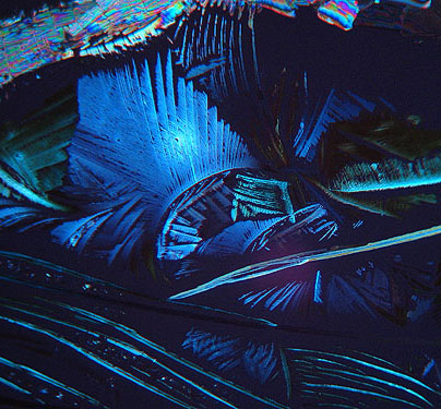
A tiny crystal of Vitamin C under crossed-polars and blue filter, 400x.
Slide prepared by Mike Samworth.
All images taken with the Leica CME microscope and Canon Ixus 400 digital camera using the remote control software, all images best viewed in screen resolutions greater than 1024x768 pixels.
|
|
|
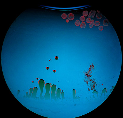
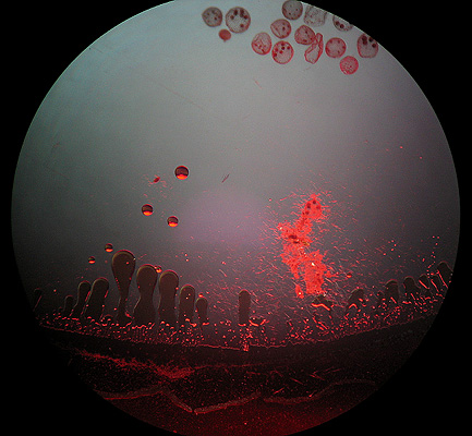
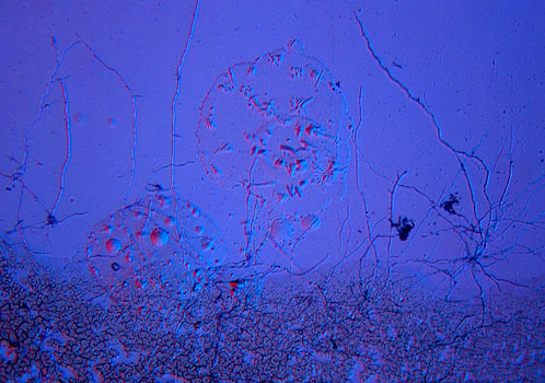
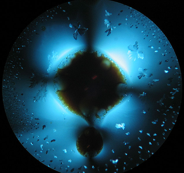
Looking at the very edge of a slide preparation, the tiny creatures at the top are Volvox [the main subject matter of the slide] with the left image capturing part of a bubble where the slide has dried out. I am not certain what the 'globules' are but another Volvox is entombed in a network of filamentous debris. The two images of the same scene show the dramatic changes that different lighting and contrast techniques can create.
Liquid mount, unknown mounter.
|
|
|
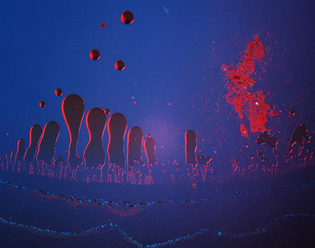
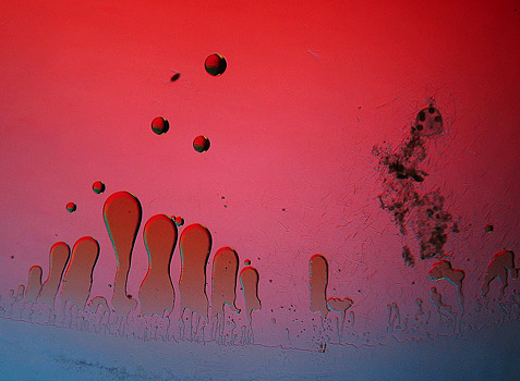
Two more variations using different lighting techniques, this time with the Canon Ixus 400 at a higher zoom level.
|
|
The edge of a 'jewel sand' slide under dark-field and Rheinberg illumination.
Slide prepared by J. Percival Yates.
|
|
A micro-bubble! 400x.
|
|
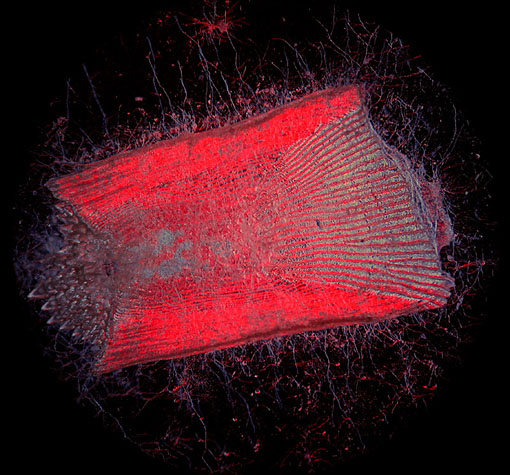
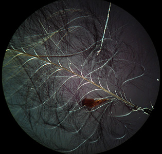
This old slide of the scale of a sole fish attacked by fungi looks particularly poor under bright-field illumination and could easily be put to the bottom of a slide collection, but it is these very slides where I seek out the unusual. Here the slide comes to life with Rheinberg illumination. I took several of these images using different filters and contrast techniques and each has its own unique character.
Old blue paper covered slide by an unknown mounter.
|
|
|
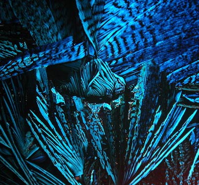
Glycine, 40x.
Glycine is the simplest naturally occurring amino acid, it is a sweet tasting crystalline compound found in proteins and is used in the food industry. There are more than 100 amino acids occurring naturally but only about twenty of these are used in building the proteins found in living organisms, we all share these - from the simplest forms of life to plants and animals. Although Glycine is one of the non-essential amino acids required by humans [it is not required from our diet and may be synthesized within the body] it shares the properties of the organic molecules that make up the group and seems fitting that such complex and beautiful crystals seen under the microscope should reflect the diversity of life we see around us.
A Biosil slide.
|
|
Another fungal attack on the same series of blue paper covered slides as the scale of a sole fish, this time on a slide marked 'Blood Globules', Rheinberg illumination.
Old paper covered slide by an unknown mounter.
And now for some more conventional images...
|
|
|
|
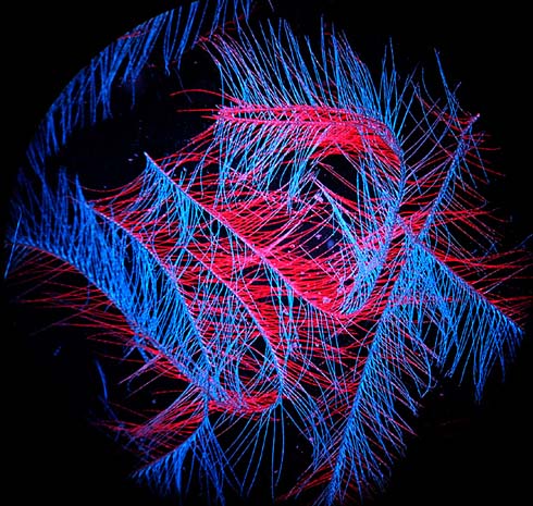
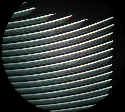
Three feathers. On the left robin, in the centre cockatiel and on the right cardinal bird, all crossed-polars at 40x.
Biosil slides.
|
|
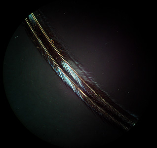
Feathers from the eider duck
Old blue paper covered slide by an unknown mounter.
|
|
|
On the left Sugar [Bush]? seed hairs under dark-field illumination plus Rheinberg filter and right, the same image under crossed-polars, both 40x.
A Biosil slide.
|
|
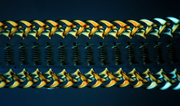
Palate of a whelk, 40x.
Slide mounted by Wheeler.
|
|
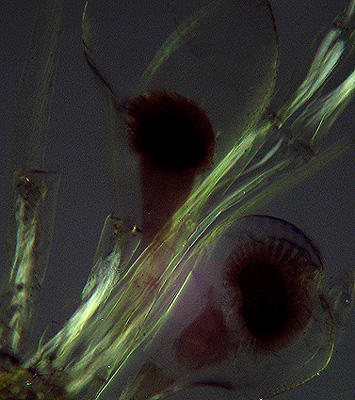
Head section of the larval stage of a marine crustacean, crossed-polars, 100x.
Mounted by John Atkinson.
|
|
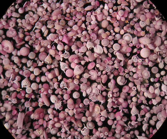
Reflected light image of Polycistina from Barbados, a composite from four images using Combine Z4.2 stacking software by Alan Hadley, 40x.
As with a lot of these older opaque slides the subjects are covered with a cover slip, unfortunately these tend to 'fog' and/or crack over many years and together with reflections from the upper surface when using my lamp, reduces the contrast of the image.
Unknown mounter.
|
|
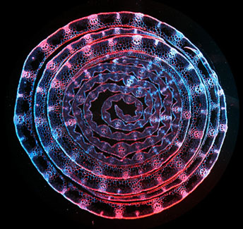
Reflected light image of Isthmia nervosa diatoms, 40x.
An excellent but damaged slide mounted by Richard Suter, careful use of Photoshop Elements has brought it back to life!
|
|
A small fossil shell from Easter Island using reflected light. A composite from five images using Combine Z4.2 stacking software by Alan Hadley, 40x.
Easter Island also known as Rapa Nui is a remote island in the Pacific Ocean about 2,200 miles west of Chile, formed by a series of underwater volcanic eruptions. It has an area of about 63 square miles and is famous for its massive monolithic stone statues of which some weigh more than 50 tons.
Slide prepared by Dr. R. S. Pyne.
|
|
|
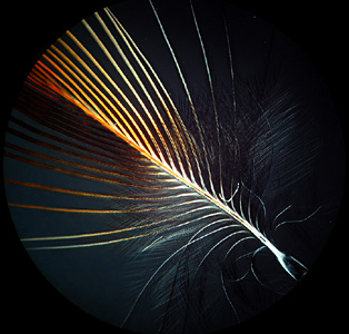
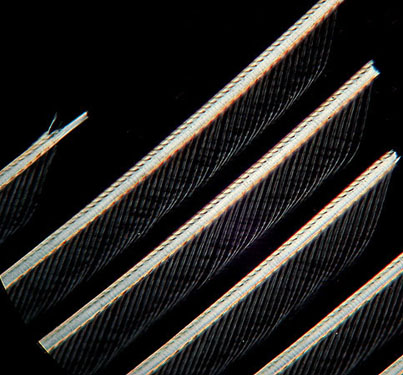
The rigid patterns of feathers like this one from a house martin can create a striking effect, left image at 40x, right image at 100x.
A Biosil slide.
|
|
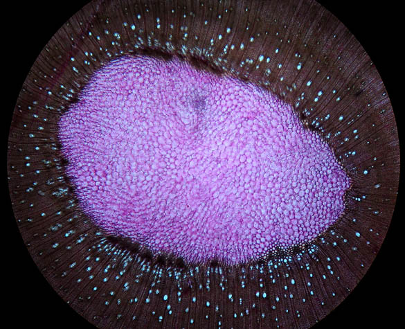
Section through the leaf-bud of wheat, dark-field illumination plus Rheinberg filter, 40x.
A slide prepared by Hornell, Biological Station Jersey.
|
|
Dark-field illumination plus blue filter, 40x.
Slide by an unknown mounter, cross-section of the stem of Fraxinus excelsior, European Ash tree.
|
|
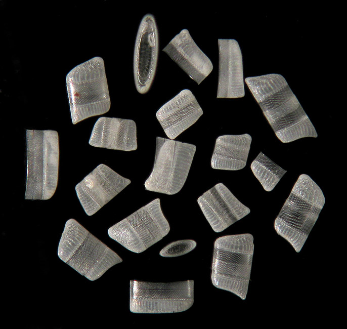
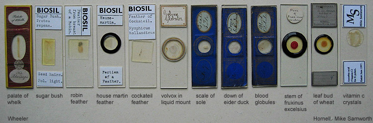
Some of the slides used in this article, the spelling of Fruxinus excelsius third slide from the right is incorrect [see the last image above], it's how I read the mounters writing!
If you enjoyed the images in this article, the author has presented a further selection in his January 2005 article 'Strange Worlds'.
The End.
Comments to the author, Ian Walker, are welcomed.
Please report any Web problems or offer general comments to the Micscape Editor.
Micscape is the on-line monthly magazine of the Microscopy
UK web
site at Microscopy-UK