The fascinating world of rotifers

(PAGE 1)

By Jean-Marie Cavanihac

France


When we first start to observe microscopic life, freshwater critters are easy to find and convenient to study because slide preparation is minimal. The liquid medium gives perfect observation conditions, particularly if you make 'micro aquarium' slides (see article in the Micscape library). You can find freshwater life in ponds, small quiet streams, aquariums, fountains in public parks...and even in an old flower jar full of rainwater long forgotten in a corner of your garden!

The most frequently encountered inhabitants of fresh water are protozoa which move rapidly and are a little difficult for beginners to observe. But we have too, other little friends: e.g. rotifers which are often attached to filamentous algae or plantdebris and allow us to admire their delicate and transparent bodies! Maybe it's a little coquetry because rotifers are mostly parthenogenic females, which produce eggs, giving... females too. Males appear only when their environment becomes adverse; probably to mix genetic material.

Rotifers are fascinating because, (as the origin of their name suggests), they have two rotating crowns of cilia on their head. Their nickname is 'wheel animalcules' which describes well their appearance. Not all species have this crown and some have reduced cilia and appear to have a sort of moustache. The crowns are also used for locomotion. A rotifer's foot has a gland which secretes a sort of glue which allows them to adhere to algae for example. Another surprising organ is the "mastax" used to chew food particles and which is in continuously motion. The shape of the trophi (jaws of the mastax) is used to help identify species.

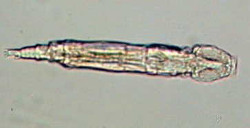
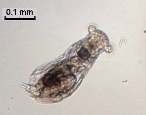
Shown right are two animated gifs. The first is a little 'family' of Philodina. The second shows the mastax in a Brachionus.


Click HERE if you want to see a video clip showing Brachionus eating a diatom! (2 Mbytes avi file.)

Rotifers have the ability to slow down their metabolism in severe conditions, and to retract to form a sort of cyst which is very resistant to long dry periods! If you add water to a cyst and some nutriments, a rotifer begins its life again!

Don't try to make permanent slides! Rotifers contract strongly with fixative agents and become hardly recognizable, and, thus it would be a pity to kill such attractive critters!


But you are probably impatient to see what these creatures look like, so below is a small gallery of a selection of the most common types. Note that marine rotifers also exist, but there are fewer species than in fresh water.

One of the most frequently encountered rotifers in moss: Rotifer vulgaris. It moves like a caterpillar! (Picture taken with x15 objective.)

Another common rotifer: Philodina showing its 'wheels'. The mastax is the clear spot at the middle of the body.

Taken with a x15 objective.


Another two common but small rotifers, Lecane and Lepidella (x40 objective).


Rotifers in the Brachionus genus occur in various shapes. It is often 'cultivated' in fish farms to feed young fishes. This specimen is shown carrying its three eggs. Note the red spot which is a light sensitive organ (X15 objective).


A common rotifer Euchlanis; it's quiet and easy to observe (x15 objective).

A less common rotifer in the genus Synchaeta: shown here are two marine specimens (left and below). Note the transparency of the second individual and sensitive 'bristle' on their heads.

(A marine specimen taken with x15 objective.)


Mytilina showing its 'horns' (X15 objective).


Keratella, a less common genus with species of different shapes. The left hand rotifer is probably Keratella hiemalis.

I am not sure of the identification of this rotifer; probably Monostyla (X15 objective) on the left hand side and Adineta vaga on the right hand side.

A predator for other rotifers: Cupelopagis vorax! (x6,3 objective)

GO TO PAGE 2 : for other intriguing species !


Comments to the author Jean-Marie Cavanihac are welcomed.

Microscopy UK Front Page
Micscape Magazine
Article Library


All drawings and photographs © Jean-Marie Cavanihac 2004

Published in the March 2004 edition of Micscape Magazine.

Please report any Web problems or offer general comments to the Micscape Editor,
via the contact on current Micscape Index.

Micscape is the on-line monthly magazine of the Microscopy UK web
site at http://www.microscopy-uk.org.uk