A
personal review of the Moticam 1000 digital microscope camera.
by David Walker, UK
Additional stills and video added March 16th 2005.
March
2008: Please note that this review refers to the 2005 model. See Conclusion for
an update.
November
2008: Updated
with some images to clarify assembly procedure. See footnote.
I recently had an opportunity to assess the Moticam 1000 digital microscope camera and share my observations below as a hobbyist. The Moticam 1000 is apparently one of the cheapest dedicated cameras with >1 Megapixel sensor (uncooled) and in a price bracket of many consumer digicams, so may be of interest to fellow enthusiasts who are considering the pros and cons of purchasing a dedicated camera/software cf adapting a consumer digicam or using a webcam.
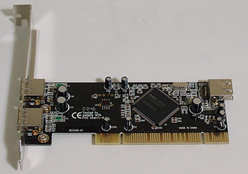
The maker's specifications and other details can be seen on the Motic (N. America) website which states that it uses a 1.3 megapixel (1240x1024) 1/2 inch 'progressive scan' CMOS sensor with a minimum illumination of 3 lux. It interfaces to a PC and is powered by a USB 2.0 interface. A USB 2.0 PCI card is supplied if PC doesn't have USB (it must be USB 2.0). It typically costs £290+VAT in the UK and $399 in the US.
The author's assessments were with a desktop PC with 1.6GHz AMD Sempron CPU, 512MB RAM and Windows XP. No problems were experienced in set-up or use. (See link 1 for notes on use with a laptop.)
Out of the box
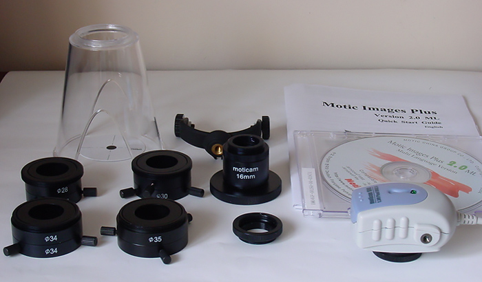
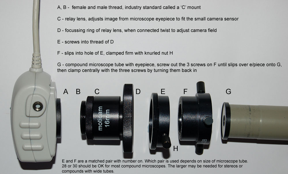
The
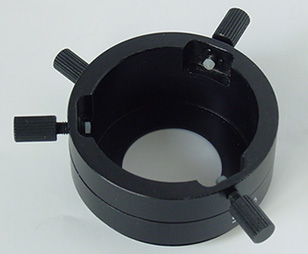
box includes: Moticam 1000 with USB 2.0 cable. Four centring mounts 28 mm, 30 mm,
34 mm, 35 mm with quick release. 16 mm focusable relay lens. Professional calibration
slide and USB 2.0 PCI card. Macro stand with calibration disc and camera support
to attach to a tripod. 'Motic Images Plus Version 2.0 ML' multilanguage software
on CD-ROM with 'Quick Start Guide' booklet.
As the pictures above show, the outfit comes well equipped with a number of items in addition to the camera / software. Some of these are quite expensive when bought separately (i.e. micrometer slide, dedicated relay lens, four metal adaptors) and must be factored in when assessing the unit's overall value for money.
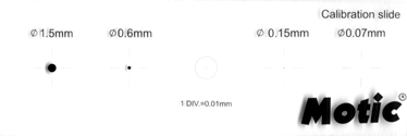
Four eyepiece tube adaptors are supplied with centring rings which are of machined anodised alloy and made to a good standard; these can cost £50+ each for an equivalent e.g. for the Coolpix series. The micrometer slide has calibrated spots and a 0.01 mm central reticule and is made to a good standard (ca. £20+ for third party examples).
|
|
|
|
|
|
|
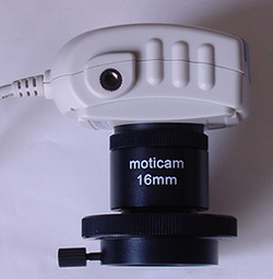
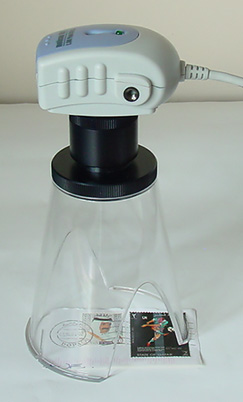
Right: Camera on the supplied macro stand with 16 mm lens. |
|
Editing and capture software
|
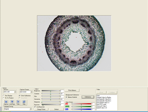
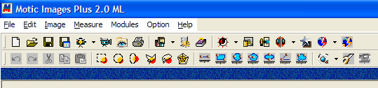
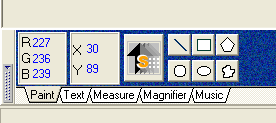
Motic Images Plus 2.0 multilingual software: Above is the main screen top menu bar and bottom left menu. Below is the full screen. This is standalone image editing software as well as the interface for image capture. Most of the editing features will be familiar to users of photo-editing software. To start image/still capture the video camera icon is clicked. This opens the separate capture screen shown at 'Image capture software' below.
|
|
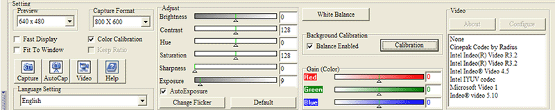
Image capture software: The main capture screen of the software is shown below. The preview is set at 640x480 here but can be set up to the full capture size of 1240x1024. A grey button bottom left allows full screen preview for larger preview settings.
A close-up of the capture parameter features is shown above. The preview and capture areas can be individually set but only certain combinations possible. e.g. for 1240x1024 capture the preview must also be set to this. Most features are self explanatory. The Fast Display box allows instant screen refresh for image focussing etc. When image set, turn off to capture. Background Calibration - this is a neat feature that allows a background e.g. of uneven lighting to be subtracted from picture before the subject is introduced into the capture area. See example below of unretouched captures, which can almost seamlessly remove the eyepiece field of view limit.
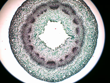
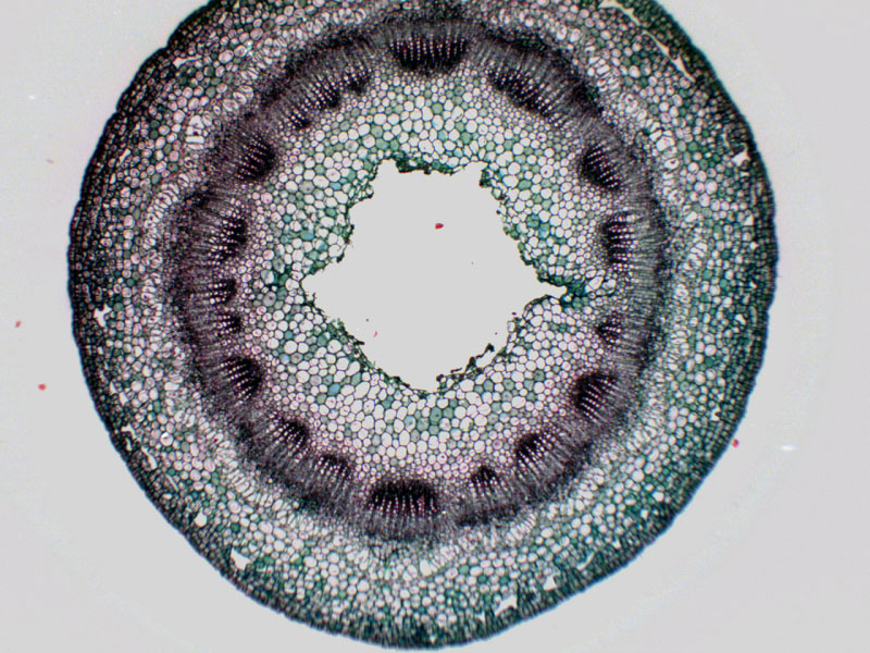
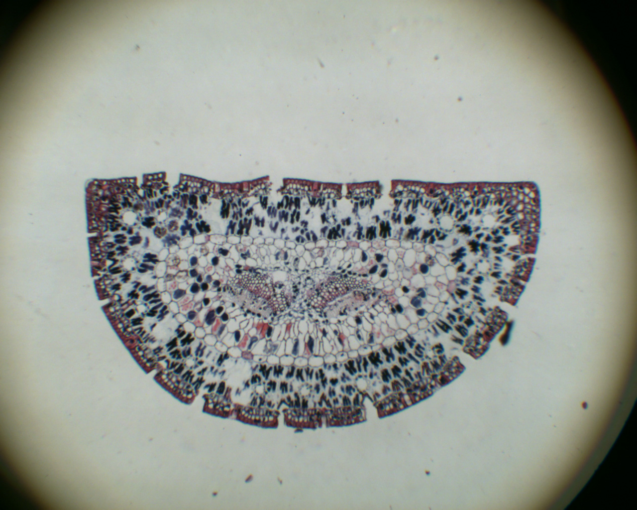
Above: T/S honeysuckle leaf, Biosil slide. LOMO 3.5x planachro, 5x Baker eyepiece, 1.5x bino' head, Moticam with its relay lens. Unretouched captures apart from resize. Capturing evenly lit images at these low mags is usually tricky on the author's scope. The background subtraction feature is particularly handy as here at the lowest mag settings. (Click r/hand image for 800x600 master). The relay lens also allow full subject capture of the eyepiece field which isn't possible with the author's consumer digicam (Sony S75). |
|
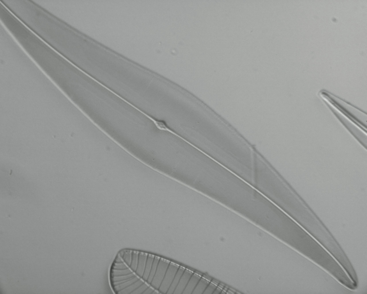
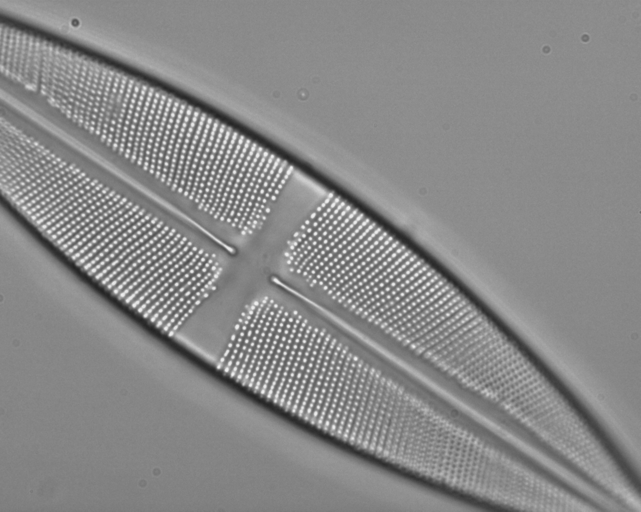
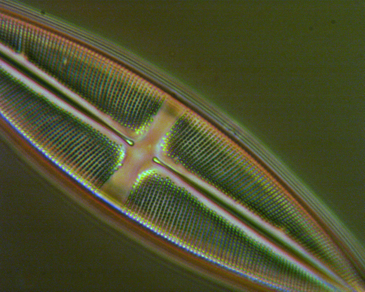
Critical
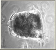
focus and fine detail capture test: Diatom Pleurosigma
angulatum. Klaus Kemp 'eight form' test slide. Taken
with LOMO 20x NA0.65 apo., no eyepiece, 5 mm oblique with LOMO aplanatic condenser.
This excellent objective can resolve the detail of this diatom but
it is on the borderline of my visual acuity with a normal eyepiece
and focus is critical, so a good test for image capture. The camera
exposure works well with these even toned type subjects. |
|
Sensitivity: The author used the HLS-1 dedicated 6V/20W halogen base lamp for the LOMO Biolam. This is a typical microscope lamp but the non-removable built-in diffuser does reduce the bulb's potential light level. For set-ups where the lighting was more demanding e.g. phase and cross-polar studies the 3 lux sensitivity of the camera did struggle when noise increased. For users with more efficient microscope lamps this may not be a factor. For higher magnification brightfield work a 6V/15W tungsten lamp without diffuser was used with 60x and 100x high NA objectives with plenty of camera sensitivity. The sensor does give noisier images than the most modern consumer digicams if the light is less than ideal, but can benefit from noise reduction programs like 'Neat Image' which the author already uses for noise reduction in images from the author's now ageing Sony S75 digicam. |
|
Selection
of images A small selection of images are shown below. This may be expanded with examples of different subjects / lighting techniques during the month when time permits.
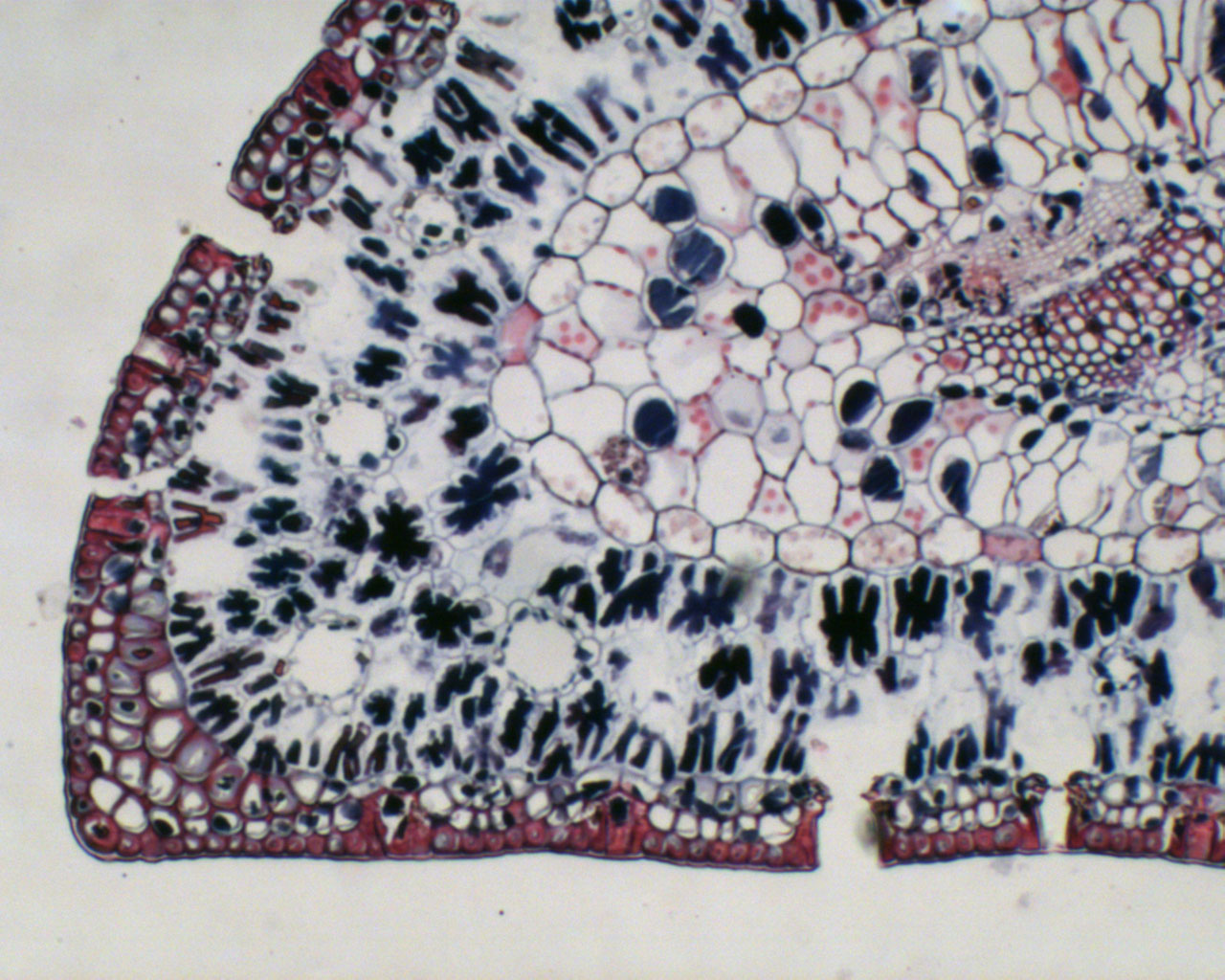
T/S stained maritime pine leaf, Biosil slide. LOMO 3.5x/0.10 planachro. Left: with 7x eyepiece and Motic relay lens, cropped from eyepiece f.o.v vignette (master shows it). Right: same objective using camera in tube without relay lens and eyepiece. The ability to rapidly change between almost full eyepiece field to some enlargement without changing objective is very useful. With the LOMO optics the Motic relay lens emphasises somewhat edge chromatic aberrations. At low mags like these the camera, like many diggies, is unforgiving of uneven field illumination. A large source 60W enlarger lamp was used. Focus / lighting / dynamic range: The ability to focus very accurately with no video lag makes the Moticam 1000 particularly suited to imaging tricky subjects like diatoms where the exact focal plane required can be chosen. The auto exposure / dynamic range copes well with even toned subjects like Stauroneis phoenocenteron below left in brightfield. But when the same objective was used in phase, below right, the highlights tend to burn out and the lighting has to be decreased to hold them with the result of more noise. But noise reduction software like 'Neat Image' can pull a usable image back. Before applying noise reduction, I've also recently tried stacking software like the free RegiStax which considerably reduces noise and increases contrast to give better results in low light. (Stills from a short avi were stacked but only applies to non live subjects).
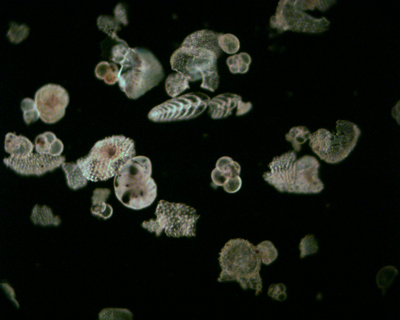
Left:
Forams from Malta. Brian Darnton slide. 9x/0.20 LOMO objective, home
made darkfield stop. |
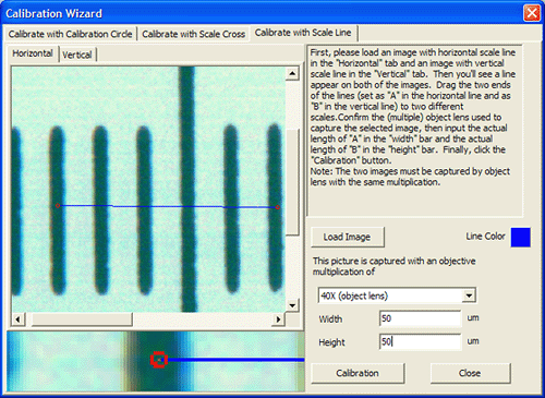
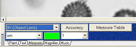
Calibration and measurement
|
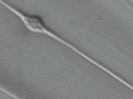
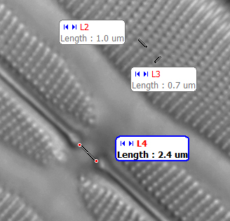
Once this sequence has been carried out for each optic combination it shouldn't need redoing. Circle calibrations are autodetected, For scale cross or lines a zoomed in area of the line set is shown to allow accurate placing of the calibration line (see right). |
|
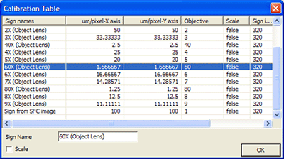
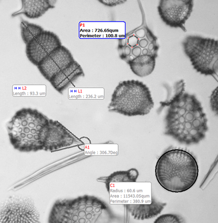
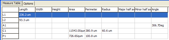
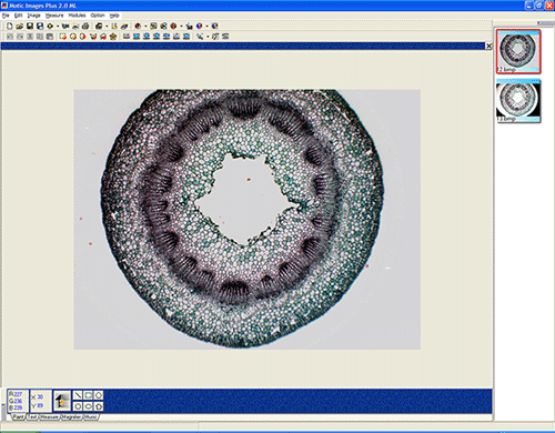
Measurements are automatically presented on the image as shown in the example above of a radiolaria strew and are retained e.g. if saved in *.bmp format. An example of the measurement of lines, angles, polygons and circles are shown, the software auto-assigns sequential codes for each measurement depending on its type. A sub-menu can alter the 'look' of the labels. All measurements are saved in a measurement table (the one for the example is below). The table data can be exported if desired e.g. in spreadsheet format.
Measurements of fine features such as diatom detail are possible e.g. in the diatom above right. When an objective is calibrated the software should give more accurate results than the micrometer slide, eyepiece reticle combination. The seamless integration of image capture and measurement features could be of particular interest e.g. to the enthusiast repetitively measuring organisms / features under the microscope, where the individual transcription of calibrated eyepiece reticule measurements or incorporation of accurate measuring scales onto say a consumer digicam image capture can involve a lot of work. |
Image analysis
|
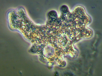
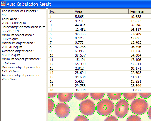
Image analysis is not an area the author is very familiar with, but could be useful for example counting discrete objects in a given field. I couldn't find a way of changing the autodetect parameters so that edges were correctly detected. e.g. in this example the edge detection is omitting the centre of some cells which would affect measurements. The analysis results can be exported in text or spreadsheet format. Third party freeware like 'ImageJ' can also offer these sort of image analysis features although calibration would need to be manual. |
Macro imaging
|
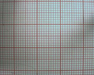
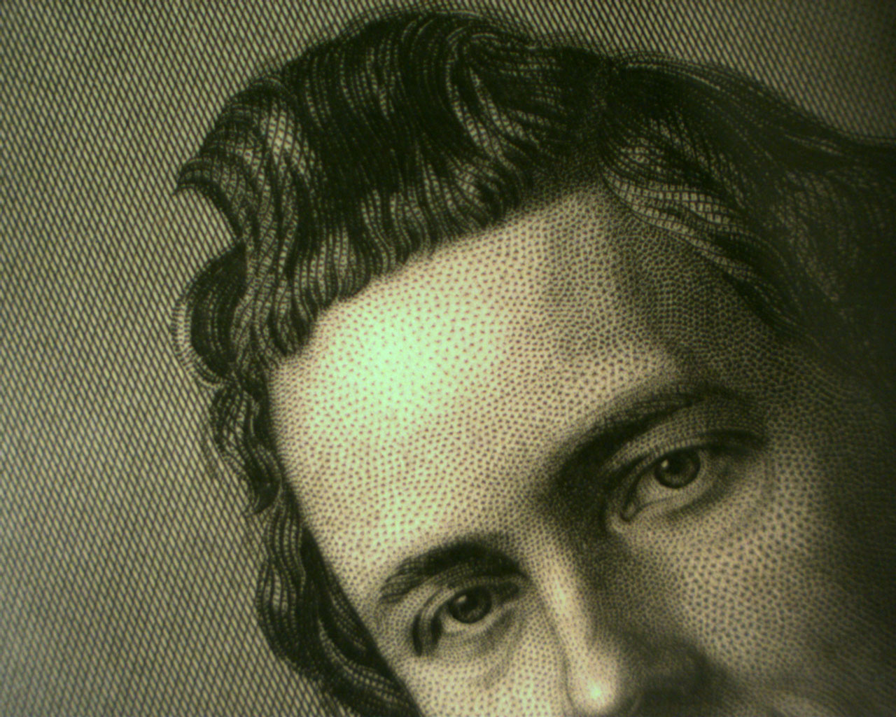
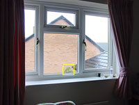
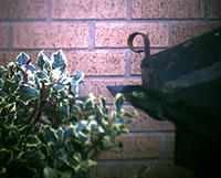
As illustrated in the 'Out of the box' section earlier, the camera with relay lens can be attached to the supplied clear plastic macro support for fixed distance imaging of flattish subjects. I found it a little fiddly to use because there's no lens focus lock so the entire mount moved when focussing, with the USB cable also tugging at unit and focus set. An image of 1 mm / 1 cm graph paper shown left above captures an area ca. 4.2 x 3.3 cm ; some barrel distortion is present with softening towards edges and corners. It is supplied with its own calibration disc for measuring macro objects so a useful way of extending still and video image capture for suitable larger subjects. Right above: detail of an original engraving of Paul Morphy by D J Pound. Click image for master 1.3 Mpixel capture. The four adaptors and relay lens supplied enable it to be readily connected to other optical items. A quick try of the webcam with relay lens on 10x40 binoculars gave no vignetting. It shows some promise for 'digiscoping' although the limited dynamic range may not cope with contrasty scenes. Left - visual field with Sony S75 digicam, yellow box shows binocular / Moticam view on right. May be useful for moon shots, but clear nights are few in this neck of the woods in winter!
|
Video or time lapse capture
|
Motic's stated maximum frame rates for the Moticam 1000 are '10fps @ 1240x1024 , 30fps @ 640x480'. Video is captured by pressing the button in the capture screen. Videos are saved as avi with either no compression or one of the compression codecs shown can be chosen. The codecs can be configured where appropriate. Needless to say, full frame video stored uncompressed will fill a hard drive rather quickly!
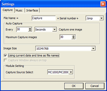
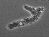
'Auto Capture' is also offered which is configured in a Motic Images Plus menu shown right. Seconds, minutes or hours between image captures can be selected. Saving in bmp format would allow third party software to assemble captures into a time lapse video if desired. A short real time video of a pond life critter is below left, a telotroch I believe, the motile stage of a vorticella. Cropped and compressed from master, phase contrast. Click to view, 970kbyte avi. The limitations of the dynamic range is evident on the bright edges of this phase image where the highlights tend to burn out. Below right shows the usefulness of the Auto Capture function. Stills were auto captured of an amoeba every 6 seconds (LOMO 20x NA0.4 phase, no eyepiece) which are autosaved as bitmaps. Third party software like 'bmp2avi' can assemble them into an avi video (440 kbyte compressed) which in this case is 5 frames per sec. This allows much less space to be taken up cf full 15fps video as well as speeding up slowish movement, in this case by a factor of 30x. The file is resized from the 800x600 master. Free editing software like VirtualDub offers plenty of facilities for resizing, cropping and compressing.
|
Other features of this versatile camera / software package include:
'Distant
Imaging Sharing Module': Allows
owners of the same Moticam camera / software to share captured video or images
in real time over the Internet via Microsoft Net Meeting. So, for example, a
friend the other side of the world could help identify a live pond critter while
it's under the microscope. (Users of a webcam on a microscope with video
conferencing software could provide a similar capability.)
'Motic
Report': Captured and edited images
can be ported into a Motic Report screen which allows text addition and basic
page layout to create a report which are saved in a *.mp Motic Report format
(unsure if exportable). Could be useful for e.g. quickly preparing a report
with images and text for sharing in the
above module with others without the need for external editing software.
Menu
choice: the menus shown above are 'Standard' a simpler fun 'Plus' screen
can be chosen ... music can be played in the background as well.
Conclusions
This is the first dedicated compound microscope camera the author has used and have only tried it on a LOMO Biolam so image quality is dependent on the quality of the LOMO optics/illumination. Its performance was compared with the author's/brother's impressions of past/current imaging routes on the same type of microscope with a webcam, video cameras with 'C' mount, consumer digicams and 35mm film work/scanning. On this basis ....
Strengths:
-
good value for money overall for a dedicated camera/software as the relay lens,
micrometer slide and four couplings are also supplied
- useful for microscopes with no trinocular head as the camera is small, light and
compact
- quick interchange between eyepiece capture and in-tube capture
-
zero lag critical focussing at 1240x1024 a joy cf. consumer digicams
- quite
good
quality captures when subject well lit and if not too contrasty
- camera
integrates well with supplied software, rapid multiple measurements possible
Weaker
aspects:
- no focus lock on relay lens; can lose focus in micro and macro
imaging as unit swings round
- dynamic range seems less than consumer digicam,
can struggle with highlights on contrasty subjects
- maker's quoted sensitivity of
3 lux can be limiting for e.g. phase, polar etc. if the microscope isn't fitted with a high power lamp
-
not a TWAIN camera so must be used with supplied software. Unsure if Mac versions.
(Update: see footnote on TWAIN and Mac.)
The Moticam 1000 is convenient and easy to use; the well specified outfit allowing it to be used with a variety of microscopes, with or without eyepiece at no extra expense. If the author's (often frustrating!) experiences of digital photomicrography to date are typical, convenience can often be the key feature rather than ultimate quality. During a session studying pond life for example, if an interesting critter is spotted, the camera can be attached in seconds and stills or video taken. Measurements can be taken at leisure afterwards on the image capture. A USB webcam (<$100) can provide similar convenience much more cheaply and is a popular option; the Moticam's main benefits for the extra price would arguably be 4x the resolution of a native VGA webcam (0.3 Mpixels), the integrated software/measurement facility, relay lens and adaptors.
In the digital microscope camera market it seems very competitively priced at ca. $399. The Moticam 2000 (ca. $899) offers 2 megapixels with the same 3 lux sensitivity of the Moticam 1000. The PAXcam EDU 1.3 megapixel model is more expensive (ca. $1200 although with a higher spec. chip, higher video frame rates and more sensitive i.e. 1.2 lux, PAXcam's spec) and supplied with the powerful 'PAX-it' software.
The Moticam 1000 is perhaps more likely to suit the user requiring images for e.g. the web, small prints and especially where the image capture convenience with critical focussing is needed for recording and measurement. For higher quality larger digital images or prints the 1.3 Mpixel Moticam 1000 can't be expected to match the multi-megapixel consumer digicams available if a model is chosen that can be interfaced with and works well with a microscope. The author's own requirement for multi-megapixel photomicrographs are quite infrequent and currently uses 35mm film with slide scanning to give up to 10 Mbyte images when needed.
Update May 2008 The review is three years old now and progress is very rapid in digital imaging technology. As remarked in the review, the Moticam 1000 2005 model has limited sensitivity and also dynamic range compared with some consumer digicams. Motic have kindly told me that 'the chips inside the Moticam 1000 are updated from time to time, either on our input or from the chip manufacturer'. ... 'The most current chip is still a 1.3MP chip but the size has changed to 1/3" '. The camera is XP/Vista as well as OSX compatible. The dynamic range and sensitivity to light have been vastly improved since 2005'. So the reviewer's comments on the imaging quality refer only to the 2005 model.
As an aside I use constantly the excellent adapters and the relay lens for other purposes. The spare adapters can be temporarily glued end to end to attach small bore consumer digicams (shown in use here) like a Sony P200 to either a stereo or compound microscope with microscope eyepiece in place. I use the relay lens with other 'C' mount cameras like the good value USB monochrome astronomy type for highest mag studies where colour isn't crucial.
As an alternative to a Moticam style camera, in eyepiece tube USB microscope cameras are readily available now and good value, but from user's reviews perhaps best to keep to modest 1.3 Mpixels designs and ensure they have a relay lens, e.g. see Paul Wilton's Micscape review of two eBay sourced microscope cameras.
If measuring software with calibration features are required there are also excellent freeware or shareware software packages like Marien van Westen's Micam software which seem to work with a variety of USB cameras.
Comments to the author David Walker are welcomed.
Footnote: Thank you to Reto Wieland of Motic who kindly told me that TWAIN software for the Moticam 1000/2000 should be available/downloadable from the manufacturer's 'new digital web site' probably after April. Also for his comments on how the Moticam 1000 has been updated since this review.
Links
National Optical offers a useful downloadable supplement on using the Moticam and notes problems interfacing with some computer equipment/software combinations. Provides advice on laptop specs.
Martin Microscope Company maintains an invaluable resource which compares a variety of consumer and dedicated still/video camera options for photomicroscopy.
Acknowledgement: All diatom images were taken of Klaus Kemp's invaluable 'Test plate 8 forms', the diatoms are mounted in hyrax. See Klaus Kemp's website for the range of slides available to purchase.
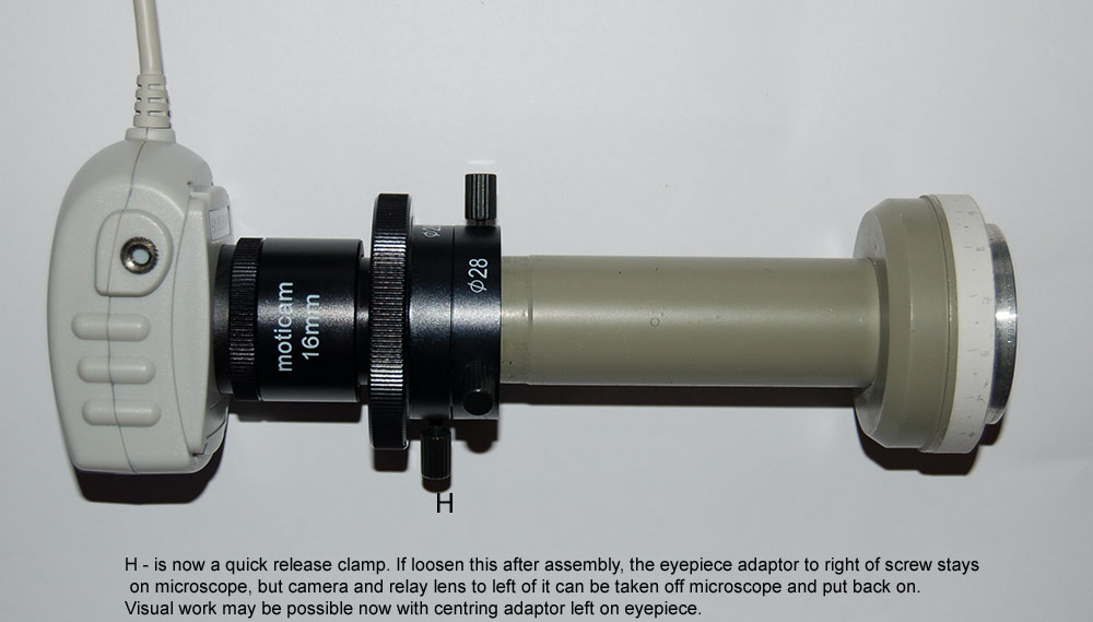
Footnote November 2008: To clarify the assembly procedure, the unit before and after assembly are shown below.
In this example, the eyepiece has little depth and adaptor slips past eyepiece to clamp on the microscope tube. Some eyepieces, especially modern ones, can be much deeper in which case the adaptor clamps onto eyepeice. The adaptor should sit flush on top of eyepiece before clamping.
The so-called 'C mount' on camera and relay lens is an industry standard, so if desired a simple adaptor can be bought from most microscope dealers to insert camera directly into eyepiece tube as shown earlier. This gives a sensor crop of the full eyepiece field but can be useful for studying fine detail in some subjects such as diatoms.
Published in the March 2005 edition of Micscape.
Please report any Web problems or offer general comments to the Micscape Editor .
Micscape is the on-line monthly magazine of the Microscopy UK web site at Microscopy-UK
© Onview.net Ltd, Microscopy-UK, and all contributors 1995
onwards. All rights reserved.
Main site is
at www.microscopy-uk.org.uk
with full mirror
at www.microscopy-uk.net
.
 Full
screen of Motic Images Plus 2.0. Captured images appear in the right
hand vertical box. Each can be clicked to bring into the image editing
area.
Full
screen of Motic Images Plus 2.0. Captured images appear in the right
hand vertical box. Each can be clicked to bring into the image editing
area.