One of the pleasures of studying the
macroscopic and microscopic world, is that even the most
common plants can provide a wealth of interesting features
when viewed in 'close-up'.
Just out of interest, I chose one of the commonest wild flowers in bloom at the time of writing - a dandelion, and spent a few hours examining and imaging the plant on both the macroscopic and microscopic scale. It's an interesting exercise as the most familiar plants can often be dismissed as too commonplace for detailed study.
Macroscopic tour
The first two images were taken with a Fuji DX-10 digital camera. The larger macro images were taken with a security video camera fitted with a 50mm SLR lens and extension tubes.
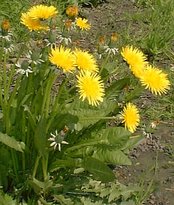
The grasslands around the author's north of England home have been drained and 'improved' for sheep and cattle. One of the few flowers that does grow with impunity in these 'green deserts' is the dandelion, and the splash of colour is a welcome sight. The flowerheads are ca. 35-50mm across. It grows all year but April-June in particular. It also grows well in garden borders and lawns, so is often viewed as a weed and is hard to eradicate with its deep tap roots. |
|
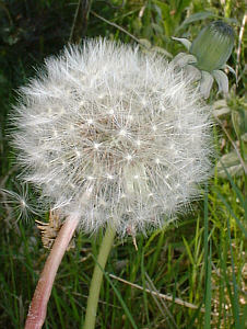
The dandelion is possibly most well known for its attractive seedhead. An unopened flower bud is shown in the background. |
|
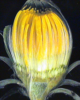
The dandelion (genus Taraxacum) is a member of the Family Compositae - the 'daisy family'. So-called because the flowerheads are not a single flower but a composite of flowers called florets. Cutting vertically through an unopened bud shows this clearly, to show the immature individual florets. For amateur naturalists like the author, identifying flowers to species in the Taraxacum genus is difficult, as its a variable group and there are many microspecies. |
|
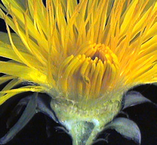
Cutting vertically through an opened flowerhead shows the florets in more detail. These are called ray florets as they each have a single petal. In some Compositae which have a central disc (e.g. the common daisy), there are two type of florets; the outer ray florets each with a petal and disc florets without a petal, the latter which occupy the centre of the flower. Note the milky latex which exudes from the stem when cut. |
|
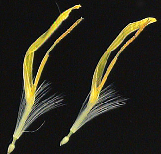
If the flowerhead base is opened out, individual florets can be gently pulled out. Two are shown right. They are attractive subjects for the stereo microscope and have a beauty of their own. Note the bi-lobed stigma covered in pollen (Shown magnified below). The florets are ca. 18mm long. The immature single seeds are seen at the floret bases. |
|
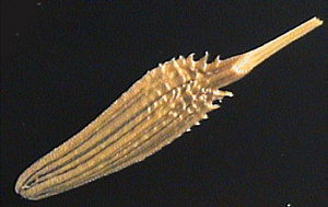
| The flowerhead with all the seeds removed shows the arrangement of how the seeds are attached. |
|
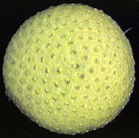
| Close inspection of a single seed (or achene) reveals interesting surface features. The seed is ca. 4mm long. The stalk with 'parachute' (the pappus) has been removed. |
|
Microscopic tour
Imaging:
A security video camera was attached without eyepiece to a
Russian Biolam microscope.
Achromatic 9x, 20x objectives were used.
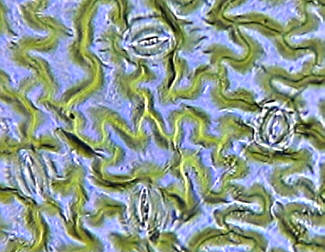
In the Micscape article 'Comparison of leaf epidermis in different species', Anne Bruce describes a neat tip; painting clear nail varnish on plant leaves can form a cast of the leaf surface. This cast when dry can be peeled off and mounted under a coverslip. This was done by the present author with the upper side of a dandelion leaf. Four stomata are shown in the leaf cast shown right as well as the cellular detail of the leaf epidermis. (Objective 9x, brightfield). Stomata ca. 35mm long. |
|
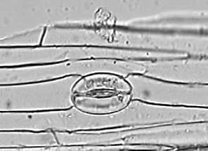
The dandelion flower stem is hollow and quite brittle. Splitting a stem and gently breaking it back on itself can allow a small section of the stem epidermis to be peeled off. If a piece is cut off and mounted under a coverslip in water, the thin section is only a few cells thick and shows the long thin cells of the stem epidermis. Even the occasional stomata can be found, shown right. (Objective 20x, brightfield). Stomata ca. 37mm long. |
|
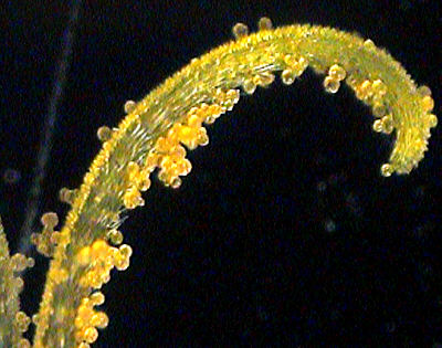
One of the two lobes of the stigma with pollen. Compound microscope with 3.5x objective (no eyepiece) using incident light. |
|
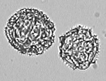
Dandelion pollen washed in isopropanol then hydrated and mounted in 50% water/glycerine. The 3D structure of the pollen is fascinating but hard to convey in this still image. The depth of field is small and focusing through the grains is required to appreciate the structure. Objective 20x. Pollen ca. 46mm in diameter. Click here for a Micscape article on studying pollen. |
|
So I hope the above image gallery demonstrates that even the humblest 'weed' or wild flower can provide a wealth of macroscopic and microscopic detail worth studying; either just for fun and/or for learning more about the structure of plant groups.
Why not try a study of a common flowering plant/'weed' local to you ... the ordinary may prove extraordinary in close-up!
Comments to the author Dave Walker are welcomed.
Microscopy UK Front Page
Micscape Magazine
Article Library
© Microscopy UK or their contributors.
Published in the May 2000 edition of Micscape Magazine.
Please report
any Web problems or offer general comments to the Micscape Editor,
via the contact on current Micscape Index.
Micscape is
the on-line monthly magazine of the Microscopy UK web
site at Microscopy-UK